. 2012 Jun 12;109(24):9366-71.
doi: 10.1073/pnas.1206840109. Epub 2012 May 24.

In vitro loading of human cohesin on DNA by the human Scc2-Scc4 loader complex

Affiliations

In vitro loading of human cohesin on DNA by the human Scc2-Scc4 loader complex

Vladimir P Bermudez et al. Proc Natl Acad Sci U S A. .

Abstract

The loading of cohesin onto chromatin requires the heterodimeric complex sister chromatid cohesion (Scc)2 and Scc4 (Scc2/4), which is highly conserved in all species. Here, we describe the purification of the human (h)-Scc2/4 and show that it interacts with h-cohesin and the heterodimeric Smc1-Smc3 complex but not with the Smc1 or Smc3 subunit alone. We demonstrate that both h-Scc2/4 and h-cohesin are loaded onto dsDNA containing the prereplication complex (pre-RC) generated in vitro by Xenopus high-speed soluble extracts. The addition of geminin, which blocks pre-RC formation, prevents the loading of Scc2/4 and cohesin. Xenopus extracts depleted of endogenous Scc2/4 with specific antibodies, although able to form pre-RCs, did not support cohesin loading unless supplemented with purified h-Scc2/4. The results presented here indicate that the Xenopus or h-Scc2/4 complex supports the loading of Xenopus and/or h-cohesin onto pre-RCs formed by Xenopus high-speed extracts. We show that cohesin loaded onto pre-RCs either by h-Scc2/4 and/or the Xenopus complex was dissociated from chromatin by low salt extraction, similar to cohesin loaded onto chromatin in G(1) by HeLa cells in vivo. Replication of cohesin-loaded DNA, both in vitro and in vivo, markedly increased the stability of cohesin associated with DNA. Collectively, these in vitro findings partly recapitulate the in vivo pathway by which sister chromatids are linked together, leading to cohesion.


Conflict of interest statement

The authors declare no conflict of interest.

Figures
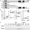
Fig. 1.
Fig. 1.
Isolation of full-length h-Scc2/4 and derivatives. (A) Schematic diagram of h-Scc2 showing conserved residues among species and h-Scc2 derivatives expressed and purified in insect cells. (B) h-Scc2 derivatives and h-Scc4 expressed and purified from insect cells. Shown is Coomassie blue-stained SDS/10% polyacrylamide gels containing 0.3 μg of protein from glycerol gradient (GG) peak fractions of His-Flag Scc2 (N500)/Scc4 (GG#28), His-Flag Scc2 (N1500)/Scc4 (GG#22), and Scc2 (N500 + 1,742–2,376 aa/HEAT)/His-Flag Scc4 (GG#24). The roman numerals listed above the lanes correspond to the schematic diagram in A. (C) HeLa-expressed Scc2/His-Flag Scc4 (0.5 μg) eluted from Heparin column was analyzed by SDS/6.5% PAGE, followed by silver staining (Left) and Western blotting with h-αScc2 and h-αScc4 (Right). (D) Heparin-eluted Scc2/His Flag Scc4 (20 μg) was loaded onto 15–40% (vol/vol) glycerol gradient as described in SI Materials and Methods. Shown is the Western blot (to facilitate visualization of the Scc2 and Scc4 bands) of the glycerol gradient fractions.
Fig. 2.
Fig. 2.
Interactions between Scc2/4, cohesion, and cohesin derivatives. (A) SDS/PAGE analysis of the cohesin complex and various subunits isolated as described in the SI Materials and Methods. Lanes 1–3 contained 0.5 μg of the indicated protein, whereas lanes 4 and 5 contained 0.75 μg of protein. Following SDS/10% PAGE separation, gels were stained with Coomassie blue. (B) Full-length h-Scc2/4 interacts with h-cohesin. The indicated Scc2/4 derivatives and h-cohesin (1 pmol of each) were mixed and incubated at 25 °C for 30 min, and the mixture immunoprecipitated as described in SI Materials and Methods. Protein complexes precipitated with the indicated antibodies (IgG, αScc4, and αSmc3) were separated by SDS/PAGE and then Western blotted with h-αScc1 antibodies. The level of protein pulled down by the antibodies was quantified as indicated. (C) Full-length Scc2/4 binds to the Smc1/Smc3 heterodimer. In vitro-transcribed and -translated [35S]methionine-labeled cohesin subunits were mixed with purified full-length Scc2/4 and incubated at 25 °C, followed by IP with antibodies (IgG and αScc2). The Scc2/4-bound material was eluted with SDS loading buffer and separated by SDS/10% PAGE, followed by autoradiography to visualize radiolabeled cohesin subunits.
Fig. 3.
Fig. 3.
h-Scc2/4 and h-cohesin are loaded onto X-pre-RCs. (A) Reaction mixtures (150 μL), as described in SI Materials and Methods, containing Xenopus HSS extract, 4 pmol of h-cohesin, 1 pmol of full-length h-Scc2/4, and 400 ng of biotinylated dsDNA bound to magnetic beads in the presence or absence of geminin (400 nM) were incubated for 30 min at 23 °C, and the dsDNA beads were collected and washed as described in the SI Materials and Methods. Proteins associated with the biotinylated dsDNA magnetic beads were eluted with 20 μL of SDS loading buffer and applied to SDS/7% PAGE gel and subjected to Western blotting with the indicated antibodies. (B) Purified full-length h-Scc2/4 supports loading of h-cohesin onto X-pre-RCs. X-Scc2 was depleted from Xenopus HSS extracts using X-Scc2 antibodies as described previously (4). The X-Scc2-depleted HSS extract was supplemented with 1 pmol of full-length h-Scc2/Scc4 and used to assemble the pre-RC in the presence or absence of h-cohesin. Antibodies against h-Scc2, FLAG (for Scc1), and Mcm2 were used to visualize proteins after Western blotting.
Fig. 4.
Fig. 4.
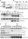
Salt stability of proteins loaded onto pre-RCs by Xenopus HSS extracts. (A) Loading of h-cohesin and h-Scc2/4 onto high-salt-washed X-pre-RC on dsDNA magnetic beads pre-RCs that were washed with 100 mM KCl buffer (ELB) were incubated with geminin-treated HSS and 4 pmol of h-cohesin or 1 pmol of h-Scc2/Scc4 for 30 min at 23 °C. The dsDNA magnetic beads were then washed with ELB, and the proteins bound to DNA were analyzed by SDS/PAGE and Western blotting. (B) X-pre-RC was assembled on dsDNA magnetic beads as described previously (21). The bead-associated complex was washed with 50, 75, or 100 mM KCl in ELB buffer, and the proteins that remained bound to the beads were analyzed by Western blotting. (C) Replication catalyzed by Xenopus NPE leads to the stabilization of loaded cohesin. Reactions (40 μL) were incubated with Xenopus HSS extracts in the presence of a biotinylated-circular 2.5-kb plasmid at 22 °C or 30 min to assemble pre-RCs and load X-cohesin onto the DNA. Mixtures were then supplemented with NPE and [32P]dATP. Kinase interacting protein (Kip)1 (p27, 0.1 μM) was added to inhibit DNA synthesis, where indicated. DNA synthesis, the determination of level of DNA present in each reaction with SYBR Gold staining, autoradiography, KCl extraction, and immunoblotting were carried out as described previously (24). (D) Singly biotinylated 3.0-kb plasmid was immobilized on Sepharose beads and incubated in HSS at 40 ng/μL for 30 min at 22 °C. NPE supplemented with [α-32P]dCTP in the presence of 50 μg/mL aphidicolin (lanes 1–5) or DMSO (control: lanes 6–10) was added into the reaction mixtures and incubated for 2 h. The DNA beads were recovered and washed with buffers containing the indicated KCl concentrations. Bound proteins were eluted in SDS loading buffer and separated in SDS/PAGE, and the protein bands were visualized by Western blotting using the indicated antibodies. DNA was detected by using SYBR Gold, and nucleotide incorporation [α-32P]dCMP was measured by BAS2000 phosphoimager (Fujifilm).
Fig. 5.
Fig. 5.
Stability of cohesin associated with chromatin during the cell cycle. (A) Proteins extracted from the chromatin fraction at different salt (NaCl) concentrations and phase of the cell cycle were immunoblotted with antibodies specific for Scc1 (cohesin), Smc3, Ac-Smc3, Scc4 (Scc2/4), or Orc2 (ORC). The preparation of cells and conditions used were as described in the SI Materials and Methods. (B) The level of chromatin bound cohesin (Scc1) detected in the Western blot shown in A was quantified and normalized to the amount of ORC (0.1 M NaCl extract). The percentage of cohesin (Scc1), designated as weak (extracted with 0.1 plus 0.4 M NaCl), and stably associated with chromatin (extracted with 1 M NaCl plus sonication), indicated as strong, are plotted as a function of the cell cycle.

References

    1. Onn I, Heidinger-Pauli JM, Guacci V, Unal E, Koshland DE. Sister chromatid cohesion: a simple concept with a complex reality. Annu Rev Cell Dev Biol. 2008;24:105–129. - PubMed
    1. Gruber S, Haering CH, Nasmyth K. Chromosomal cohesin forms a ring. Cell. 2003;112:765–777. - PubMed
    1. Nasmyth K, Haering CH. Cohesin: its roles and mechanisms. Annu Rev Genet. 2009;43:525–558. - PubMed
    1. Takahashi TS, Yiu P, Chou MF, Gygi S, Walter JC. Recruitment of Xenopus Scc2 and cohesin to chromatin requires the pre-replication complex. Nat Cell Biol. 2004;6:991–996. - PubMed
    1. Nasmyth K. Cohesin: a catenase with separate entry and exit gates? Nat Cell Biol. 2011;13:1170–1177. - PubMed

Publication types