

Comment
doi: 10.1161/CIRCRESAHA.112.268961.
Biased DNA segregation during stem cell division
Affiliations
- PMID: 22628572
- PMCID: PMC3407569
- DOI: 10.1161/CIRCRESAHA.112.268961
Item in Clipboard
Comment
Biased DNA segregation during stem cell division
Circ Res.
.
Erratum in
-
Correction.Circ Res. 2015 Dec 4;117(12):e130. doi: 10.1161/RES.0000000000000085. Circ Res. 2015. PMID: 26635384 No abstract available.
Abstract
Adult skeletal muscle stem cells are a heterogeneous cell population characterized by a small subset of undifferentiated cells that express at high level the paired/homeodomain gene Pax7. This category of satellite cells divides predominantly by asymmetric chromatid segregation generating a daughter cell that carries the mother DNA and retains stem cell property, and a daughter cell that inherits the newly-synthesized DNA and acquires the myocyte lineage.
Figures
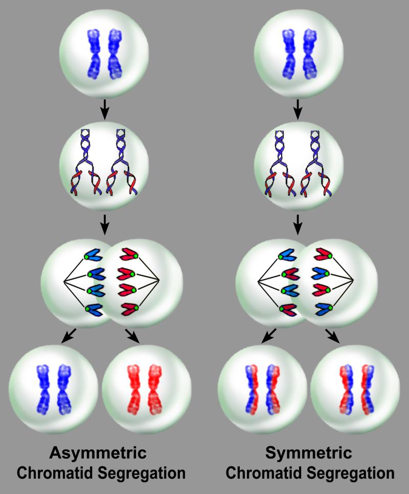
With asymmetric chromatid segregation, a dividing mother stem cell synthesizes new DNA during S-phase and generates two daughter stem cells, one carrying only the mother DNA, which is the true stem cell, and the other only the newly-synthesized DNA. With symmetric chromatid segregation, a dividing stem cell synthesizes new DNA and generates two daughter stem cells, each carrying the mother DNA and the newly-synthesized DNA.
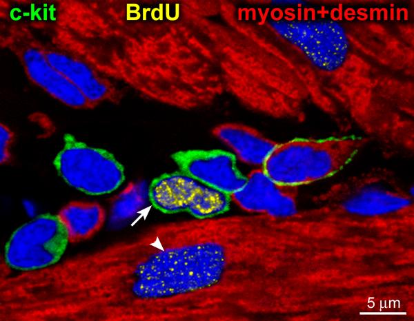
Five c-kit positive cells (green) surrounded by cardiomyocytes (myosin and desmin, red). One of the 5 c-kit-positive cells is brightly labeled by BrdU (yellow, arrow). A dimly labeled cardiomyocyte nucleus (arrowhead) is also present. The brightly BrdU-labeled c-kit-positive CSC may reflect a parent stem cell derived from a BrdU-negative grandparent stem cell that incorporated the halogenated nucleotide during the first division. Subsequently, the BrdU-labeled parent stem cell entered the G0/G1 phase. Adapted from reference .
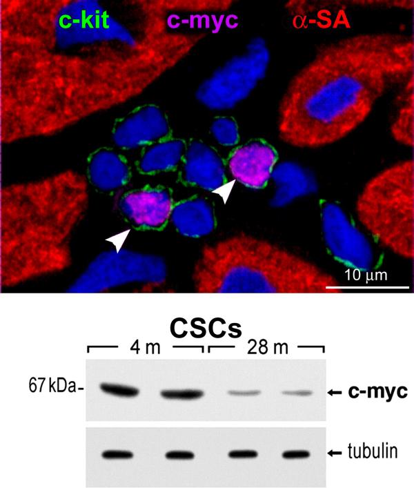
Cardiac stem cell niche in a young rat heart contains eight c-kit-positive CSCs (green); two express c-myc (magenta, arrowheads). Nuclei are stained by DAPI. The attenuation in the expression of c-myc in CSCs with age is apparent by Western blotting. m, months. Tubulin, loading conditions.
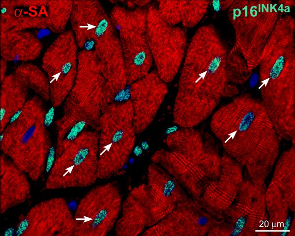
The myocardium of a 79 years old man is characterized by cardiomyocytes (α-sarcomeric actin, α-SA, red) that express the senescence-associated protein p16INK4a (green, arrows).
Comment on
-
A subpopulation of adult skeletal muscle stem cells retains all template DNA strands after cell division.Cell. 2012 Jan 20;148(1-2):112-25. doi: 10.1016/j.cell.2011.11.049. Cell. 2012. PMID: 22265406
References
-
- Rocheteau P, Gayraud-Morel B, Siegl-Cachedenier I, Blasco MA, Tajbakhsh S. A subpopulation of adult skeletal muscle stem cells retains all template DNA strands after cell division. Cell. 2012;148:112–125. - PubMed
-
- Rando TA. The immortal strand hypothesis: segregation and reconstruction. Cell. 2007;129:1239–1243. - PubMed
-
- Lansdorp PM. Immortal strands? Give me a break. Cell. 2007;129:1244–1247. - PubMed
-
- Merok JR, Lansita JA, Tunstead JR, Sherley JL. Cosegregation of chromosomes containing immortal DNA strands in cells that cycle with asymmetric stem cell kinetics. Cancer Res. 2002;62:6791–6795. - PubMed
Publication types
Grants and funding
- R01 AG017042/AG/NIA NIH HHS/United States
- R01 HL111183/HL/NHLBI NIH HHS/United States
- R01 HL075480/HL/NHLBI NIH HHS/United States
- R01 HL114346/HL/NHLBI NIH HHS/United States
- P01 HL092868/HL/NHLBI NIH HHS/United States
- R01 AG037490/AG/NIA NIH HHS/United States
- R01 HL039902/HL/NHLBI NIH HHS/United States
- R01 HL105532/HL/NHLBI NIH HHS/United States
- R01 AG037495/AG/NIA NIH HHS/United States
- R01 AG026107/AG/NIA NIH HHS/United States
- R01 HL065577/HL/NHLBI NIH HHS/United States
- R37 HL081737/HL/NHLBI NIH HHS/United States
- P01 AG023071/AG/NIA NIH HHS/United States
- R01 HL065573/HL/NHLBI NIH HHS/United States
LinkOut - more resources
Full Text Sources
