Review
. 2012 May 25;110(11):1525-37.
doi: 10.1161/CIRCRESAHA.111.260026.

Gene regulatory networks in cardiac conduction system development

Affiliations
Review

Gene regulatory networks in cardiac conduction system development

Nikhil V Munshi. Circ Res. .

Abstract

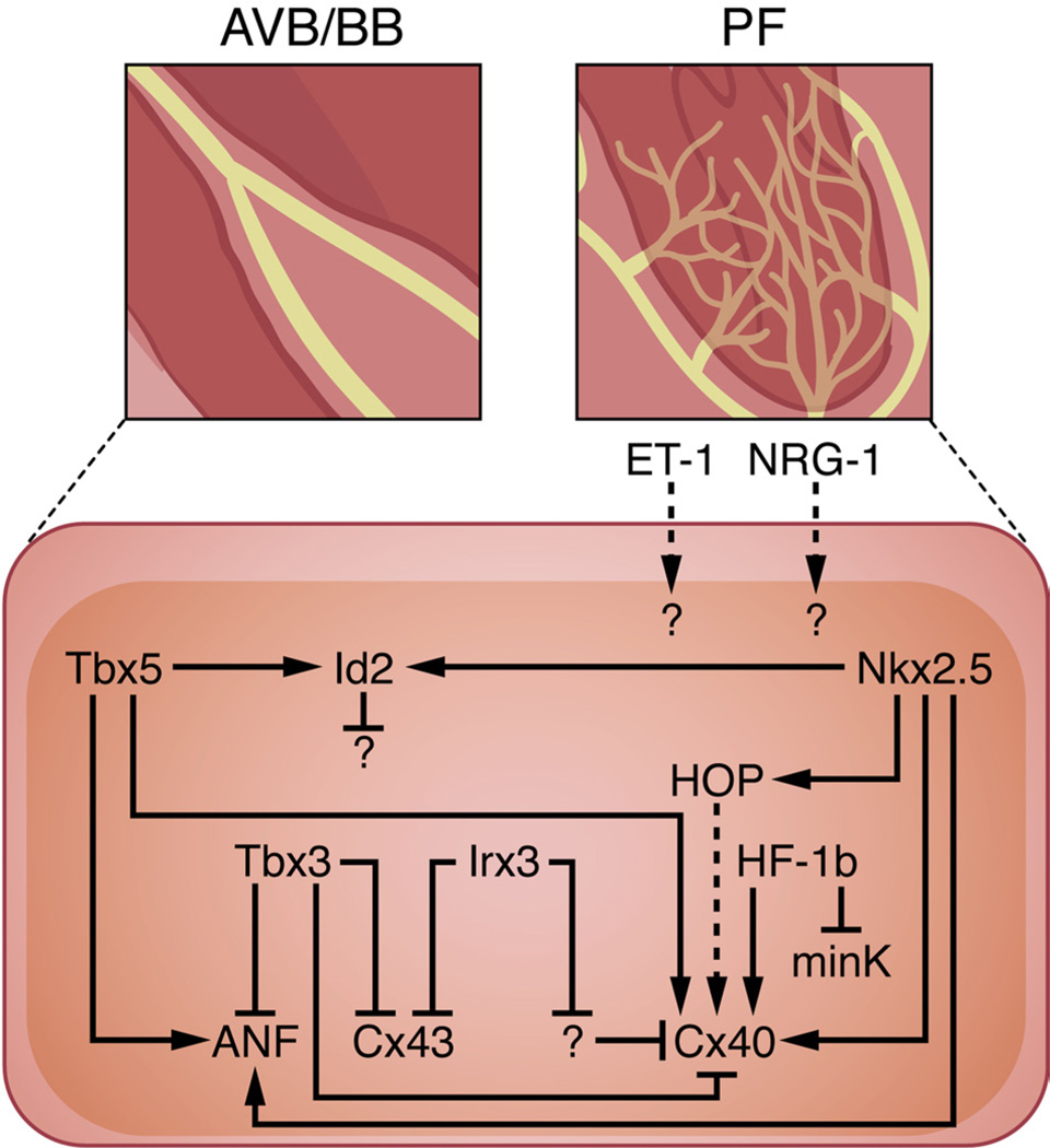
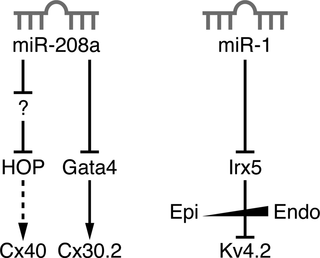
The cardiac conduction system is a specialized tract of myocardial cells responsible for maintaining normal cardiac rhythm. Given its critical role in coordinating cardiac performance, a detailed analysis of the molecular mechanisms underlying conduction system formation should inform our understanding of arrhythmia pathophysiology and affect the development of novel therapeutic strategies. Historically, the ability to distinguish cells of the conduction system from neighboring working myocytes presented a major technical challenge for performing comprehensive mechanistic studies. Early lineage tracing experiments suggested that conduction cells derive from cardiomyocyte precursors, and these claims have been substantiated by using more contemporary approaches. However, regional specialization of conduction cells adds an additional layer of complexity to this system, and it appears that different components of the conduction system utilize unique modes of developmental formation. The identification of numerous transcription factors and their downstream target genes involved in regional differentiation of the conduction system has provided insight into how lineage commitment is achieved. Furthermore, by adopting cutting-edge genetic techniques in combination with sophisticated phenotyping capabilities, investigators have made substantial progress in delineating the regulatory networks that orchestrate conduction system formation and their role in cardiac rhythm and physiology. This review describes the connectivity of these gene regulatory networks in cardiac conduction system development and discusses how they provide a foundation for understanding normal and pathological human cardiac rhythms.


Figures

Figure 1
Figure 1. Functional anatomy of the cardiac conduction system
The cardiac impulse is initiated in the SAN and propagates through the atria. The electric impulse is then delayed in the AVN before rapid conduction through the right and left bundle branches before terminating in the Purkinje fiber network, which directly interfaces with ventricular cardiomyocytes. Distinct time-voltage relationships are noted in the SAN, atria, AVN, PFs, and ventricles. RA indicates right atrium; LA, left atrium; TV, tricuspid valve; MV, mitral valve; RV, right ventricle; LV, left ventricle.
Figure 2
Figure 2. Models for cardiac conduction system development
Two theoretical models are shown that provide a conceptual framework for understanding the mechanisms by which distinct components of the conduction system develop. In the recruitment model (left), a cardiac progenitor cell has the potential to give rise to a conduction cell or a working myocyte. As development proceeds, working myocytes retain bipotential characteristics and can continue to give rise to conduction cells, leading to the concept of ongoing recruitment. In the prespecification model (right), a cardiac progenitor cell can differentiate into a conduction cell or a myocyte, similar to the recruitment model. However, once a cell becomes committed to one of these fates, its progeny remain distinct from each other. During development of the SAN and AVN in particular, Tbx2 and Tbx3 are restricted to the primary myocardium (ie, inflow tract, AVC, outflow tract) and act to locally repress working myocyte differentiation while simultaneously reinforcing the nodal gene expression program. Note that the two models are not necessarily mutually exclusive.
Figure 3
Figure 3. Gene networks in the SAN
Tbx18 is specifically expressed in cells of the SAN “head” region and is required for its morphogenesis in part by repressing Cx43, although the downstream effectors of this pathway remain largely uncharacterized. While Tbx5 activates chamber-specific genes in atrial cells along with Nkx2.5, Tbx5 likely acts as a competence factor for the SAN by activating Shox2 expression, which in turn directly represses Nkx2.5 transcription within this cell type. Nkx2.5 is typically expressed in a mutually exclusive manner in surrounding atrial myocytes, where it represses HCN4 and Tbx3 expression and activates transcription of the chamber-specific genes ANF, Cx40, and Cx43. Tbx3 is also specifically expressed in the SAN and works by preventing expression of chamber-specific genes. Left sinus horn (LSH) pacemaker tissue is prevented from forming by Pitx2, which inhibits Shox2 expression.
Figure 4
Figure 4. Gene networks in the AVN
BMP and Notch signaling are known effectors of AVC and AVN development. BMP2 signals via the Alk3 receptor to activate Smad-dependent genes such as Tbx2; Foxn4 independently activates Tbx2 expression in zebrafish. Tbx5, which is expressed in the developing AVC, upregulates expression of Tbx3, which represses the chamber-specific genes ANF, Cx40, and Cx43 in conjunction with Tbx2, Msx1, and Msx2. While Tbx5 and Nkx2.5 coactivate expression of ANF and Cx40, Tbx5 functions in concert with Gata4 to activate Cx30.2 expression. Notch functions within the AVN to activate Cx30.2 expression either through a direct or indirect mechanism. Notch signaling also acts in a reciprocal manner in atrial and ventricular myocardium adjacent to the AVC by activating transcription of the Hey1 and Hey2 repressors, respectively, which inhibit Tbx2 expression. Moreover, Tbx20, which is expressed specifically in chamber myocardium, directly represses Tbx2 transcription.
Figure 5
Figure 5. Gene networks in the AVB, BB, and PF network
While Nkx2.5 orchestrates morphogenesis of the entire ventricular conduction system, Tbx5 regulates development of the AVB and BB. Both Nkx2.5 and Tbx5 synergistically activate Id2 expression in the developing AVB and BB to mediate cell-cycle exit. Whereas Tbx3 inhibits expression of Cx43, Cx40, and ANF during early AVB development, Tbx5 activates Cx40 and ANF expression during maturation of the AVB and BB. In addition, Nkx2.5 increases Cx40 expression directly by binding to the Cx40 promoter and indirectly by activating HOP in the AVB, BB, and PF network. HF-1b is also expressed within the ventricular conduction system and impacts its electrical properties by simultaneously upregulating Cx40 and downregulating minK levels. Similarly, Irx3 indirectly activates Cx40 expression while directly repressing Cx43 transcription in the developing AVB, BB, and PF network. ET-1 and NRG-1 have been shown to stimulate PF formation in avian and murine systems, respectively, although the downstream effectors of this process remain unknown.
Figure 6
Figure 6. MicroRNA regulation of cardiac conduction
Two miRs have been implicated in regulating cardiac conduction: miR-208a and miR-1. miR-208a directly targets the 3′UTR of Gata4 (a direct activator of Cx30.2), but it activates HOP expression by inhibiting a putative HOP inhibitor. miR-1 directly targets the 3′UTR of Irx5, which is responsible for the normal ventricular repolarization sequence by establishing an epicardial-to-endocardial gradient of Kv4.2 expression.

References

    1. Rubart M, Zipes DP. Mechanisms of sudden cardiac death. J Clin Invest. 2005;115:2305–2315. - PMC - PubMed
    1. Echt DS, Liebson PR, Mitchell LB, et al. Mortality and morbidity in patients receiving encainide, flecainide, or placebo: the cardiac arrhythmia suppression trial. N Engl J Med. 1991;324:781–788. - PubMed
    1. Splawski I, Tristani-Firouzi M, Lehmann MH, Sanguinetti MC, Keating MT. Mutations in the hmink gene cause long QT syndrome and suppress Iks function. Nat Genet. 1997;17:338–340. - PubMed
    1. Keating MT, Sanguinetti MC. Pathophysiology of ion channel mutations. Curr Opin Genet Dev. 1996;6:326–333. - PubMed
    1. Keating MT, Sanguinetti MC. Molecular genetic insights into cardiovascular disease. Science. 1996;272:681–685. - PubMed

Publication types

MeSH terms

Substances