. 2012 Aug;53(8):1522-34.
doi: 10.1194/jlr.M025692. Epub 2012 May 24.

Glycosylphosphatidylinositol anchors regulate glycosphingolipid levels

Affiliations

Glycosylphosphatidylinositol anchors regulate glycosphingolipid levels

Ursula Loizides-Mangold et al. J Lipid Res. 2012 Aug.

Abstract

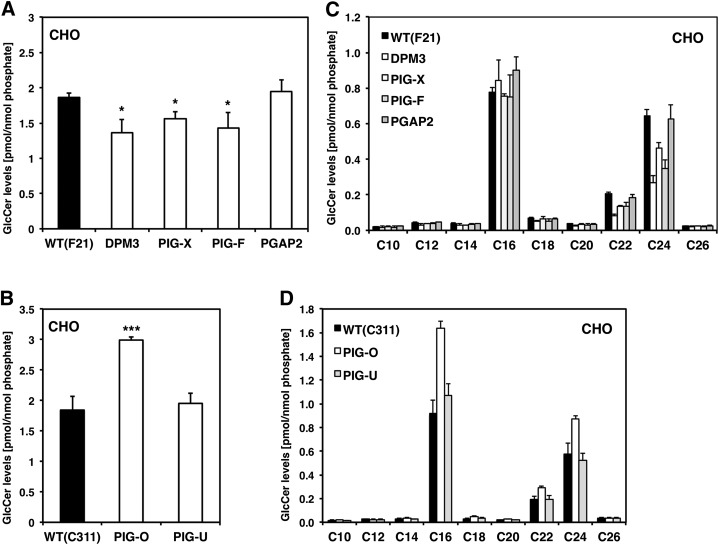
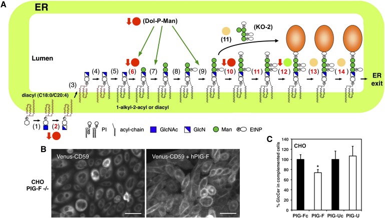
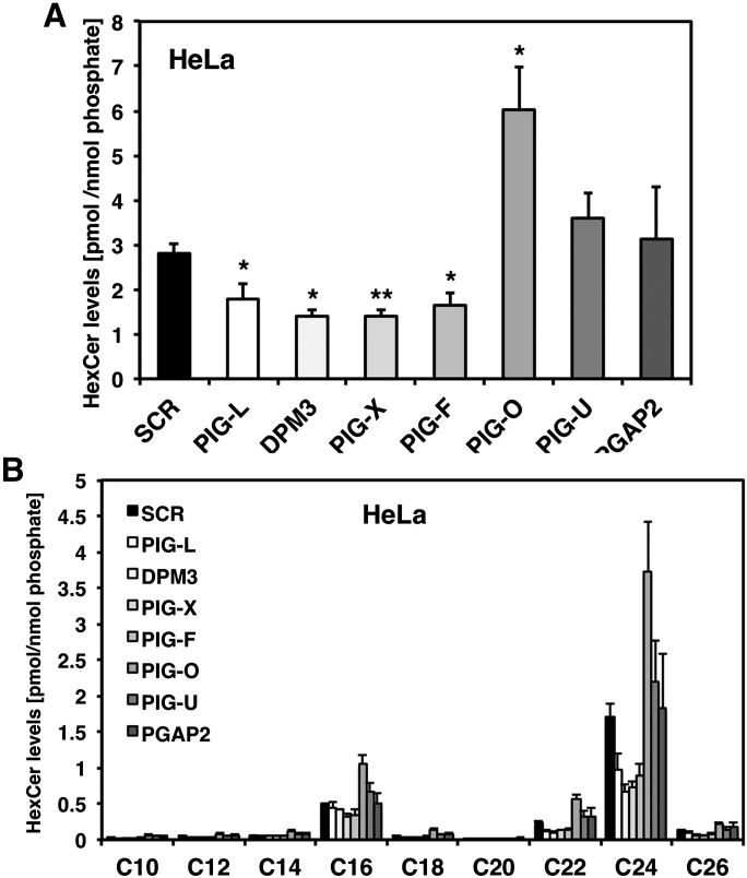
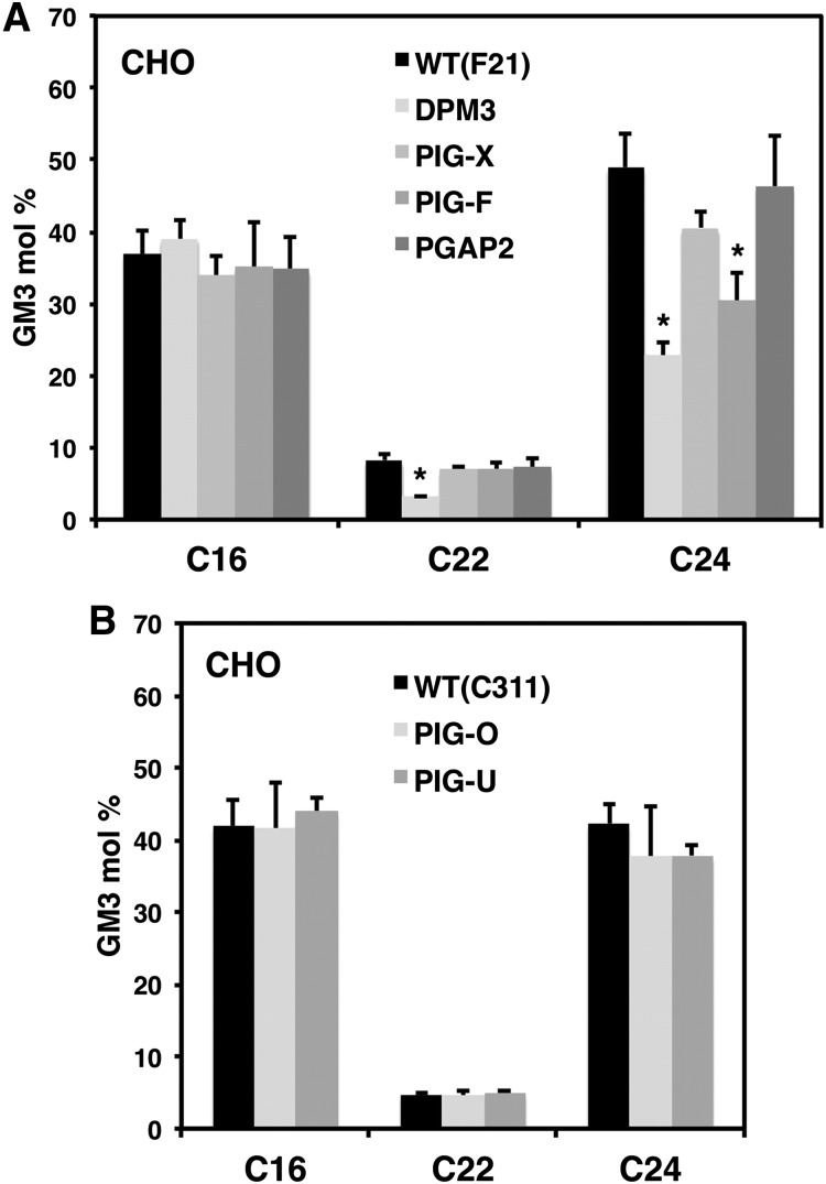
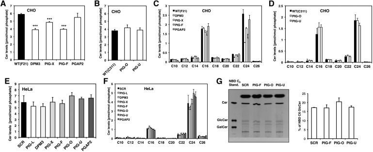
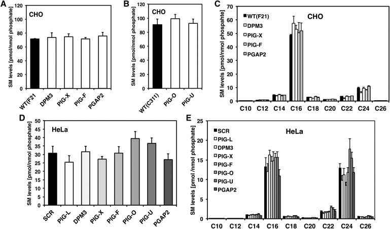
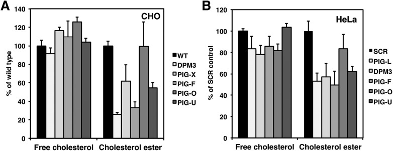
Glycosylphosphatidylinositol (GPI) anchor biosynthesis takes place in the endoplasmic reticulum (ER). After protein attachment, the GPI anchor is transported to the Golgi where it undergoes fatty acid remodeling. The ER exit of GPI-anchored proteins is controlled by glycan remodeling and p24 complexes act as cargo receptors for GPI anchor sorting into COPII vesicles. In this study, we have characterized the lipid profile of mammalian cell lines that have a defect in GPI anchor biosynthesis. Depending on which step of GPI anchor biosynthesis the cells were defective, we observed sphingolipid changes predominantly for very long chain monoglycosylated ceramides (HexCer). We found that the structure of the GPI anchor plays an important role in the control of HexCer levels. GPI anchor-deficient cells that generate short truncated GPI anchor intermediates showed a decrease in very long chain HexCer levels. Cells that synthesize GPI anchors but have a defect in GPI anchor remodeling in the ER have a general increase in HexCer levels. GPI-transamidase-deficient cells that produce no GPI-anchored proteins but generate complete free GPI anchors had unchanged levels of HexCer. In contrast, sphingomyelin levels were mostly unaffected. We therefore propose a model in which the transport of very long chain ceramide from the ER to Golgi is regulated by the transport of GPI anchor molecules.


Figures

Fig. 1.
Fig. 1.
Glucosylceramide profile of GPI anchor deficient CHO cells. A: Total GlcCer levels in F21 (wild-type) and GPI anchor mutants DPM3, PIG-X, PIG-F, and PGAP2. B: Total GlcCer levels in C311 (wild-type) and GPI anchor mutant cell lines PIG-O and PIG-U. C: Individual GlcCer profile of GPI anchor mutants from the F21 series and (D) mutants from the C311 series.
Fig. 2.
Fig. 2.
Complementation of the GPI anchor mutants PIG-F and PIG-U. (A) Biosynthetic pathway of mammalian GPI-APs (1). GPI-N-acetylglucosamine transferase (2), GlcNAc-PI deN-acetylase (PIG-L) (3), Flippase (4), Inositol acyltransferase (5), PI remodeling enzyme (6), GPI-MT I (PIG-M/PIG-X) (7), GPI-MT II (8), EtN-P transferase I (9), GPI-MT III (10), EtN-P transferase III (PIG-O/PIG-F) (11), EtN-P transferase II (PIG-G/PIG-F) (12), GPI transamidase (PIG-K, GAA1, PIG-S, PIG-T, PIG-U) (13), Inositol deacylase (PGAP1) (14), EtN-P Phosphoesterase (PGAP5). Red circles, decreased levels of HexCer; green circles, unchanged levels of HexCer; yellow circles, elevated levels of HexCer. Red arrows indicate a reduction in cholesterol ester levels. B: Left panel: PIG-F mutant CHO cells (CHO PIG-F −/−) stably expressing Venus-CD59. Right panel: PIG-F mutant CHO cells (CHO PIG-F −/−) stably expressing Venus-CD59 and human PIG-F. Bar = 10 μm. C: Relative GlcCer levels in the GPI anchor mutants PIG-F and PIG-U and in the complemented PIG-Fc and PIG-Uc cells. GlcCer levels were calculated as percentage of PIG-Fc and PIG-Uc respectively. *P < 0.05 uncomplemented versus complemented cells.
Fig. 3.
Fig. 3.
HexCer profile of GPI anchor deficient HeLa cells. A: Total HexCer levels of HeLa cells treated with siRNA against PIG-L, DPM3, PIG-X, PIG-F, PIG-O, PIG-U, or PGAP2, respectively. Scrambled (SCR) siRNA was used as control. Cells were harvested 72 h after transfection. B: Individual HexCer levels of the GPI anchor-deficient HeLa cells. *P < 0.05 and **P < 0.01 SCR versus siRNA treated cells.
Fig. 4.
Fig. 4.
GM3 levels of GPI anchor mutant CHO cells. A: Individual GM3 profile of wild-type (F21) and GPI anchor mutant cell lines DPM3, PIG-X, PIG-F and PGAP2. B: Individual GM3 profile of wild-type (C311) and GPI anchor mutant PIG-O and PIG-U cells. *P < 0.05 wild-type versus GPI anchor mutant.
Fig. 5.
Fig. 5.
Ceramide profile of GPI anchor-deficient cells. A: Total ceramide (Cer) levels in F21 wild-type and GPI anchor mutants DPM3, PIG-X, PIG-F, and PGAP2. B: Total Cer levels in C311 wild-type and GPI anchor mutant cell lines PIG-O and PIG-U. C: Individual Cer profile of GPI anchor mutants from the F21 series and (D) mutants from the C311 series. E: Total Cer and (F) individual Cer levels of HeLa cells treated with siRNA against PIG-L, DPM3, PIG-X, PIG-F, PIG-O, PIG-U, or PGAP2, respectively. Scrambled (SCR) siRNA was used as control. ***P < 0.005 wild-type versus GPI anchor mutant. G: Ceramide glycosylation assay: HeLa cells were treated with siRNA against PIG-F, PIG-O, and PIG-U, respectively. After 70 h, cells were incubated with NBD C6-Cer for 2 h after which sphingolipids were extracted. Lipid extracts were resolved on borate impregnated HPTLC plates and visualized under UV exposure. Standards were NBD C6-Cer, NBD C6-GlcCer, and NBD C6 GalCer. Bands were quantified with Image J software and values were calculated as percentage of input (NBD C6-Cer).
Fig. 6.
Fig. 6.
Sphingomyelin levels do not change in GPI anchor deficient cells. A: Total sphingomyelin (SM) levels of F21 wild-type and GPI anchor mutant cell lines DPM3, PIG-X, PIG-F, and PGAP2. B: Total SM levels of C311 wild-type and GPI anchor mutant cell lines PIG-O and PIG-U. C: Individual SM levels in the F21 series of GPI anchor mutants. D: Total SM levels of Hela cells treated with siRNA gainst PIG-L, DPM3, PIG-X, PIG-F, PIG-O, PIG-U, and PGAP2. Scrambled (SCR) siRNA was used as control. E: Individual SM profile of GPI anchor-deficient HeLa cells.
Fig. 7.
Fig. 7.
CERT knockdown does not lead to a decrease in HexCer levels. A: Total sphingomyelin and HexCer levels of Hela cells treated with siRNA against the ceramide transport protein CERT. Scrambled (SCR) siRNA was used as control. B: Individual sphingomyelin profile of HeLa cells treated with siRNA against CERT. **P < 0.01 SCR versus siRNA knockdown.
Fig. 8.
Fig. 8.
HexCer levels are affected by GPI glycan remodeling and involve the p24 family members. A: Total HexCer levels of Hela cells treated with siRNA against PGAP1, PGAP5, p23 or p24, respectively. Scrambled (SCR) siRNA was used as control. B: Detailed HexCer profile of the corresponding cells. C: Real time RT-PCR analysis of genes involved in UPR induction. HeLa cells were treated with siRNA against PIG-F, PIG-O, PGAP1, PGAP5, p23 and p24. SCR was used as control. Cells were harvested 72 h after transfection and total RNA was isolated. Relative levels of CHOP and BiP were assessed by qRT-PCR.
Fig. 9.
Fig. 9.
Sterol levels in the GPI anchor deficient cells. A: Free cholesterol and cholesterol ester levels in in the GPI anchor deficient CHO cell lines DPM3, PIG-X, PIG-F, PIG-O, and PIG-U. Values were calculated as percentage of wild-type (F21 or C311 respectively). B: Free cholesterol and cholesterol ester levels in HeLa cells after a transient knockdown of the GPI anchor biosynthesis genes PIG-L, DPM3, PIG-F, PIG-O, and PIG-U respectively. Scrambled (SCR) siRNA was used as control.

References

    1. Maeda Y., Ashida H., Kinoshita T. 2006. CHO glycosylation mutants: GPI anchor. Methods Enzymol. 416: 182–205 - PubMed
    1. Bütikofer P., Malherbe T., Boschung M., Roditi I. 2001. GPI-anchored proteins: now you see ‘em, now you don't. FASEB J. 15: 545–548 - PubMed
    1. Kanzawa N., Maeda Y., Ogiso H., Murakami Y., Taguchi R., Kinoshita T. 2009. Peroxisome dependency of alkyl-containing GPI-anchor biosynthesis in the endoplasmic reticulum. Proc. Natl. Acad. Sci. USA. 106: 17711–17716 - PMC - PubMed
    1. Tanaka S., Maeda Y., Tashima Y., Kinoshita T. 2004. Inositol deacylation of glycosylphosphatidylinositol-anchored proteins is mediated by mammalian PGAP1 and yeast Bst1p. J. Biol. Chem. 279: 14256–14263 - PubMed
    1. Fujita M., Maeda Y., Ra M., Yamaguchi Y., Taguchi R., Kinoshita T. 2009. GPI glycan remodeling by PGAP5 regulates transport of GPI-anchored proteins from the ER to the Golgi. Cell. 139: 352–365 - PubMed

Publication types