Peripheral osteoma of the hard palate
- PMID: 22628981
- PMCID: PMC3357024
- DOI: 10.4103/0972-124X.94623
Peripheral osteoma of the hard palate
Abstract
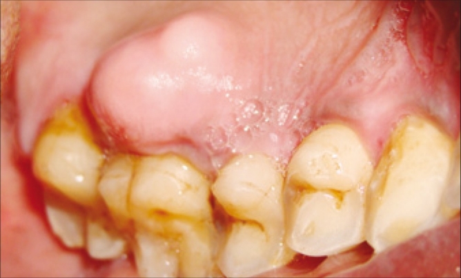
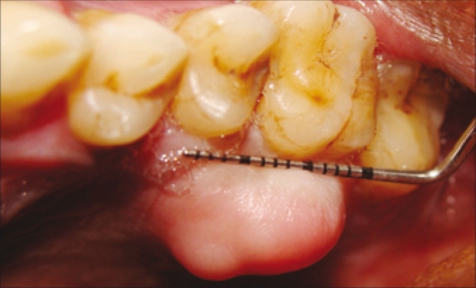
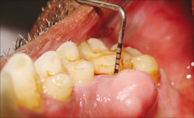
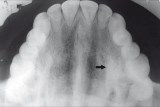
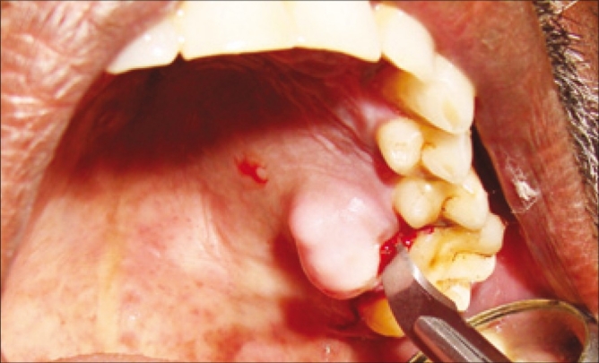
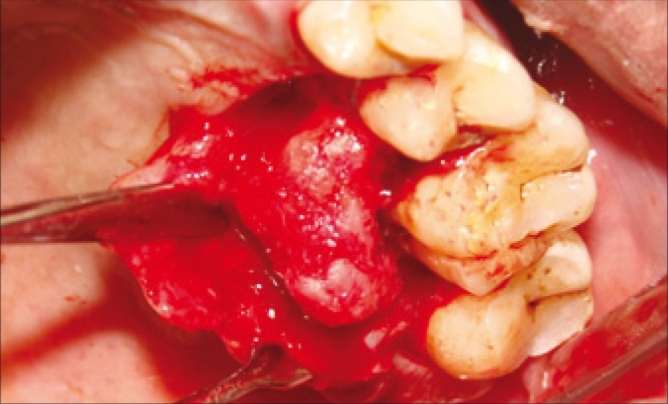
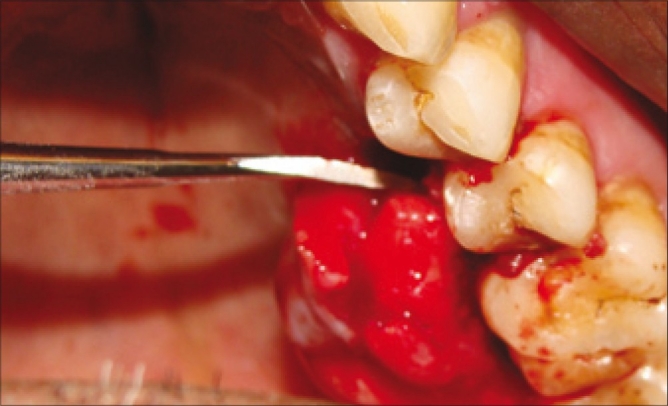
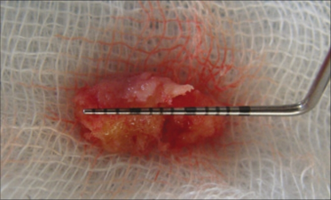
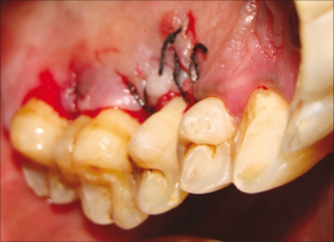
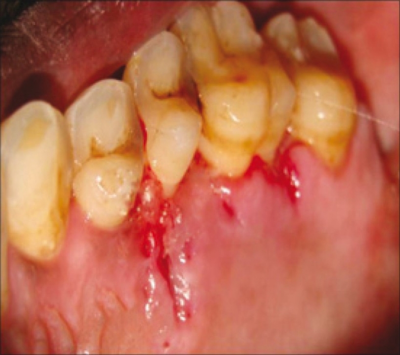
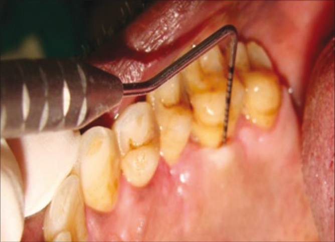
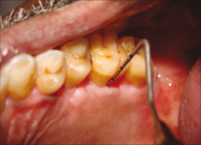
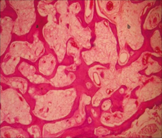
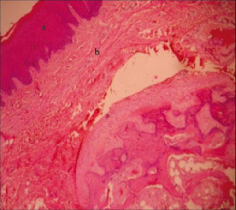
Osteomas are benign slow growing, osteogenic lesions which may arise from proliferation of either cancellous or compact bone. They are usually sessile tumours composed of dense sclerotic, well formed bone projecting out from the cortical surface, most often of the skull and facial bones. This paper reports a case of a peripheral osteoma in the hard palate of a 45-year-old man, which was treated by periodontal flap surgery with surgical excision of the bony lesion. Peripheral osteomas of jaw bone are uncommon and usually associated with Gardner's syndrome. Histological examination confirmed the clinical impression of a peripheral osteoma. Patient was reviewed after one year and was asymptomatic with no recurrence of the lesion.
Keywords: Hard palate; maxilla; osteoma; periodontal pocket.
Conflict of interest statement
Figures
References
-
- Nabeshima K, Marutsuka K, Shimao Y, Uehara H, Kodama T. Osteoma of the frontal sinus complicated by intracranial mucocele. Pathol Int. 2003;53:227–30. - PubMed
-
- Chaudhry SI, Tappuni AR, Challacombe SJ. Multiple maxillary and mandibular exostoses associated with multiple dermatofibromas: A case report. Oral Surg Oral Med Oral Pathol Oral Radiol Endod. 2000;89:319–22. - PubMed
-
- Neville BW, Damm D. Patologia oral e maxillofacial. Rio de Janeiro: GuanabaraKoogan; 1998.
-
- Longo F, Califano L, De Maria G, Ciccarelli R. Solitary osteoma of the mandibular ramus: Report of a case. J Oral Maxillofac Surg. 2001;59:698–700. - PubMed
-
- Lin CJ, Lin YS, Kang BH. Middle turbinate osteoma presenting with ipsilateral facial pain, epiphora, and nasal obstruction. Otolaryngol Head Neck Surg. 2003;128:282–3. - PubMed