. 2012;7(5):e37349.
doi: 10.1371/journal.pone.0037349. Epub 2012 May 21.

Lack of PPARγ in myeloid cells confers resistance to Listeria monocytogenes infection

Affiliations

Lack of PPARγ in myeloid cells confers resistance to Listeria monocytogenes infection

Zeinab Abdullah et al. PLoS One. 2012.

Abstract

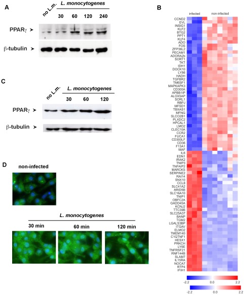
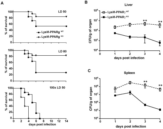
The peroxisomal proliferator-activated receptor γ (PPARγ) is a nuclear receptor that controls inflammation and immunity. Innate immune defense against bacterial infection appears to be compromised by PPARγ. The relevance of PPARγ in myeloid cells, that organize anti-bacterial immunity, for the outcome of immune responses against intracellular bacteria such as Listeria monocytogenes in vivo is unknown. We found that Listeria monocytogenes infection of macrophages rapidly led to increased expression of PPARγ. This prompted us to investigate whether PPARγ in myeloid cells influences innate immunity against Listeria monocytogenes infection by using transgenic mice with myeloid-cell specific ablation of PPARγ (LysMCre×PPARγ(flox/flox)). Loss of PPARγ in myeloid cells results in enhanced innate immune defense against Listeria monocytogenes infection both, in vitro and in vivo. This increased resistance against infection was characterized by augmented levels of bactericidal factors and inflammatory cytokines: ROS, NO, IFNγ TNF IL-6 and IL-12. Moreover, myeloid cell-specific loss of PPARγ enhanced chemokine and adhesion molecule expression leading to improved recruitment of inflammatory Ly6C(hi) monocytes to sites of infection. Importantly, increased resistance against Listeria infection in the absence of PPARγ was not accompanied by enhanced immunopathology. Our results elucidate a yet unknown regulatory network in myeloid cells that is governed by PPARγ and restrains both listeriocidal activity and recruitment of inflammatory monocytes during Listeria infection, which may contribute to bacterial immune escape. Pharmacological interference with PPARγ activity in myeloid cells might represent a novel strategy to overcome intracellular bacterial infection.


Conflict of interest statement

Competing Interests: The authors have declared that no competing interests exist.

Figures

Figure 1
Figure 1. PPARγ expression is induced in bone-marrow derived monocytes after infection with Listeria monocytogenes.
(A) Time course of PPARγ expression in human monocytes following Listeria monocytogenes infection detected by Western blot. (B) PPARγ target genes (n = 80) with the most significant differential expression in human macrophages post infection. Gene expression differences (log scale) are visualized as a heat map following hierarchical clustering of rows and columns (red = increased expression). (C,D) Time course of PPARγ expression in bone marrow derived macrophages (BMDM) post infection detected by Western blot or immunohistochemistry. (blue = DAPI staining the cell nucleus and Listeria DNA; green = PPARγ).
Figure 2
Figure 2. Enhanced resistance of LysM- PPARγKO mice to lethal infection with Listeria monocytogenes.
(A) Survival of LysM- PPARγKO mice and LysM- PPARγWT littermates after intraperitoneal infection with 2×10⁴ (LD 50) 5×10⁵ (LD 80), 2×10⁶ (100× LD50) CFU of Listeria. (n = 10, one representative out of three experiments is shown ). LD 50: p = 0.04; LD 80; 0.01; 100× LD50: not significant difference. (B, C) Colony forming units of Listeria monocytogenes in liver (B) and spleen (C) of LysM- PPARγWT or LysM- PPARγKO mice infected i.p. with 2×10⁴ CFU. (n = 9); significance ** p = 0.005.
Figure 3
Figure 3. Similar phagocytic capacity in myeloid cells from LysM- PPARγWT and LysM- PPARγKO mice.
Bone marrow derived macrophages (BMDM) and peritoneal exudates cells (PEC) from LysM- PPARγWT and LysM- PPARγKO mice were infected with FITC-labelled Listeria at MOI 2 or MOI 5). (A) Mean fluorescence intensity (MFI) of FITC positive cells was assessed by flow cytometry. and (B) Percentage of Listeria containing macrophages (FITC positive). (C) BMDM or PEC from LysM- PPARγWT and LysM- PPARγKO mice were infected with Listeria (MOI 10) and intracellular Listeria growth was determined as CFU/ml in the lysates of infected cells. The p values for titres of WT vs. KO at 4 hrs p.i. were <0.05 and <0.01 for BMDM and PEC, respectively. (D–F) BMDM from LysM- PPARγWT and LysM- PPARγKO mice were infected with Listeria (MOI of 10). The capacity to produce reactive oxygen species (ROS) in Listeria infected macrophages was determined by the OxyBURST reagent (D). At 24 hrs p.i. the concentration of NO2- as surrogate marker for the production of reactive nitrogen intermediates was determined with Griess reagent (E). Expression of iNOS was analyzed by quantitative RT-PCR (F).
Figure 4
Figure 4. Enhanced expression of inflammatory cytokines in LysM-PPARγKO mice during infection with Listeria in vivo.
(A and B) LysM- PPARγWT and LysM- PPARγKO mice were infected i.p. with 2×10⁴ CFU of Listeria. At indicated time points, expression of IFNγ, TNF, IL-1β, IL-6 and IL-12 was determined in the liver (A) and spleen (B) of mice (n = 5) by qRT-PCR. The data shown are means ± SE of three independent experiments.
Figure 5
Figure 5. Enhanced effector functions following abelation of PPARγ from myeloid cells.
(A) BMDM from LysM- PPARγWT and LysM- PPARγKO mice were infected in vitro with L.monocytogenes at a MOI 10. Expression of IFNγ, TNF, IL-1β, IL-6 and IL-12 was determined 6 hrs post infection in cell culture supernatant by ELISA. The data shown are means ± SE of three independent experiments. (B) Looking at the paracrine effects of PPARγ ablation in myeloid cells, we infected BMDM from wild type C57BL/6 mice with Listeria at an MOI of 10 and incubated them with sterile filtered conditioned medium from PPARγWT or PPARγKO macrophages infected previously for 18 hrs with L. monocytogenes; at the indicated time points intracellular Listeria growth was determined as CFU/ml in the lysates of infected cells. Experiments were performed in triplicates. One representative out of four experiments is shown. The p value for titers of wt vs. ko at 4 hrs post infection were <0.05.
Figure 6
Figure 6. Enhanced recruitment of inflammatory monocytes to the site of infection in LysM-PPARγKO mice.
LysM- PPARγWT and LysM- PPARγKO mice were infected i.p. with Listeria (2×10⁴ CFU). (A) at indicated time points total numbers of CD11b+ Ly6Chigh monocytes in the peritoneal cavity (PEC), liver and spleen were determined by FACS analysis (B–C) Expression of CCL2, CCL7 and CCR2 in the liver (B) and spleen (C) of infected mice as determined by qRT-PCR. (D) LSEC isolated from wild type C57BL/6 mice were incubated in vitro with sterile filtered supernatant peritoneal macrophages from LysM- PPARγWT and LysM- PPARγKO mice that were either infected (18 hrs) or left non-infected. The fold increase in expression of CD54 on the surface of LSEC determined by increase in mean fluorescence intensity by flow cytometry in response to contact with supernatant from infected vs non-infected peritoneal macrophages was determined after 24 hrs.
Figure 7
Figure 7. Transfer of PPARγ-deficient macrophages increases the resistance of IFNγ−/−, TNF−/− and CCR2−/− mice to Listeria infection.
5×10⁶ BMDM from LysM- PPARγWT or LysM- PPARγKO mice were adoptively transferred into the peritoneal cavity of IFNγ−/−, TNF−/− or CCR2−/− mice and two hours later mice (n = 10) were infected with 2×10⁴ CFU of Listeria. (A) Survival of IFNγ−/− and TNF−/− mice after infection with Listeria. Knockout mice not receiving any cells by adoptive transfer served as controls. Significance for results in IFNγ−/− and TNF−/− mice were p<0.0001. (B and C) On day 2 after infection, CFU were determined in the homogenates of spleen and liver. One representative out of three independent experiments is shown.

References

    1. Glass CK, Saijo K. Nuclear receptor transrepression pathways that regulate inflammation in macrophages and T cells. Nature reviews Immunology. 2010;10:365–376. - PubMed
    1. Marx N, Sukhova G, Murphy C, Libby P, Plutzky J. Macrophages in human atheroma contain PPARgamma: differentiation-dependent peroxisomal proliferator-activated receptor gamma(PPARgamma) expression and reduction of MMP-9 activity through PPARgamma activation in mononuclear phagocytes in vitro. Am J Pathol. 1998;153:17–23. - PMC - PubMed
    1. Clark RB, Bishop-Bailey D, Estrada-Hernandez T, Hla T, Puddington L, et al. The nuclear receptor PPAR gamma and immunoregulation: PPAR gamma mediates inhibition of helper T cell responses. J Immunol. 2000;164:1364–1371. - PubMed
    1. Pascual G, Fong AL, Ogawa S, Gamliel A, Li AC, et al. A SUMOylation-dependent pathway mediates transrepression of inflammatory response genes by PPAR-gamma. Nature. 2005;437:759–763. - PMC - PubMed
    1. Jiang C, Ting AT, Seed B. PPAR-gamma agonists inhibit production of monocyte inflammatory cytokines. Nature. 1998;391:82–86. - PubMed

Publication types