. 2012;7(5):e37890.
doi: 10.1371/journal.pone.0037890. Epub 2012 May 22.

Muscle fiber type-dependent differences in the regulation of protein synthesis

Affiliations

Muscle fiber type-dependent differences in the regulation of protein synthesis

Craig A Goodman et al. PLoS One. 2012.

Abstract

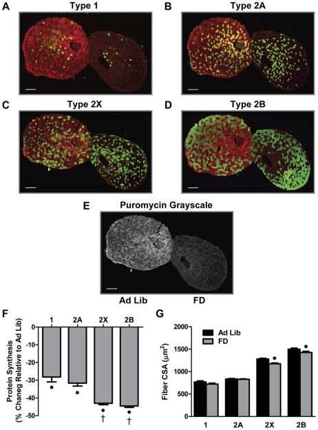
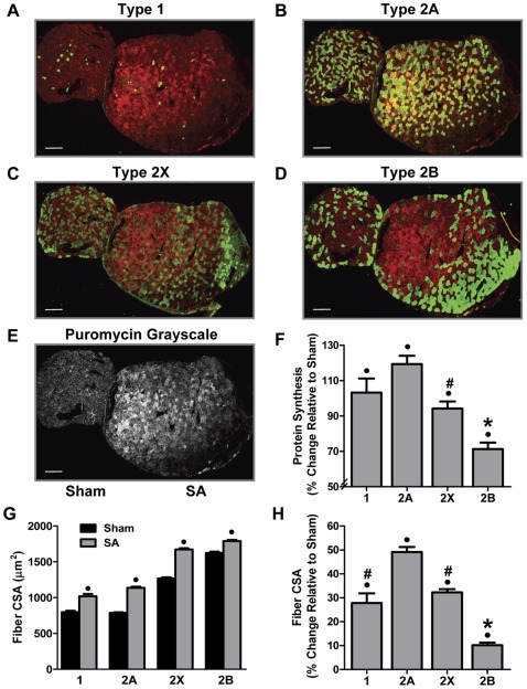
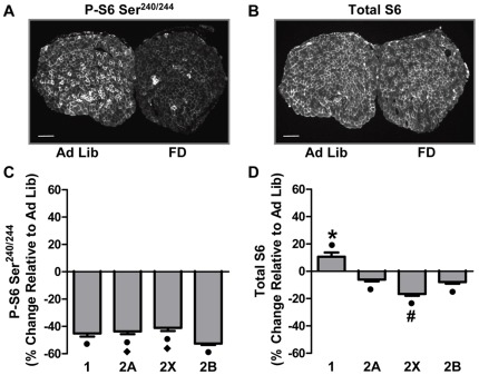
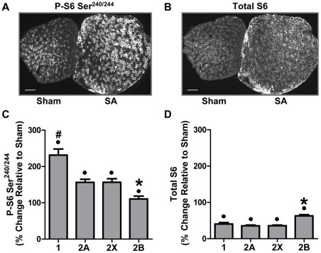
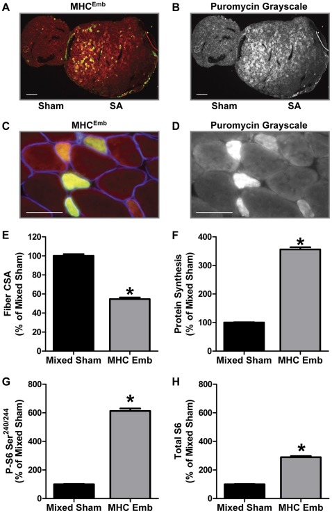
This study examined fiber type-dependent differences in the regulation of protein synthesis in individual muscle fibers found within the same whole muscle. Specifically, the in vivo SUrface SEnsing of Translation (SUnSET) methodology was used to measure protein synthesis in type 1, 2A, 2X and 2B fibers of the mouse plantaris muscle, in response to food deprivation (FD), and mechanical overload induced by synergist ablation (SA). The results show that 48 h of FD induced a greater decrease in protein synthesis in type 2X and 2B fibers compared to type 1 and 2A fibers. Type 2X and 2B fibers also had the largest FD-induced decrease in total S6 protein and Ser(240/244) S6 phosphorylation, respectively. Moreover, only type 2X and 2B fibers displayed a FD-induced decrease in cross-sectional area (CSA). Ten days of SA also induced fiber type-dependent responses, with type 2B fibers having the smallest SA-induced increases in protein synthesis, CSA and Ser(240/244) S6 phosphorylation, but the largest increase in total S6 protein. Embryonic myosin heavy chain (MHC(Emb)) positive fibers were also found in SA muscles and the protein synthesis rates, levels of S6 Ser(240/244) phosphorylation, and total S6 protein content, were 3.6-, 6.1- and 2.9-fold greater than that found in fibers from control muscles, respectively. Overall, these results reveal differential responses in the regulation of protein synthesis and fiber size between fiber types found within the same whole muscle. Moreover, these findings demonstrate that changes found at the whole muscle level do not necessarily reflect changes in individual fiber types.


Conflict of interest statement

Competing Interests: The authors have declared that no competing interests exist.

Figures

Figure 1
Figure 1. Food Deprivation Induces Fiber Type-Dependent Changes in Protein Synthesis and Cross-Sectional Area.
Plantaris muscles obtained from control (Ad Lib) and 48 h food deprived (FD) mice were frozen adjacent to one another, cross-sectioned, and then subjected to immunohistochemistry for rates of protein synthesis (puromycin, red) and fiber type via the identification of (A) type 1, (B) type 2a, (C) type 2x or (D) type 2b, myosin heavy chain isoforms (green). (E) Grayscale image of the puromycin signal from the same pair of muscles shown in A–D. (F) The effect of FD on the relative rate of protein synthesis and (G) cross-sectional area (CSA), within each fiber type. The bars in A–E indicate a length of 200 μm. All values are presented as the mean + SEM (n  =  60–250 fibers / group from 5 independent pairs of muscles). • Significant effect of FD within a given fiber type, † significantly different from type 1 and 2A fibers, (P<0.05).
Figure 2
Figure 2. Synergist Ablation Induces Fiber Type-Dependent Changes in Protein Synthesis and Cross-Sectional Area.
Plantaris muscles obtained from control (Sham) and 10 d synergist ablated (SA) mice were frozen adjacent to one another, cross-sectioned, and then subjected to immunohistochemistry for rates of protein synthesis (puromycin, red) and fiber type via the identification of (A) type 1, (B) type 2a, (C) type 2x or (D) type 2b, myosin heavy chain isoforms (green). (E) Grayscale image of the puromycin signal from the same pair of muscles shown in A–D. (F) The effect of SA on the relative rate of protein synthesis within each fiber type. (G) The absolute and (H) relative effect of SA on the cross-sectional area (CSA) of each fiber type. The bars in A–E indicate a length of 200 μm. All values are presented as the mean + SEM (n  = 84–500 fibers / group from 6 independent pairs of muscles). • Significant effect of SA within a given fiber type, * significantly different from type 1, 2A and 2X fibers, # significantly different from type 2A fibers, (P<0.05).
Figure 3
Figure 3. Food Deprivation Induces Fiber Type-Dependent Changes in Ser240/244 Phosphorylated and Total Ribosomal S6 Protein.
Plantaris muscles obtained from control (Ad Lib) and 48 h food deprived (FD) mice were frozen adjacent to one another, cross-sectioned, and then subjected to immunohistochemistry for different fiber types as described in Figure 1, and Ser240/244 phosphorylated S6 (P-S6 Ser240/244) or total S6. (A) Representative images of P-S6 Ser240/244 and (B) total S6. (C) The effect of FD on the relative amount of P-S6 Ser240/244 and (D) total S6, within each fiber type. The bars in A and B indicate a length of 200 μm. All values are presented as the mean + SEM (n  = 60–250 fibers / group from 5 independent pairs of muscles). • Significant effect of FD within a given fiber type, significantly different from type 2B fibers, * significantly different from type 2A, 2X and 2B fibers, # significantly different from type 1, 2A and 2B fibers, (P<0.05).
Figure 4
Figure 4. Synergist Ablation Induces Fiber Type-Dependent Changes in Ser240/244 Phosphorylated and Total Ribosomal S6 Protein.
Plantaris muscles obtained from control (Sham) and 10 d synergist ablated (SA) mice were frozen adjacent to one another, cross-sectioned, and then subjected to immunohistochemistry for different fiber types as described in Figure 2, and Ser240/244 phosphorylated S6 (P-S6 Ser240/244) or total S6. (A) Representative images of P-S6 Ser240/244 and (B) total S6. (C) The effect of SA on the relative amount of P-S6 Ser240/244 and (D) total S6, within each fiber type. The bars in A and B indicate a length of 200 μm. All values are presented as the mean + SEM (n  = 84–500 fibers / group from 6 independent pairs of muscles). • Significant effect of SA within a given fiber type, ∗ significantly different from type 1, 2A and 2X fibers, # significantly different from type 2A and 2X fibers, (P<0.05).
Figure 5
Figure 5. Cross-Sectional Area, Protein Synthesis, Ser240/244 Phosphorylated and Total Ribosomal S6 Protein in MHCEmb Positive Fibers.
Plantaris muscles obtained from control (Sham) and 10 d synergist ablated (SA) mice were frozen adjacent to one another, cross-sectioned, and then subjected to immunohistochemistry for MHCEmb and rates of protein synthesis (puromycin), Ser240/244 phosphorylated S6 (P-S6 Ser240/244), or total S6, as described in Figures 2 and 4. (A) Representative image of the signals for puromycin (red) and MHCEmb (green). (B) Grayscale image of the puromycin signal shown in A. (C) Higher magnification image from a SA muscle that was tripled stained for puromycin (red), MHCEmb (green) and laminin (blue). (D) Grayscale image of the puromycin signal shown in C. (E) The relative cross-sectional area (CSA), (F) rate of protein synthesis, (G) amount of P-S6 Ser240/244 and (H) total amount of S6 in MHCEmb positive fibers of SA muscles expressed relative to randomly selected fibers from sham muscles (Mixed Sham). The bars in A and B indicate a length of 200 μm, and the bars in C and D indicate 50 μm in length. All values are presented as the mean + SEM (n  = 152–360 fibers / group from 6 independent pairs of muscles). ∗ Significantly different from mixed sham, (P<0.05).

References

    1. Pette D, Staron RS. Mammalian skeletal muscle fiber type transitions. Int Rev Cytol. 1997;170:143–223. - PubMed
    1. Talmadge RJ. Myosin heavy chain isoform expression following reduced neuromuscular activity: potential regulatory mechanisms. Muscle Nerve. 2000;23:661–679. - PubMed
    1. Bottinelli R, Reggiani C. Human skeletal muscle fibres: molecular and functional diversity. Prog Biophys Mol Biol. 2000;73:195–262. - PubMed
    1. Stephenson GM. Hybrid skeletal muscle fibres: a rare or common phenomenon? Clin Exp Pharmacol Physiol. 2001;28:692–702. - PubMed
    1. Schiaffino S, Reggiani C. Fiber Types in Mammalian Skeletal Muscles. Physiol Rev. 2011;91:1447–1531. - PubMed

Publication types

Substances