. 2012 May 25:9:43.
doi: 10.1186/1742-4690-9-43.

Default in plasma and intestinal IgA responses during acute infection by simian immunodeficiency virus

Affiliations

Default in plasma and intestinal IgA responses during acute infection by simian immunodeficiency virus

Nada Chaoul et al. Retrovirology. .

Abstract

Background: Conflicting results regarding changes in mucosal IgA production or in the proportions of IgA plasma cells in the small and large intestines during HIV-infection have been previously reported. Except in individuals repeatedly exposed to HIV-1 but yet remaining uninfected, HIV-specific IgAs are frequently absent in mucosal secretions from HIV-infected patients. However, little is known about the organization and functionality of mucosal B-cell follicles in acute HIV/SIV infection during which a T-dependent IgA response should have been initiated. In the present study, we evaluated changes in B-cell and T-cell subsets as well as the extent of apoptosis and class-specific plasma cells in Peyer's Patches, isolated lymphoid follicles, and lamina propria. Plasma levels of IgA, BAFF and APRIL were also determined.

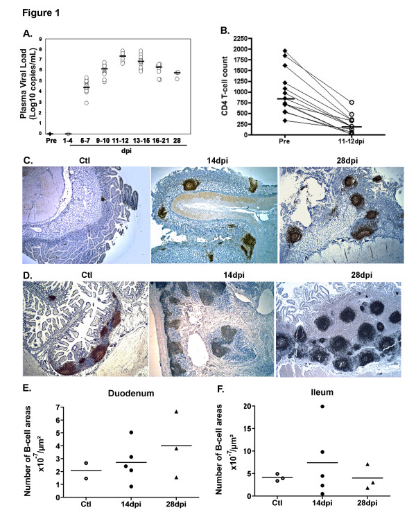
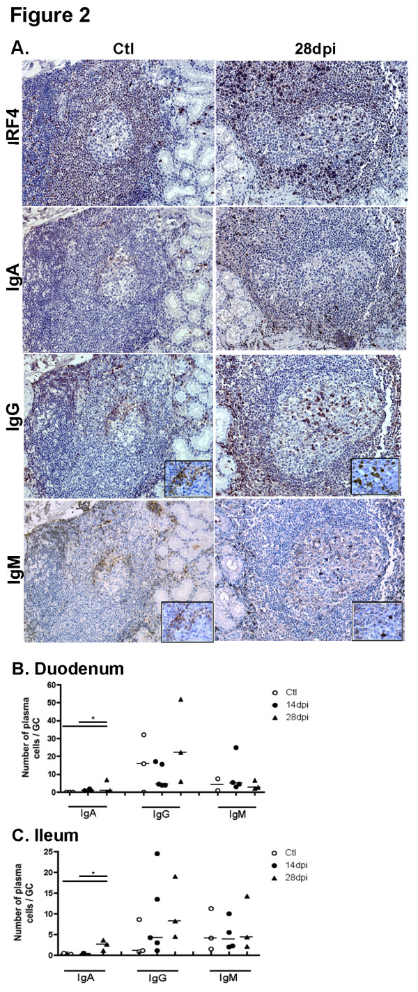
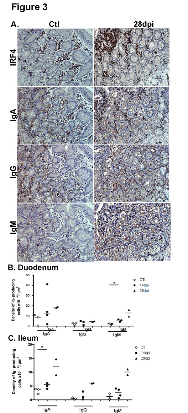
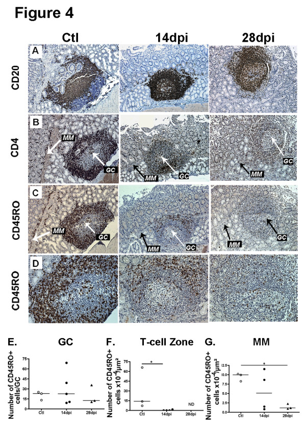
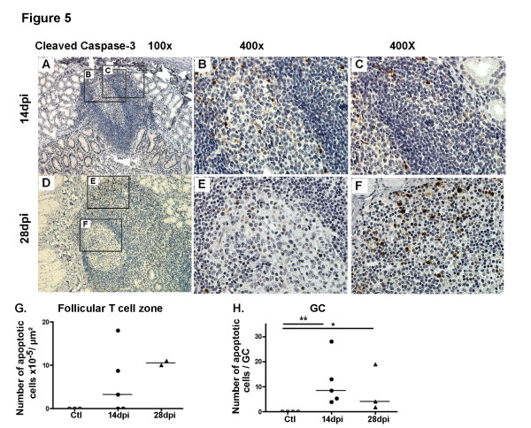
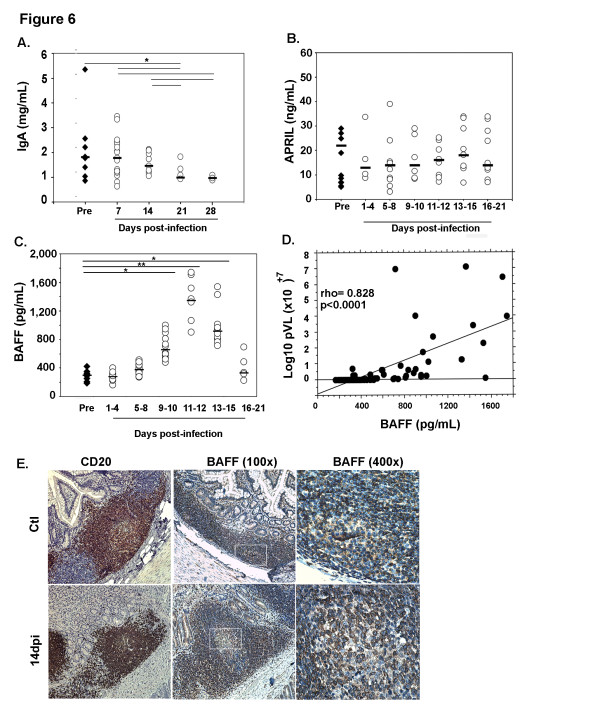
Results: Plasma IgA level was reduced by 46% by 28 days post infection (dpi), and no IgA plasma cells were found within germinal centers of Peyer's Patches and isolated lymphoid follicles. This lack of a T-dependent IgA response occurs although germinal centers remained functional with no sign of follicular damage, while a prolonged survival of follicular CD4+ T-cells and normal generation of IgG plasma cells is observed. Whereas the average plasma BAFF level was increased by 4.5-fold and total plasma cells were 1.7 to 1.9-fold more numerous in the lamina propria, the relative proportion of IgA plasma cells in this effector site was reduced by 19% (duodemun) to 35% (ileum) at 28 dpi.

Conclusion: Our data provide evidence that SIV is unable to initiate a T-dependent IgA response during the acute phase of infection and favors the production of IgG (ileum) or IgM (duodenum) plasma cells at the expense of IgA plasma cells. Therefore, an early and generalized default in IgA production takes place during the acute of phase of HIV/SIV infection, which might impair not only the virus-specific antibody response but also IgA responses to other pathogens and vaccines as well. Understanding the mechanisms that impair IgA production during acute HIV/SIV infection is crucial to improve virus-specific response in mucosa and control microbial translocation.


Figures

Figure 1
Figure 1
Increased number of B-cell areas in the intestinal mucosa in acutely SIV-infected macaques. (A) For each of the 13 SIV-infected macaques, viral load was measured in plasma before infection (Pre; filled circles) and every 3 dpi (open circles). Results are expressed as Log10 copies/mL and bars represent median values. (B) CD4+ T-cells were quantified in whole blood collected from each macaque before infection and every 3 dpi. For each animal, CD4+ T-cell counts before infection (filled circles; n=13) and at 11-12 dpi (open circles, n=13) are shown. Results are expressed as cells/μL and bars represent median values. *** p=0.0004 (Wilcoxon test) (C-D) Staining by CD20 mAb of sections from duodenum (C) and terminal ileum (D) of one representative non-infected macaque (Ctl, n=3) and macaques infected for 14 (14 dpi, n=5) or 28 days (28 dpi, n=3). Magnification x25 for all panels (E-F) The number of CD20+ B-cell areas per section was divided by the surface of the total mucosa. Results are expressed as B-cell areas x 10-7/μm2. Each symbol corresponds to one animal. Bars represent median values for each group.
Figure 2
Figure 2
Acute SIV infection induces a dominant IgG/M over IgA response in GC. (A) Duodenal sections from controls (Ctl left panels) and macaques infected for 28 days (28 dpi, right panels) were stained with IRF4 mAb, IgA, IgG and IgM polyclonal Ab. Staining of one representative section for each group of macaques is shown. Magnification x200 and x1000 for enlarged images. (B-C) Each symbol represents the mean number of IgA, IgG and IgM plasma cells per GC in duodenum (B) and terminal ileum (C) in one macaque. Bars represent median values for each group.
Figure 3
Figure 3
Acute SIV infection induces an increase in total plasma cells in the intestinal LP with a dominant IgG/M over IgA response. (A) Duodenal sections from controls (Ctl left panels) and macaques infected for 28 days (28 dpi, right panels) were stained with IRF4 mAb, IgA, IgG and IgM polyclonal Ab. Staining of one representative section for each group of macaques is shown. Magnification x200 and x1000 for enlarged images. (B, C) Each symbol corresponds to the mean number of IgA, IgG and IgM plasma cells in LP of duodenum (B) and terminal ileum (C) from one macaque. Bars represent median values for each group. * p values <0.05 (Mann–Whitney, one tailed).
Figure 4
Figure 4
Prolonged survival of CD4 + CD45RO + T-cells in GC as compared to follicular T-cell zones and lamina propria. (A-D) Duodenal sections from controls (Ctl) and macaques infected at 14dpi and 28dpi were stained with CD20 mAb (A), CD4 mAb (B), CD45RO mAb (C-D). In A to D, data from one representative macaque per group are shown. Original magnification x100 for panels A to C, x200 for panel D. (E-G) Each symbol corresponds to the mean density of CD45RO+ cells per μm2 of duodenal muscularis mucosae (MM,G), of follicular T-cell zone (F) and to the mean number of CD45R0+ cells per GC (E) per macaque. Bars represent median values for each group. * p values <0.05 (Mann–Whitney).
Figure 5
Figure 5
Apoptosis progresses more rapidly in LP than in GC. (A-F) Duodenal sections from one representative macaque at 14 dpi and 28 dpi were stained with anti-cleaved caspase-3 Ab (A-F). Inserts from left panels are shown in middle (B and E; showing GC) and right panels (C and F; showing follicular T-cell zones). Original magnification x100 in left panels and x400 in middle and right panels. (G-H) Each symbol corresponds to the mean density of cleaved caspase3+ cells per μm2 of follicular T-cell Zone (G) and to the mean number of cleaved caspase-3+ cells per GC (H) per macaque. Bars represent median values for each group. *p value < 0.05 (Mann–Whitney).
Figure 6
Figure 6
Decreased plasma IgA levels despite a strong increase in BAFF levels. (A) Plasma IgA levels were measured in SIV-infected macaques before infection (Pre; filled diamonds) and every 7 dpi (open circles). Results are expressed in mg/mL and bars represent median levels. Significant differences between groups are shown (*p < 0.05). (B-C) APRIL and BAFF levels were measured in plasma from SIV-infected macaques, before (Pre: filled diamonds) and every 3 dpi (open circles). Results are expressed in pg/mL (BAFF) and ng/mL (APRIL), and bars represent median levels. Significant differences between groups are shown (*p < 0.05, **p < 0.01). (D) Correlation between BAFF levels and plasma viral load (Log10 pVL) is shown. (E) Ileum sections from control macaques (upper panel) and macaques infected for 14 days (lower panel) were stained with anti-CD20 (B-cells, left panels) or anti-BAFF (Buffy2, middle and right panels) mAb. Original magnification: x200 for CD20, x100 and x400 for Buffy2.

Similar articles

Cited by

References

    1. Brenchley JM, Schacker TW, Ruff LE, Price DA, Taylor JH, Beilman GJ, Nguyen PL, Khoruts A, Larson M, Haase AT. et al.CD4+ T cell depletion during all stages of HIV disease occurs predominantly in the gastrointestinal tract. J Exp Med. 2004;200(6):749–759. doi: 10.1084/jem.20040874. - DOI - PMC - PubMed
    1. Veazey RS, DeMaria M, Chalifoux LV, Shvetz DE, Pauley DR, Knight HL, Rosenzweig M, Johnson RP, Desrosiers RC, Lackner AA. Gastrointestinal tract as a major site of CD4+ T cell depletion and viral replication in SIV infection. Science. 1998;280(5362):427–431. doi: 10.1126/science.280.5362.427. - DOI - PubMed
    1. Suzuki K, Fagarasan S. How host-bacterial interactions lead to IgA synthesis in the gut. Trends Immunol. 2008;29(11):523–531. doi: 10.1016/j.it.2008.08.001. - DOI - PubMed
    1. Fagarasan S, Honjo T. Intestinal IgA synthesis: regulation of front-line body defences. Nat Rev Immunol. 2003;3(1):63–72. doi: 10.1038/nri982. - DOI - PubMed
    1. Ozaki K, Spolski R, Feng CG, Qi CF, Cheng J, Sher A, Morse HC, Liu C, Schwartzberg PL, Leonard WJ. A critical role for IL-21 in regulating immunoglobulin production. Science. 2002;298(5598):1630–1634. doi: 10.1126/science.1077002. - DOI - PubMed

Publication types

MeSH terms

Substances