. 2012 May 24;74(4):706-18.
doi: 10.1016/j.neuron.2012.02.034.

Balance of activity between LN(v)s and glutamatergic dorsal clock neurons promotes robust circadian rhythms in Drosophila

Affiliations

Balance of activity between LN(v)s and glutamatergic dorsal clock neurons promotes robust circadian rhythms in Drosophila

Ben Collins et al. Neuron. .

Erratum in

  • Neuron. 2012 Jun 21;74(6):1138

Abstract

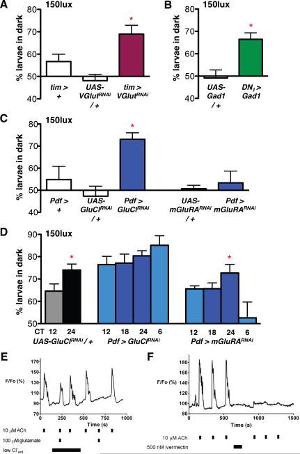
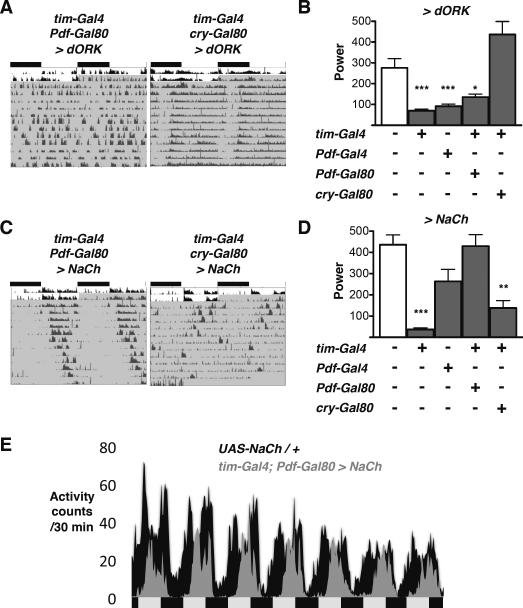
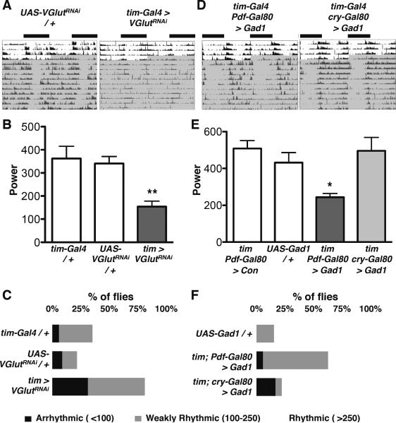
Circadian rhythms offer an excellent opportunity to dissect the neural circuits underlying innate behavior because the genes and neurons involved are relatively well understood. We first sought to understand how Drosophila clock neurons interact in the simple circuit that generates circadian rhythms in larval light avoidance. We used genetics to manipulate two groups of clock neurons, increasing or reducing excitability, stopping their molecular clocks, and blocking neurotransmitter release and reception. Our results revealed that lateral neurons (LN(v)s) promote and dorsal clock neurons (DN(1)s) inhibit light avoidance, these neurons probably signal at different times of day, and both signals are required for rhythmic behavior. We found that similar principles apply in the more complex adult circadian circuit that generates locomotor rhythms. Thus, the changing balance in activity between clock neurons with opposing behavioral effects generates robust circadian behavior and probably helps organisms transition between discrete behavioral states, such as sleep and wakefulness.


Figures

Figure 1
Figure 1. Pre- and post-synaptic DN1 terminals are located close to LNv axonal termini
(A) The nuclei of larval clock neurons were marked with the circadian transcription factor Par Domain Protein 1 (PDP1, red). LNvs were co-labeled with PDF (blue). cry13-Gal4; Pdf-Gal80 driven expression of the Dscam17.1-GFP post-synaptic marker (green) labels DN1 projections very close to LNv axons. The 5th LNv was identified by lack of PDF and GFP staining and its location. (B) cry16-Gal4; Pdf-Gal80 driven expression of UAS-CD8-GFP (green) and the pre-synaptic marker UAS-Syt-HA (anti-HA, red) co-localize to DN1 projections adjacent to LNv axons (labeled with PDF, blue). (C) Same image as (B) with GFP channel removed to show Syt expression in DN1 projections adjacent to LNv axons. This image shows a 20μm stack but single 4μm sections also show DN1 projections adjacent to LNv axons.
Figure 2
Figure 2. LNvs promote and DN1s inhibit larval light avoidance
Larval light avoidance was measured by counting the number of larvae on the dark sides of a Petri dish after 15min. Transgenes were targeted to either LNvs using Pdf-Gal4 (Pdf >), or DN1s using cry-Gal4; Pdf-Gal80 (DN1 >). Control lines are either the Gal4 line crossed to the non-conducting UAS-dORKΔNC transgene (Con) or the relevant UAS-transgene crossed to y w (transgene / +). Error bars show SEM. All statistical comparisons to the relevant control line were made using the students t-test. *p<0.05, **p<0.01. (A) Light avoidance was assayed between ZT 3–6 at 150lux. Hyperpolarizing LNvs with UAS-dORKΔC (Pdf > dORK) or ablating LNvs with UAS-Dti (Pdf > Dti) had no significant effect on light avoidance vs control larvae. Hyperexciting LNvs via NaChBac (Pdf > NaCh, p<0.005) increased larval light avoidance. (B) Light avoidance was assayed as in (A). Hyperpolarizing DN1s with UAS-dORKΔC or UAS-mKir2.1 (DN1 > dORK, p<0.05 and DN1 > Kir, p<0.005), or DN1-ablation (DN1 > Dti, p<0.01) significantly increased larval light avoidance. Hyperexciting DN1s (DN1 > NaCh) had no significant effect on light avoidance.
Figure 3
Figure 3. Altering CLK/CYC activity has opposite effects on LNv and DN1 excitability
All Statistical comparisons were made by ANOVA with Tukey's post-hoc test. *p<0.05, **p<0.01, ***p<0.001 (A) Light avoidance was assayed between ZT 3–6 in LD at 150lux. Expressing ClkDN (p<0.05) or cycDN (p<0.001) in LNvs increased larval light avoidance at 150lux compared to controls (Pdf-Gal4 or UAS-transgene crossed to y w). Hyperpolarization of LNvs expressing cycDN (Pdf > cycDN + dORK) restored light avoidance to wild-type levels while hyperexcitation of LNvs expressing cycDN (Pdf > cycDN + NaCh) did not. See also Fig S1. (B) Light avoidance was assayed as in (A). Expressing ClkDN (p<0.01) and cycDN (p<0.01) in DN1s increased larval light avoidance at 150lux compared to controls (DN1 > Con, reproduced from Fig 2B, or UAS-transgenes crossed to y w, reproduced from Fig 3A). Hyperexcitation of DN1s expressing cycDN (DN1 > cycDN + NaCh) restored light avoidance to wild-type levels. (C) Light avoidance was assayed between ZT 3–6 in LD at 750lux. Hyperexciting LNvs (per01; Pdf > NaCh, p<0.01) rescued the low levels of light avoidance of per01 larvae whilst hyperpolarizing LNvs did not (per01; Pdf > dORK). Hyperpolarizing DN1s (per01; DN1 > dORK, p<0.01) also increased light avoidance whilst hyperexciting DN1s (per01; DN1 > NaCh) did not.
Figure 4
Figure 4. A signal from DN1s is necessary and sufficient for light avoidance rhythms
All Statistical comparisons are as specified below. *p<0.05, **p<0.01, (A) Light avoidance was assayed on day 2 (CT12, 18, 24) or day 3 (CT6) of DD after prior LD entrainment. Control UAS-Dti / + larvae (grey) show time-dependent light avoidance at 150lux (CT12 vs CT24, t-test p<0.01). DN1-ablated larvae (green) show no time-dependent light avoidance, (ANOVA p=0.79). 2-Way ANOVA between control and DN1-ablated larvae for CT12 and 24 reveals a significant Genotype × Time interaction (F1,11=8.53, p<0.05). No time-dependent differences in light avoidance were observed in control or DN1-ablated larvae at 50lux (t-test). (B) Light avoidance was assayed as in (A) at 150lux. All statistical comparisons by students t-test. Light avoidance scores were higher at CT24 than CT12 in control (per+ UAS-per, p<0.05) but not in per01UAS-per larvae. Rhythms were rescued by restoring per expression to LNvs (blue, p<0.005) or DN1s (green, p<0.05) in per01 mutants. See also Fig S2. (C) Light avoidance was assayed as in (A). Light avoidance scores were lower at CT12 than at CT24 in control UAS-CLKDN / + larvae at 150lux (t-test p<0.001). DN1 > CLKDN increased light avoidance compared to controls (2 Way ANOVA F1,31=5.81, p<0.05), with no time-dependent differences in light avoidance observed at either 150lux (ANOVA) or 50lux (t-test). (D) Light avoidance was assayed on day 2 in DD at 150lux using larvae reared at 20°C. Light avoidance scores were lower at CT12 than at CT24 in DN1 > TrpA1 larvae when assayed at 20°C (t-test p<0.01) but not 26°C. At 26°C, temperature-induced activation of DN1s via TrpA1 reduces light avoidance at CT24 to CT12 levels (2 Way ANOVA, Temperature × Time interaction F1,12=5.73, p<0.05). See also Fig S3
Figure 5
Figure 5. DN1s release glutamate to inhibit light avoidance
For all RNAi experiments UAS-dcr-2 was co-expressed to improve efficacy. The Gal4 control lines shown also express UAS-dcr-2. Statistical comparisons are as stated below. *p<0.05 (A–C) Larval light avoidance was measured as in Fig 2. (A) Expression of a VGlut-RNAi transgene (GD2574) in all clock neurons (tim > VGlutRNAi increased light avoidance at 150lux compared to control larvae. These data are significantly different (ANOVA p<0.005). Tukey's post-hoc comparison gives a significant difference only between tim > VGlutRNAi and UAS-VGlutRNAi / +. However, light avoidance in tim > VGlutRNAi is higher than tim > + by t-test (p<0.05) and tim > VGlutRNAi larvae also lose circadian rhythms in light avoidance (Fig S4A). (B) Expression of Glutamate decarboxylase (UAS-Gad1) in DN1s (DN1 > Gad1) significantly increased light avoidance at 150lux compared to UAS-Gad1 / + control larvae (Gad1 / +, t-test p<0.05). See also Fig S4B. (C) A GluCl-RNAi transgene expressed in LNvs (Pdf > GluClRNAi) significantly increased light avoidance at 150lux compared to control larvae (ANOVA p<0.05). An mGluRA-RNAi transgene expressed in LNvs (Pdf > mGluRARNAi) had no effect on light avoidance compared to controls (ANOVA). See also Fig S4C. (D) Light avoidance was assayed in DD at 150lux as in Fig 4. Light avoidance is higher at CT24 than CT12 in control larvae (UAS-GluClRNAi / +, which also contain a UAS-dcr-2 transgene, t-test p<0.05). No rhythms in light avoidance were detectable when GluCl-RNAi was expressed in LNvs (Pdf > GluClRNAi, ANOVA). Rhythmic light avoidance was still detectable in larvae expressing mGluRA-RNAi in LNvs (Pdf > mGluRARNAi, ANOVA p<0.05). By 2 Way ANOVA comparison of CT12 and 24 time points, Pdf > GluClRNAi is different to control (F1,22=9.17, p<0.01) whilst Pdf > mGluRARNAi is not (F1,24=0.00, p=0.9547) (E) Glutamate-mediated inhibition of ACh-stimulated Ca2+ transients in dissociated larval LNvs. Representative relative fluorescence (F/Fo) recordings are shown from dissociated larval LNvs expressing UAS-GCaMP1.6. Solution changes, including neurotransmitter applications, are indicated by black bars. Lowering extracellular Cl to 13.6mM completely relieved glutamate-dependent inhibition. Glutamate completely blocked ACh-stimulated transients when physiological Cl was restored. (F) A 2min incubation of a larval LNv with 500nM ivermectin irreversibly blocks subsequent ACh-induced Ca2+ transients.
Figure 6
Figure 6. Adult CRY+ non-LNvs are required for robust locomotor activity rhythms
Locomotor activity was recorded from flies entrained to 12:12 LD (white area of actogram), then transferred to DD (shaded). Comparisons to UAS-Control / + are by ANOVA with Tukey's post-hoc test. *p<0.05, **p<0.01, ***p<0.001 (A) Representative, double-plotted, normalized actograms are shown for tim-Gal4; Pdf-Gal80 > dORK and tim-Gal4; cry-Gal80 > dORK flies. (B) The power of rhythms is plotted for flies expressing UAS-dORK under the control of tim-Gal4, Pdf-Gal4, tim-Gal4; Pdf-Gal80 and tim-Gal4; cry-Gal80 drivers. Power is significantly reduced in Pdf > dORK, tim > dORK and tim-Gal4; Pdf-Gal80 > dORK flies compared to UAS-dORK / + control flies. (C) Representative, double-plotted, normalized actograms are shown for tim-Gal4; Pdf-Gal80 > NaCh and tim-Gal4; cry-Gal80 > NaCh flies. (D) The power of rhythms is plotted for flies expressing UAS-NaCh under the control of tim-Gal4, Pdf-Gal4, tim-Gal4; Pdf-Gal80 and tim-Gal4; cry-Gal80 drivers. Power is significantly reduced in tim-Gal4; cry-Gal80 > NaCh flies compared to UAS-NaCh / + control flies. (E) Average locomotor activity over the first 7 days in DD plotted for UAS-NaCh / + control (black) and tim-Gal4; Pdf-Gal80 > NaCh flies (grey). tim-Gal4; Pdf-Gal80 > NaCh fly activity is substantially reduced during the subjective morning. Each genotype shows the average of 16 flies from a single experiment.
Figure 7
Figure 7. Glutamate signaling from Adult CRY+ non-LNvs is required for robust locomotor activity rhythms
Locomotor activity was recorded from flies entrained to 12:12 LD (white area of actogram), then transferred to DD (shaded). Comparisons to UAS-Control / + by ANOVA with Tukey's post-hoc test. *p<0.05, **p<0.01 (A) Representative, double-plotted, normalized actograms are shown for UAS-VGlutRNAi/ + and tim > VGlutRNAi flies. UAS-dcr-2 was co-expressed to improve RNAi efficacy and the Gal4 control line also expresses UAS-dcr-2. (B) The average power of rhythms is shown for tim > dcr2 + VGlutRNAi flies and tim > dcr2 and UAS-VGlutRNAi / + control flies. Power is significantly reduced in tim > VGlutRNAi flies compared to tim > dcr2 and UAS-VGlutRNAi control flies. Error bars represent SEM. (C) Data from (B) plotted as % of flies that are “arrhythmic” (power >100) “weakly rhythmic” (power from 100–250) or “rhythmic” (power > 250). (D) Representative, double-plotted, normalized actograms are shown for tim-Gal4; Pdf-Gal80 > Gad1 and tim-Gal4; cry-Gal80 > Gad1 flies. (E) The average power of rhythms is shown for flies expressing UAS-Gad1 with the tim-Gal4; Pdf-Gal80 and tim-Gal4; cry-Gal80 drivers. Power is significantly reduced in tim-Gal4; Pdf-Gal80 > Gad1 flies compared to UAS-Gad1 / + control or tim-Gal4; Pdf-Gal80 crossed to the UAS-dORKΔNC control (con) flies. Error bars represent SEM. (F) Data from (E) plotted as in (C)

References

    1. Allada R, Chung BY. Circadian organization of behavior and physiology in Drosophila. Annu Rev Physiol. 2010;72:605–624. - PMC - PubMed
    1. Brand AH, Perrimon N. Targeted gene expression as a means of altering cell fates and generating dominant phenotypes. Development. 1993;118:401–415. - PubMed
    1. Cao G, Nitabach MN. Circadian control of membrane excitability in Drosophila melanogaster lateral ventral clock neurons. J Neurosci. 2008;28:6493–6501. - PMC - PubMed
    1. Cully DF, Vassilatis DK, Liu KK, Paress PS, Van der Ploeg LH, Schaeffer JM, Arena JP. Cloning of an avermectin-sensitive glutamate-gated chloride channel from Caenorhabditis elegans. Nature. 1994;371:707–711. - PubMed
    1. Dahdal D, Reeves DC, Ruben M, Akabas MH, Blau J. Drosophila pacemaker neurons require g protein signaling and GABAergic inputs to generate twenty-four hour behavioral rhythms. Neuron. 2010;68:964–977. - PMC - PubMed

Publication types

LinkOut - more resources