. 2012 May 25;149(5):1023-34.
doi: 10.1016/j.cell.2012.03.043.

Cyclin F-mediated degradation of ribonucleotide reductase M2 controls genome integrity and DNA repair

Affiliations

Cyclin F-mediated degradation of ribonucleotide reductase M2 controls genome integrity and DNA repair

Vincenzo D'Angiolella et al. Cell. .

Abstract

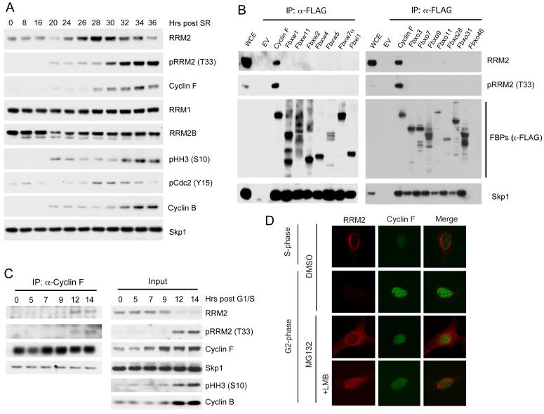
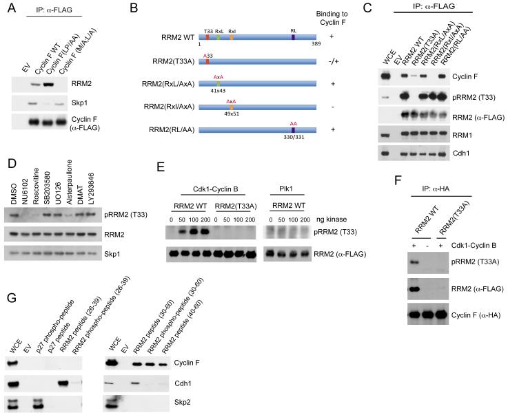
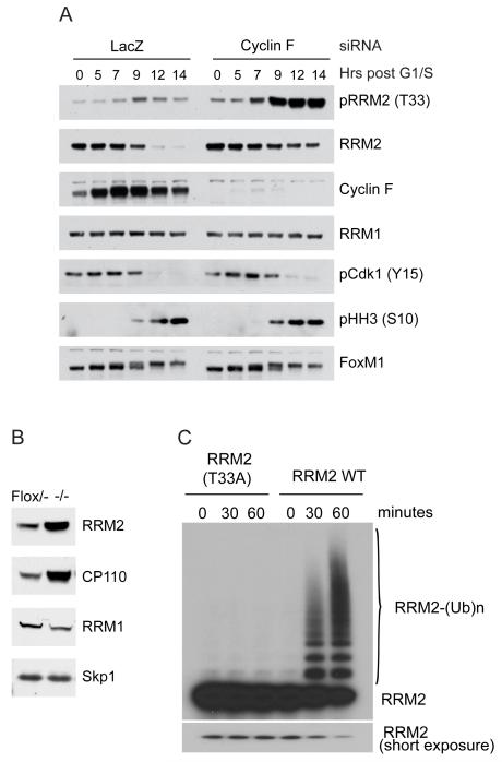
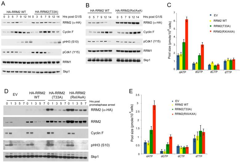
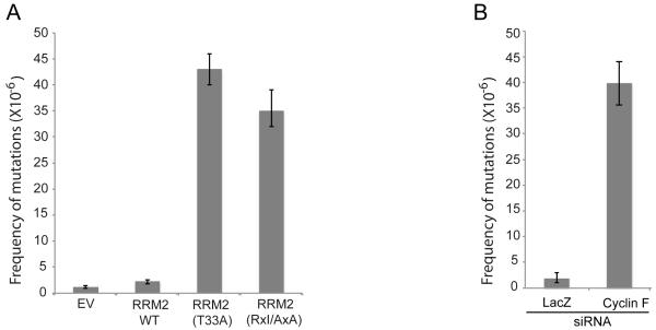
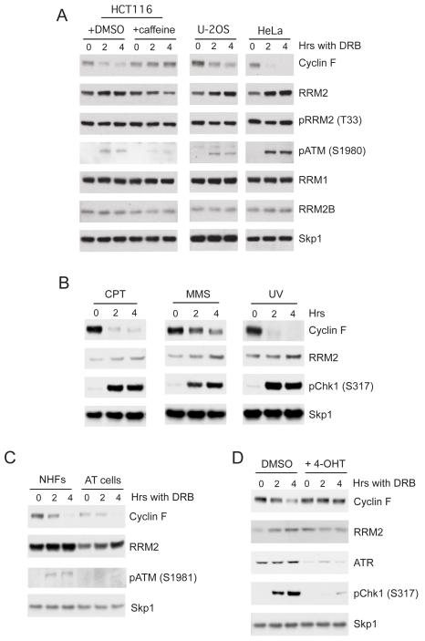
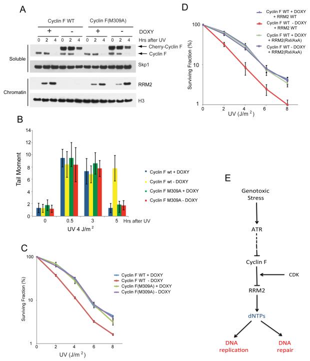
F-box proteins are the substrate binding subunits of SCF (Skp1-Cul1-F-box protein) ubiquitin ligase complexes. Using affinity purifications and mass spectrometry, we identified RRM2 (the ribonucleotide reductase family member 2) as an interactor of the F-box protein cyclin F. Ribonucleotide reductase (RNR) catalyzes the conversion of ribonucleotides to deoxyribonucleotides (dNTPs), which are necessary for both replicative and repair DNA synthesis. We found that, during G2, following CDK-mediated phosphorylation of Thr33, RRM2 is degraded via SCF(cyclin F) to maintain balanced dNTP pools and genome stability. After DNA damage, cyclin F is downregulated in an ATR-dependent manner to allow accumulation of RRM2. Defective elimination of cyclin F delays DNA repair and sensitizes cells to DNA damage, a phenotype that is reverted by expressing a nondegradable RRM2 mutant. In summary, we have identified a biochemical pathway that controls the abundance of dNTPs and ensures efficient DNA repair in response to genotoxic stress.


Figures

Figure 1
Figure 1. Cyclin F and RRM2 physically interact and colocalize to the nucleus in G2
A) RPE1-hTERT cells were synchronized in G0/G1 by 72 hours of serum starvation before release into fresh medium containing serum. Cells were collected at the indicated time points after serum re-addition (SR), lysed, and immunoblotted as indicated. B) HEK-293T cells were transfected with an empty vector (EV) or the indicated FLAG-tagged F-box protein constructs (FBPs). Whole cell extracts were immunoprecipitated (IP) with anti-FLAG resin, and immunoprecipitates were immunoblotted as indicated. C) HeLa cells were synchronized at G1/S using a double-thymidine block before release into fresh medium. Cell lysates were generated at the indicated time points, immunoprecipitated with an antibody to Cyclin F, and immunoblotted as indicated. Ten percent of the material used for immunoprecipitation (input) is shown on the right panels. D) U-2OS cells were synchronized at G1/S using a double-thymidine block before release into fresh medium. Cells were fixed at five hours (S-phase) and nine hours (G2-phase) after release from the block and stained with an antibody to Cyclin F (green) or RRM2 (red). Where indicated, cells where pretreated for two hours with Leptomycin B (LMB) before fixation. Confocal microscopy was used to visualize stained cells.
Figure 2
Figure 2. Both a CY motif and the phosphorylation of Thr33 in RRM2 are required for RRM2 binding to Cyclin F
A) HEK-293T cells were transfected with either an empty vector (EV), FLAG-tagged Cyclin F, or the indicated FLAG-tagged Cyclin F mutants. Whole cell extracts were immunoprecipitated (IP) with anti-FLAG resin, and immunocomplexes were immunoblotted as indicated. B) Schematic representation of RRM2 mutants highlighting putative cyclin F binding motifs in RRM2. RRM2 mutants that interacted with endogenous Cyclin F are designated with the symbol (+). C) HEK-293T cells were transfected with either an empty vector (EV), FLAG-tagged RRM2, or the indicated FLAG-tagged RRM2 mutants. Whole cell extracts were immunoprecipitated (IP) with anti-FLAG resin, and immunocomplexes were immunoblotted as indicated. D) HeLa cells were treated with the indicated kinase inhibitors. Cells were collected four hours later, lysed, and immunoblotted as indicated. E) In vitro-transcribed/translated RRM2 was incubated at 30°C with the indicated amounts of either Cdk1-Cyclin B complex (upper panels) or Plk1 (bottom panels). After 30 minutes, reactions were stopped, and samples were immunoblotted as indicated. F) In vitro-transcribed/translated RRM2 or RRM2(T33A) were incubated at 30°C in the presence or absence of Cdk1-Cyclin B. After 30 minutes, in vitro-transcribed/translated HA-Cyclin F was added to the reaction and incubated for an additional 30 minutes. Cyclin F was then immunoprecipitated with an anti-HA antibody, and immunocomplexes were immunoblotted as indicated. G) HeLa cell extracts were incubated with beads coupled to the following peptides: RRM2 (26-39) (SLVDKENTPPALSG), phospho-RRM2 (26-39) (SLVDKENTp-PPALSG), RRM2 (30-60) (KENTPPALSGTRVLASKTARRIFQEPTEPK), phospho-RRM2 (30-60) (KENTp-PPALSGTRVLASKTARRIFQEPTEPK), RRM2 (40-60) (TRVLASKTARRIFQEPTEPK), p27 (180-198) (NAGSVEQTPKKPGLRRRQT), or phospho-p27 (180-198) (NAGSVEQTp-PKKPGLRRRQT). Beads were extensively washed, and bound proteins were immunoblotted as indicated. (See also Figure S1 and S2)
Figure 3
Figure 3. RRM2 is targeted for ubiquitylation and degradation by SCFCyclin F
A) HeLa cells were transfected with siRNAs to either a non-relevant mRNA (LacZ) or Cyclin F mRNA. Cells were synchronized at G1/S by a double-thymidine block and collected at the indicated time points after release from the block. Cell lysates were immunoblotted as indicated. B) Cyclin F−/− and parental Cyclin FFlox/− MEFs were lysed and immunoblotted as indicated. C) 35[S]-in vitro-translated RRM2 or RRM2(T33A) were incubated at 30°C with Cdk1-Cyclin B and then, for the indicated times, with a ubiquitylation mix containing purified, recombinant SCFCyclin F. Reactions were analyzed by autoradiography. The bracket indicates a ladder of bands corresponding to polyubiquitylated RRM2. (See also Figure S3)
Figure 4
Figure 4. Expression of stable RRM2 mutants induces an increase in the concentration of dATP and dGTP
A) HeLa cells infected with either a retrovirus expressing HA-tagged RRM2 or HA-tagged RRM2(T33A) were synchronized using a double-thymidine block. Samples were collected at the indicated times after release, lysed and immunoblotted as indicated. B) HeLa cells infected with either a retrovirus expressing HA-tagged RRM2 or HA-tagged RRM2(RxI/AxA) were analyzed as in (A). C) RPE1-hTert cells infected with either a retrovirus expressing HA-tagged RRM2, HA-tagged RRM2(T33A), or HA-tagged RRM2(RxI/AxA) were enriched for the G2 and M populations and lysed to asses the concentration of dNTPs. Each data point represents the mean ± S.D. of three separate experiments. D) RPE1-hTert cells infected with either an empty virus (EV) or a retrovirus expressing HA-tagged wild type (WT) RRM2, HA-tagged RRM2(T33A), or HA-tagged RRM2(RxI/AxA) were incubated for 16 hours with nocodazole and subjected to a mitotic shake-off to isolate round, prometaphase cells, which were subsequently released into fresh medium. Samples were collected at the indicated times after release from the block, lysed, and immunoblotted as indicated. E) RPE1-hTert cells were treated as in (D). Three hours after release from the prometaphase arrest, cells were lysed to quantify dNTP concentrations. Each data point represents the mean ± S.D. of three separate experiments. (See also Figure S4)
Figure 5
Figure 5. Cyclin F-mediated degradation of RRM2 prevents genome instability
A) The frequency of mutations at the HPRT locus was determined in U-2OS cells infected with a retrovirus expressing either RRM2 or the indicated HA-tagged RRM2 mutants. Each data point represents the mean ± S.D. of three separate experiments. B) The frequency of mutations at the HPRT locus was determined in U-2OS cells transfected with siRNAs to either a non-relevant mRNA (LacZ) or Cyclin F mRNA. Each data point represents the mean ± S.D. of three separate experiments.
Figure 6
Figure 6. Upon genotoxic stress, Cyclin F is downregualated and RRM2 accumulates in a ATR-dependent manner
A) HCT116, U-2OS, and HeLa cells were treated with doxorubicin (DRB) for the indicated times. After DRB treatment, cells were collected, lysed, and immunoblotted as indicated. B) HeLa cells were treated with the indicated DNA damaging agents: or (camptothecin), MMS (methyl methanesulfonate), UV (8 J/m2). Cells were collected at the indicated times after treatment, lysed, and immunoblotted as indicated. C) Normal Human Fibroblasts immortalized with hTert (NHFs) and GM0252A-hTert fibroblasts from an ataxia-teleangectasia patient (AT cells) were treated with DRB. Cells were collected at the indicated times after treatment, lysed, and immunoblotted as indicated. D) HCT116 ATR Flox/− cells were incubated with 4-OHT (4-hydroxytamoxifen) for 24 hours (to induce ATR ablation) and then with doxorubicin (DRB). Cells were collected at the indicated times after DRB treatment, lysed, and immunoblotted as indicated. (See also Figures S5 and S6)
Figure 7
Figure 7. Cyclin F downregulation is required for efficient DNA repair
A) HeLa cells infected with a pLVX Tet-Off lentivirus and either pLVX-Tight-puro Cherry-Cyclin F or pLVX-Tight-puro Cherry-Cyclin F(M309A) lentiviruses were treated with or without doxycyclin (DOXY) for 48 hours. Next, cells were treated with UV (4 J/m2) for the indicated times. After treatment, cell pellets were divided into chromatin and soluble fractions and immunoblotted as indicated. B) HeLa cells treated as in (A) were subjected to alkaline comet assays. Each data point represents the mean ± S.D. of three separate experiments in which at least 100 cells per sample were counted. C) HeLa cells were treated as in (A), except that the indicated doses of UV were used. A colony formation assay was performed 10 days after treatment. Each data point represents the mean ± S.D. of three separate experiments. D) HeLa cells pre-infected with either pBabe HA-tagged RRM2 or pBabe HA-tagged RRM2(RxI/AxA) were infected as described in (A), and exposed to the indicated UV doses. A colony formation assay was performed 10 days after treatment. Each data point represents the mean ± S.D. of three separate experiments. E) A model of the regulation of DNA replication and repair by the Cyclin F-RRM2 axis. During G2, after the last majority of DNA replication has occurred, Cyclin F accumulates, thereby promoting RRM2 degradation in collaboration with a G2 CDK. DNA damage induces a ATR-dependent downregulation of Cyclin F to allow accumulation of RRM2 for efficient DNA repair. (See also Figure S7)

Similar articles

Cited by

References

    1. Andersson B, Hou SM, Lambert B. Mutations causing defective splicing in the human hprt gene. Environmental and molecular mutagenesis. 1992;20:89–95. - PubMed
    1. Bai C, Sen P, Hofmann K, Ma L, Goebl M, Harper JW, Elledge SJ. SKP1 connects cell cycle regulators to the ubiquitin proteolysis machinery through a novel motif, the F-box. Cell. 1996;86:263–274. - PubMed
    1. Balczon R, Bao L, Zimmer WE, Brown K, Zinkowski RP, Brinkley BR. Dissociation of centrosome replication events from cycles of DNA synthesis and mitotic division in hydroxyurea-arrested Chinese hamster ovary cells. J Cell Biol. 1995;130:105–115. - PMC - PubMed
    1. Ballabeni A, Melixetian M, Zamponi R, Masiero L, Marinoni F, Helin K. Human geminin promotes pre-RC formation and DNA replication by stabilizing CDT1 in mitosis. Embo J. 2004;23:3122–3132. - PMC - PubMed
    1. Duan S, Cermak L, Pagan JK, Rossi M, Martinengo C, di Celle PF, Chapuy B, Shipp M, Chiarle R, Pagano M. FBXO11 targets BCL6 for degradation and is inactivated in diffuse large B-cell lymphomas. Nature. 2012;481:90–93. - PMC - PubMed

Publication types