. 2012 Jun 8;90(6):1014-27.
doi: 10.1016/j.ajhg.2012.04.019. Epub 2012 May 24.

RAD21 mutations cause a human cohesinopathy

Affiliations

RAD21 mutations cause a human cohesinopathy

Matthew A Deardorff et al. Am J Hum Genet. .

Abstract

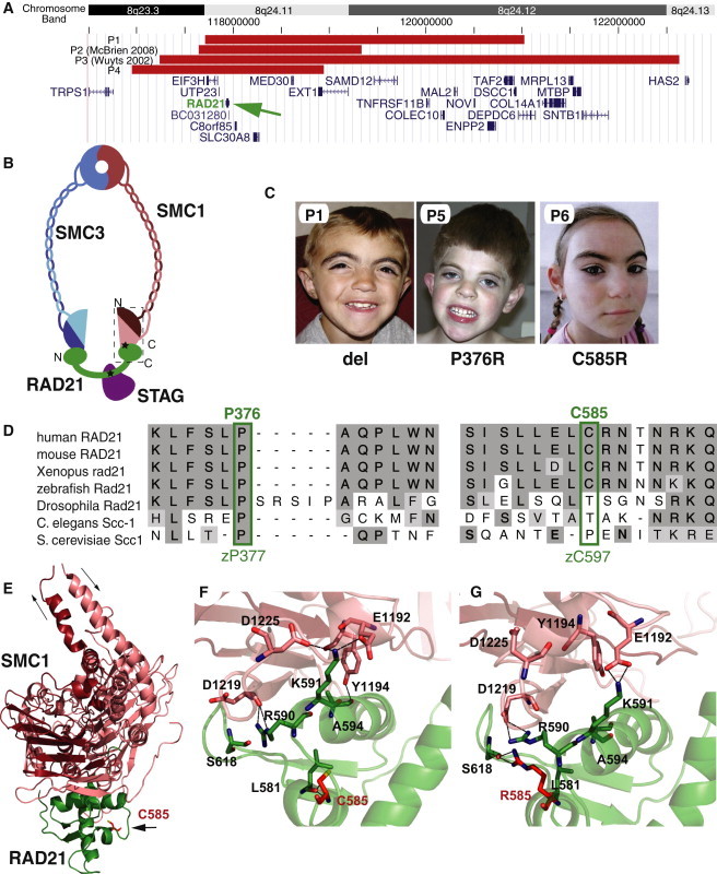
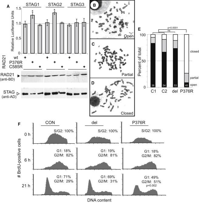
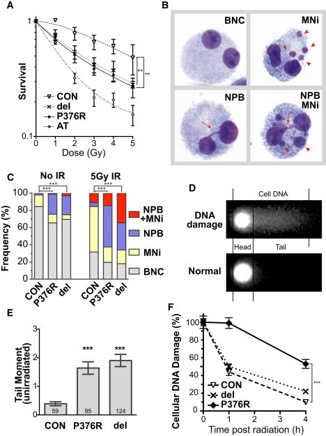
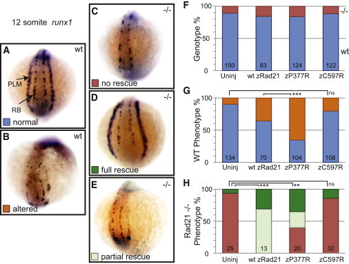
The evolutionarily conserved cohesin complex was originally described for its role in regulating sister-chromatid cohesion during mitosis and meiosis. Cohesin and its regulatory proteins have been implicated in several human developmental disorders, including Cornelia de Lange (CdLS) and Roberts syndromes. Here we show that human mutations in the integral cohesin structural protein RAD21 result in a congenital phenotype consistent with a "cohesinopathy." Children with RAD21 mutations display growth retardation, minor skeletal anomalies, and facial features that overlap findings in individuals with CdLS. Notably, unlike children with mutations in NIPBL, SMC1A, or SMC3, these individuals have much milder cognitive impairment than those with classical CdLS. Mechanistically, these mutations act at the RAD21 interface with the other cohesin proteins STAG2 and SMC1A, impair cellular DNA damage response, and disrupt transcription in a zebrafish model. Our data suggest that, compared to loss-of-function mutations, dominant missense mutations result in more severe functional defects and cause worse structural and cognitive clinical findings. These results underscore the essential role of RAD21 in eukaryotes and emphasize the need for further understanding of the role of cohesin in human development.


Figures

Figure 1
Figure 1
Mutations in RAD21 (A) Deletions including RAD21. Localization of deletions (red bars) on 8q24 is indicated by chromosome band and position (hg 18). Gene locations are indicated in blue with gene names. RAD21 is indicated by a green arrow. (B) Schematic representation of the cohesin complex. SMC1A (red) and SMC3 (blue) N- (lighter) and C- (darker) termini are indicated. Relative positions of RAD21 binding to SMC1A via its C terminus and to STAGs via its central region are noted. Black stars indicate relative positions of missense mutations. A dashed box indicates the region represented in the crystal structure in (D). (C). Facial features of children with RAD21 mutations. del = RAD21 deletion, P376R = p.Pro376Arg, and C585R = p.Cys585Arg mutations. (D) Conservation of RAD21 sequence and position of missense mutations. (E) Localization of the RAD21 p.Cys585Arg (C585) mutation near the interface with SMC1. (F) Detail of SMC1 wild-type RAD21 molecular interface. (G) Modeled effect of RAD21 p.Cys585Arg mutation on SMC1 interaction.
Figure 2
Figure 2
RAD21 Alteration Effects on the Cohesin Complex (A) RAD21 p.Pro376Arg increases binding affinity to mitotic STAG proteins. Immunoblotting of similarly expressed hybrid proteins is indicated below. Significant p values (<0.05) from two-tailed t tests are indicated (). Open (B), partially open (C) and closed (D) sister-chromatid phenotypes are demonstrated. (E) Increased prevalence of closed sister chromatids in LCLs from the boy with the p.Pro376Arg mutation and in normal control LCLs (C1, C2). The RAD21-deleted LCLs (del) show no difference from controls. The number of metaphases analyzed for each cell line is indicated in white at the base. Chi-square calculations of significance in difference of all cell phenotypes of RAD21 mutant cell lines compared with the averaged controls are indicated above the columns. (F) Flow cytometry smoothened histogram plots of BrdU-positive cells versus DNA content showing the proportions of G1, S, and G2/M cells 0, 6, and 21 hr after BrdU labeling. The proportion of cells in each cell cycle phase is expressed as the percentage of BrdU-positive cells. Two-sided Fisher's exact test is shown. Error bars indicate standard deviations. del = RAD21 deletion, P376R = p.Pro376Arg, C585R = p.Cys585Arg mutations, and CON = control cells.
Figure 3
Figure 3
RAD21 Mutations Increase Susceptibility to DNA Damage (A) Cell survival after a graded dose of ionizing radiation (IR). Radiation doses are indicated beneath the samples. Fractional survival was expressed relative to unirradiated cells. Control (CON), ATM-mutated (AT), RAD21-deleted (del), and RAD21 p.Pro376Arg-mutated (P376R) lymphoblastoid cell lines are indicated. Each data point represents the mean of three independent experiments. Error bars represent SEM. (B) Representative images of binucleated cells (BNCs, top left) with micronuclei (MNi, top right, arrowheads); nucleoplasmic bridges (NPBs, bottom left, arrows), and NPBs accompanying MNi (bottom right panel). (C) The frequencies of spontaneous and radiation-induced MNi, NPBs, and NPBs accompanying MNi, as shown in (B). The frequencies were calculated per 200 binucleated cells. (D) A diagram of the Comet assay, indicating the head and tail dimensions used for assessments. (E) The basal level of DNA damage, as measured by the tail moment in unirradiated cells via the Comet assay. A significant difference was observed between the control cell line (CON) and the RAD21 mutant cell lines. Numbers at base of column indicate number of nuclei assayed. (F) DNA damage repair kinetics after IR at 8 Gy. Cellular DNA damage was measured as the tail moment and expressed relative to 0 hr after irradiation. Also see Figure S7. Significant p values for p < 0.0001 (∗∗∗) and p < 0.01 (∗∗) are noted. del = RAD21 deletion, P376R = p.Pro376Arg, and C585R = p.Cys585Arg mutations.
Figure 4
Figure 4
RAD21 Mutations Alter Transcriptional Activity Rescue of lateral-plate-mesoderm expression of runx1 in Rad21-null zebrafish embryos with zebrafish rad21 mRNA that are either wild-type (wt zRad21) or carrying the equivalent human mutations (see Figure 1D). (A) Wild-type, uninjected 12 somite embryo viewed from the posterior. PLM = posterior lateral-plate mesoderm and RB = Rohon-Beard cells. (B) Wild-type embryo with altered runx1 expression. (C) Rad21 homozygous null embryo with loss of PLM runx1 expression. (D and E) Rad21 homozygous null embryos with full and partial rescues by mRNA injection. (F) Genotype fractions of embryos analyzed for each injection cohort. Red = Rad21-null, blue = wild-type, uninj = uninjected. (G) Frequency of altered runx1 expression in wild-type embryos. Orange = altered expression and blue = normal expression. (H) Frequency of runx1 LPM rescue. Green = full rescue, light green = partial rescue, and red = no rescue. Error bars indicate standard deviations. Significant p values for p < 0.0001 (∗∗∗) and p < 0.01 (∗∗) are noted.

References

    1. Nasmyth K., Haering C.H. Cohesin: Its roles and mechanisms. Annu. Rev. Genet. 2009;43:525–558. - PubMed
    1. Shintomi K., Hirano T. Releasing cohesin from chromosome arms in early mitosis: Opposing actions of Wapl-Pds5 and Sgo1. Genes Dev. 2009;23:2224–2236. - PMC - PubMed
    1. Watrin E., Schleiffer A., Tanaka K., Eisenhaber F., Nasmyth K., Peters J.M. Human Scc4 is required for cohesin binding to chromatin, sister-chromatid cohesion, and mitotic progression. Curr. Biol. 2006;16:863–874. - PubMed
    1. Hou F., Zou H. Two human orthologues of Eco1/Ctf7 acetyltransferases are both required for proper sister-chromatid cohesion. Mol. Biol. Cell. 2005;16:3908–3918. - PMC - PubMed
    1. Hadjur S., Williams L.M., Ryan N.K., Cobb B.S., Sexton T., Fraser P., Fisher A.G., Merkenschlager M. Cohesins form chromosomal cis-interactions at the developmentally regulated IFNG locus. Nature. 2009;460:410–413. - PMC - PubMed

Publication types

MeSH terms

Supplementary concepts