Review
. 2012 Aug;16(3-4):292-9.
doi: 10.1016/j.cbpa.2012.04.010. Epub 2012 May 25.

Light-controlled synthetic gene circuits

Affiliations
Review

Light-controlled synthetic gene circuits

Laura Gardner et al. Curr Opin Chem Biol. 2012 Aug.

Abstract

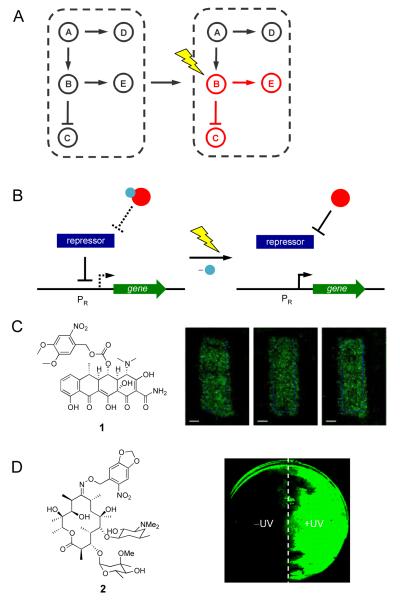
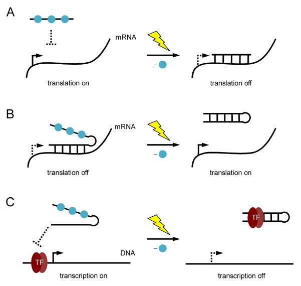
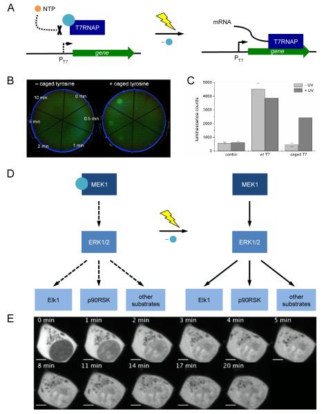
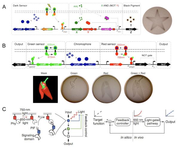
Highly complex synthetic gene circuits have been engineered in living organisms to develop systems with new biological properties. A precise trigger to activate or deactivate these complex systems is desired in order to tightly control different parts of a synthetic or natural network. Light represents an excellent tool to achieve this goal as it can be regulated in timing, location, intensity, and wavelength, which allows for precise spatiotemporal control over genetic circuits. Recently, light has been used as a trigger to control the biological function of small molecules, oligonucleotides, and proteins involved as parts in gene circuits. Light activation has enabled the construction of unique systems in living organisms such as band-pass filters and edge-detectors in bacterial cells. Additionally, light also allows for the regulation of intermediate steps of complex dynamic pathways in mammalian cells such as those involved in kinase networks. Herein we describe recent advancements in the area of light-controlled synthetic networks.


Figures

Figure 1
Figure 1
(A) Light enables precise spatial and temporal activation of genetic circuits, even the activation of specific nodes in a natural or synthetic network. (B) Control over gene expression with photocaged small molecule inducers of transcription. The photocaging group is represented by a blue sphere and the small molecule inducer is represented by a red sphere. When the small molecule is caged, the repressor protein will bind the promoter PR. After UV irradiation the caging group is removed and the small molecule will bind the repressor, which releases PR, and allows for transcription to occur. (C) NvOC-Dox (1) was used to create photolithographic images onto an NIH 3T3 monolayer expressing GFP. (D) Photocaged erythromycin (2) was used in the spatial control of a light activated logic gate; cells treated with 2 where one half of the plate was exposed to UV light. Adapted with permission from the American Chemical Society, © 2010, from [17] and the Royal Society of Chemistry, © 2011, from [18].
Figure 2
Figure 2
(A) Photochemical activation of gene silencing using a caged antisense agent. Photocaging groups (blue spheres) were placed on the antisense agent to prevent hybridization to its target mRNA. UV irradiation induces decaging and gene silencing. (B) Photochemical deactivation of an antisense agent. Hairpin formation of the antisense agent is disrupted through nucleobase caging, allowing the antisense agent to bind its target mRNA and downregulate translation. After UV irradiation, hairpin formation occurs rendering the antisense agent inactive and activating gene expression. (C) Photochemical control of transcription using a caged DNA decoy. The caged decoy is unable to form a hairpin and does not bind the transcription factor NF-κB. After UV irradiation, the DNA decoy forms a hairpin and is able to bind the transcription factor.
Figure 3
Figure 3
(A) Schematic representation of the photocaged T7RNAP system. The photocaging group is represented by a blue sphere. NTPs are blocked from entering the active site of T7RNAP when the enzyme is caged. After UV irradiation the caging group is removed, NTPs are free to enter the active site and transcription is activated. (B) Photoactivation of EGFP expression in E. coli through light-activation of a photocaged T7RNAP; EGFP expression is restored after 10 minutes exposure to localized 365 nm irradiation. (C) Light-activation of T7RNAP activity and gene expression in vivo. (D) Light-activated MAP kinase signaling pathway. The photocaging group is represented by a blue sphere. (E) Light-activated translocation of EGFP-tagged ERK into the nucleus of HEK293 cells. Adapted with permission from Wiley-VCH Verlag GmbH & Co, © 2010, from [37] and from the American Chemical Society, © 2011, from [36].
Figure 4
Figure 4
(A) Synthetic edge detector for spatiotemporal control over gene expression in E coli. Exposure to light inhibits the diffusion signal X and β-galactosidase expression Z. When cells are in the dark (NOT light), X and Y are turned on. Y functions as an inverter, thus X AND (NOT Y) generates the signal Z. (B) A multichromic light-inducible gene expression system. The green sensor turns on one β-galactosidase reporter to generate Z in response to green light. When exposed to red light, the green sensor is turned off, and the red sensor is turned on to depress Y and activate the second β-galactosidase reporter Z. (C) Light-regulated network that uses feedback to adjust input levels to create stable output patterns. The PIF/PhyB system responds to 650 nm light to produce an output from a signaling domain. This output is then fed back through a controller which will alter light input levels to maintain a desired cellular output. Adapted with permission from Elsevier, © 2009, from [42] and © 2010, from [43], and from Nature Publishing Group, © 2011, from [40].

References

    1. Agapakis CM, Silver PA. Synthetic biology: exploring and exploiting genetic modularity through the design of novel biological networks. Mol Biosyst. 2009;5:704–713. - PubMed
    1. Tanouchi Y, Pai A, You L. Decoding biological principles using gene circuits. Mol Biosyst. 2009;5:695–703. - PubMed
    1. Weber W, Fussenegger M. Molecular diversity--the toolbox for synthetic gene switches and networks. Curr Opin Chem Biol. 2011;15:414–420. - PubMed
    1. Deiters A. Light activation as a method of regulating and studying gene expression. Curr Opin Chem Biol. 2009;13:678–686. - PMC - PubMed
    1. Deiters A. Principles and applications of the photochemical control of cellular processes. Chembiochem. 2010;11:47–53. - PMC - PubMed

Publication types

LinkOut - more resources