. 2012 Sep;51(3):488-97.
doi: 10.1016/j.bone.2012.05.014. Epub 2012 May 23.

Alterations in the osteocyte lacunar-canalicular microenvironment due to estrogen deficiency

Affiliations

Alterations in the osteocyte lacunar-canalicular microenvironment due to estrogen deficiency

Divya Sharma et al. Bone. 2012 Sep.

Abstract

While reduced estrogen levels have been shown to increase bone turnover and induce bone loss, there has been little analysis of the effects of diminished estrogen levels on the lacunar-canalicular porosity that houses the osteocytes. Alterations in the osteocyte lacunar-canalicular microenvironment may affect the osteocyte's ability to sense and translate mechanical signals, possibly contributing to bone degradation during osteoporosis. To investigate whether reduced estrogen levels affect the osteocyte microenvironment, this study used high-resolution microscopy techniques to assess the lacunar-canalicular microstructure in the rat ovariectomy (OVX) model of postmenopausal osteoporosis. Confocal microscopy analyses indicated that OVX rats had a larger effective lacunar-canalicular porosity surrounding osteocytes in both cortical and cancellous bone from the proximal tibial metaphysis, with little change in cortical bone from the diaphysis or cancellous bone from the epiphysis. The increase in the effective lacunar-canalicular porosity in the tibial metaphysis was not due to changes in osteocyte lacunar density, lacunar size, or the number of canaliculi per lacuna. Instead, the effective canalicular size measured using a small molecular weight tracer was larger in OVX rats compared to controls. Further analysis using scanning and transmission electron microscopy demonstrated that the larger effective canalicular size in the estrogen-deficient state was due to nanostructural matrix-mineral level differences like loose collagen surrounding osteocyte canaliculi. These matrix-mineral differences were also found in osteocyte lacunae in OVX, but the small surface changes did not significantly increase the effective lacunar size. The alterations in the lacunar-canalicular surface mineral or matrix environment appear to make OVX bone tissue more permeable to small molecules, potentially altering interstitial fluid flow around osteocytes during mechanical loading.


Figures

Fig. 1
Fig. 1
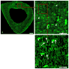
(a) A typical tibial diaphysis section from an OVX rat created from approximately 50 montaged confocal images; scale bar = 300 μm. (b) Enlarged image of square region in (a), which demonstrates the high resolution of the images; scale bar = 50 μm. (c) Enlarged image of the square region in (b), further illustrating the details of the lacunar-canalicular network; scale bar = 10 μm. Osteocyte lacunae (short arrows), and canaliculi (long arrows) are indicated in (b) and (c).
Fig. 2
Fig. 2
(a) A montaged light micrograph of the proximal rat tibia showing the cancellous regions sampled with 375 μm × 375 μm field of view images from the metaphysis (dashed line boxes) and the epiphysis (solid line boxes); scale bar = 800 μm. Typical metaphysis (b) and epiphysis (c) confocal images; osteocyte lacunae (short arrows) are indicated; scale bars = 50 μm.
Fig. 3
Fig. 3
SEM images of cancellous bone from the tibial metaphysis for (a) a CTRL specimen and (b) an OVX specimen showing canalicular openings (white arrows); loose collagen fibers are visible in the OVX specimen (black arrow). Canalicular diameter was measured as the smallest dimension of the opening (black lines). (c) TEM image of cancellous bone from the tibial metaphysis from SHAM showing canalicular cross-sections and measurement of canalicular diameter (black lines). TEM sections were counterstained with uranyl acetate for 40 minutes and lead citrate for 5 minutes.
Fig. 4
Fig. 4
(a) Schematic of cubic volume of bone (average side length L) surrounding one osteocyte lacuna. Secondary canaliculi intersect the faces of the cube. (b) Confocal scan of cubic volume of bone tissue centered on one osteocyte lacuna demonstrating the canalicular network spanning in all directions; scale bar = 5 μm. (c) Confocal scan of a 25 μm × 25 μm × 25 μm cubic volume surrounding one osteocyte lacuna with only secondary canaliculi rendered viewable on all six faces of the cube; scale bar = 5 μm. (d) Cropped scan from (c) showing primary canaliculi emanating directly from the osteocyte lacuna; scale bar = 3 μm. (e) Isolated osteocyte lacuna from same scan constructed in Mimics software; scale bar = 2 μm. (f) Higher resolution scan of canaliculi taken between two osteocytes; scale bar = 2 μm.
Fig. 5
Fig. 5
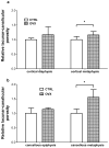
Relative lacunar-canalicular porosities measured using 2D confocal microscopy for (a) cortical diaphysis and cortical metaphysis cross-sections and (b) cancellous bone from the epiphysis and metaphysis of the proximal tibia. All measurements were scaled to the CTRL mean for each porosity; bars represent mean values ± standard deviations; *statistical difference between CTRL (n=6) and OVX (n=6) (p < 0.05).
Fig. 6
Fig. 6
(a) Confocal reconstructed image of a typical OVX tibial metaphysis cross-section (cancellous bone in the medullary region was not preserved in these unembedded sections); scale bar = 400 μm. (b) Confocal and (c) backscattered electron images of the rectangular region indicated in (a); the region of trabecular remnants is enclosed within the yellow lines and lamellar bone is marked with red asterisks. Scale bar in (c) = 200 μm. (d) Confocal and (e) backscattered electron images of the same portion of a CTRL tibial diaphysis section; a red line traces the same pathway in each image. Areas with an absent or disconnected canalicular network (black regions surrounding osteocyte lacunae in the confocal images) correspond to higher mineralization (whiter regions in the backscattered electron images), whereas greater canalicular density areas correspond to areas of lower mineralization. Scale bar in (e) = 200 μm.
Fig. 7
Fig. 7
Box plots showing 3D confocal microscopy measurements of canalicular volume (%) from cortical bone from the proximal tibial metaphysis. + indicates the mean of each group. A significant difference (34% increase) in canalicular volume was found between SHAM (n=6) and OVX (n=6) (*p < 0.05).
Fig. 8
Fig. 8
TEM images of cancellous bone from the tibial metaphysis showing (a) smooth surfaces in a SHAM osteocyte lacuna (red arrows) and (b) collagen fibers in an OVX lacuna giving the border a rough appearance (yellow arrow). (c) Smooth SHAM canaliculi (red arrow); scale bar = 500 nm and (d) OVX canaliculus with collagen fibers at the surface (yellow arrow); scale bar = 500 nm.

References

    1. McNamara LM, Majeska RJ, Weinbaum S, Friedrich V, Schaffler MB. Attachment of osteocyte cell processes to the bone matrix. Anat Rec (Hoboken) 2009;292:355–63. - PMC - PubMed
    1. Burger EH, Klein-Nulend J. Mechanotransduction in bone--role of the lacuno-canalicular network. FASEB J. 1999;13 (Suppl):S101–12. - PubMed
    1. Bonewald LF. Osteocytes as dynamic multifunctional cells. Ann N Y Acad Sci. 2007;1116:281–290. - PubMed
    1. Fritton SP, Weinbaum S. Fluid and solute transport in bone: flow-induced mechanotransduction. Annu Rev Fluid Mech. 2009;41:347–74. - PMC - PubMed
    1. Han Y, Cowin SC, Schaffler MB, Weinbaum S. Mechanotransduction and strain amplification in osteocyte cell processes. Proc Natl Acad Sci U S A. 2004;101:16689–94. - PMC - PubMed

Publication types