. 2012 Aug;236(2):283-97.
doi: 10.1016/j.expneurol.2012.05.012. Epub 2012 May 23.

Identification of radial glia-like cells in the adult mouse olfactory bulb

Affiliations

Identification of radial glia-like cells in the adult mouse olfactory bulb

Jason G Emsley et al. Exp Neurol. 2012 Aug.

Abstract

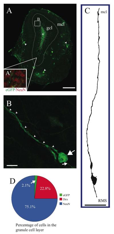
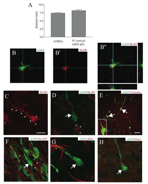
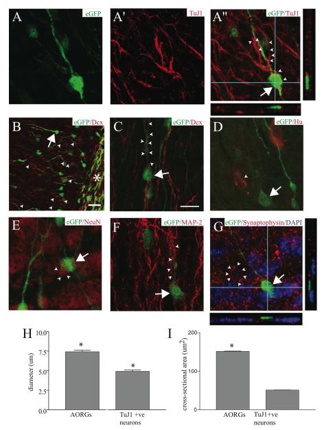
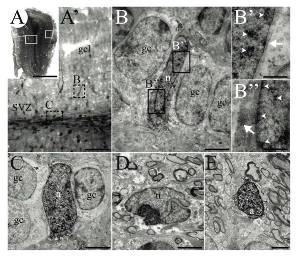
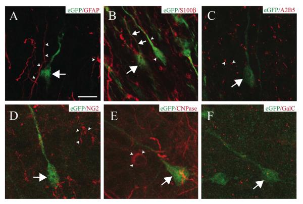
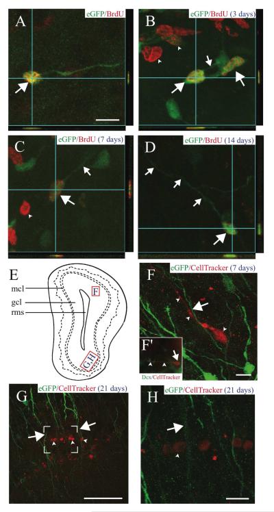
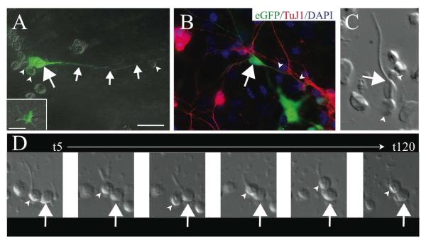
Immature neurons migrate tangentially within the rostral migratory stream (RMS) to the adult olfactory bulb (OB), then radially to their final positions as granule and periglomerular neurons; the controls over this transition are not well understood. Using adult transgenic mice with the human GFAP promoter driving expression of enhanced GFP, we identified a population of radial glia-like cells that we term adult olfactory radial glia-like cells (AORGs). AORGs have large, round somas and simple, radially oriented processes. Confocal reconstructions indicate that AORGs variably express typical radial glial markers, only rarely express mouse GFAP, and do not express astroglial, oligodendroglial, neuronal, or tanycyte markers. Electron microscopy provides further supporting evidence that AORGs are not immature neurons. Developmental analyses indicate that AORGs are present as early as P1, and are generated through adulthood. Tracing studies show that AORGs are not born in the SVZa, suggesting that they are born either in the RMS or the OB. Migrating immature neurons from the adult SVZa are closely apposed to AORGs during radial migration in vivo and in vitro. Taken together, these data indicate a newly-identified population of radial glia-like cells in the adult OB that might function uniquely in neuronal radial migration during adult OB neurogenesis.


Figures

FIGURE 1
FIGURE 1. Location, morphology, and abundance of AORGs
(A) Photomontage of a coronal section of the adult OB in an hGFAP-eGFP transgenic mouse, in which eGFP is expressed under the control of a human GFAP promoter fragment, showing the position and orientation of AORGs in the granule cell layer (gcl; box B), extending their processes from the base of the RMS (asterisk) toward the gcl. Inner white line denotes outer perimeter of the RMS (*); outer white line denotes outer perimeter of the gcl; arrowheads indicate clusters of astroglia in the mitral cell layer (mcl). Inset in A (A’) shows a magnified view of the granule cell layer, with NeuN in red, and AORGs in green). The box (B) in panel A denotes the approximate region from which panel B is derived. Scale bar, 500 ⌈m. (B) Confocal reconstruction (z-stack) of a typical AORG in the adult OB. Note the simple, elongated, and unbranched apical (arrowheads) and basal (small arrow) radial processes, and the large, round soma. Scale bar, 10 ⌈m. (C) Camera lucida drawing of a typical AORG, oriented from the RMS to the mcl. Scale bar, 50 ⌈m. (D) AORGs comprise a very small percentage of cells in the granule cell layer of the adult OB (2.1%), compared to mature, NeuN-positive granule cells (75.1%) and immature, Dcx-positive migrating neurons (22.8%). (AORG, adult olfactory radial glia-like cell; RMS, rostral migratory stream; gcl, granule cell layer; mcl, mitral cell layer).
FIGURE 2
FIGURE 2. AORGs are morphologically and immunochemically similar to developmental cortical radial glia
(A) AORGs are similar in size to radial glia present during early postnatal neocortical development (6 mice per assessment). (B, B’, and B”) A subset of AORGs expresses the radial glial marker BLBP, as shown with images of eGFP alone (B, green), BLBP alone (B’, red) and merged (B”, shown with orthogonal reconstruction in the XZ and YZ planes). (C) Some BLBP-positive cells are eGFP-negative, but have AORG-like morphology (outlined by arrowheads). (D) Some non-radial cells express BLBP (arrowheads), and some eGFP-positive AORGs (green, arrow) do not express BLBP (red). (E-H) AORGs (green) do not express any of a panel of other radial glial markers at high level, including vimentin (E, inset, indicated by an asterisk); GLAST (F, red, arrowheads); 40EC (G, arrowhead), or Hes5 (H, red). Scale bar in (C), 10 ⌈m for panels (B) through (D) and (F) through (H); scale bar in (E), 25 ⌈m.
FIGURE 3
FIGURE 3. AORGs are not neurons
High magnification three-dimensional confocal reconstructions of AORGs in the adult OB indicate that AORGs (arrows; green in all panels) do not express any of a variety of markers of progressive neuronal differentiation. (A through A”) Confocal reconstruction showing an AORG in the OB (A, green) closely apposed to cells that express the immature neuronal marker TuJ1 (A’, red, and merged A”); the arrow indicates the soma of the AORG, and arrowheads indicate TuJ1-positive fibers and their close apposition to the radially extended process of the AORG. AORGs do not express TuJ1. (B-G) AORGs also do not express a broad set of other immature and mature neuronal markers (shown in red). AORGs do not express the immature neuronal marker doublecortin, Dcx (B and C, arrowheads). (B) Low magnification view of the end of the RMS (asterisk), indicating substantial co-expression of eGFP and Dcx in many migrating immature neurons within and proximal to the RMS. Within the granule cell layer, there are numerous Dcx-positive cell bodies (arrowheads) and fine processes, as well as an eGFP-positive AORG (arrow). (C) High magnification image of an AORG (arrow) in close association with Dcx-positive processes of immature migrating neurons (arrowheads). (D-F) AORGs do not express the post-mitotic neuronal marker Hu (D, arrowhead), the mature neuronal nuclear marker NeuN (E, arrowheads), or the neuron-specific, somato-dendritic microtubule-associated protein MAP-2 (F, arrowheads). (G) AORGs are also not labeled by the presynaptic protein synaptophysin (neuron rimmed by synapses, arrowheads; DAPI nuclear counterstain is in blue). Panels (A”) and (G) include orthogonal reconstructions in the XZ and YZ planes to demonstrate lack of co-localization of eGFP with TuJ1 or synaptophysin. Scale bar in (C) for all panels, 10 ⌈m, except for (B), in which the scale bar is 25 ⌈m. (H, I) AORGs are significantly larger than TuJ1-positive immature neurons, in both soma diameter and cross-sectional area (*, p < 0.0001; 6 mice per assessment).
FIGURE 4
FIGURE 4. AORGs resemble immature glia ultrastructurally
(A) Photomontage of a coronal section of the adult OB from an hGFAP-eGFP transgenic mouse in which expression of GFP is reported via immunoperoxidase staining. White boxed areas (left, right) indicate approximate regions from which images shown in A’ and E were obtained, respectively. (A’) Higher magnification image of left boxed area in A, showing deep layers of the OB, granule cell layer, and the anterior extension of the RMS. Note that the DAB precipitate reveals cells of different intensity in the granule cell layer and SVZ. Dashed boxes indicate areas shown in B and C. (B, B’, and B”) An example of an AORG, found between mature granule cells. (B’ and B”) Higher magnification of boxed areas in B, showing the DAB immunoprecipitate (arrowheads indicate precipitate in nuclei; arrows indicate cytoplasmic precipitate). (C) Another example of an identified AORG positioned between granule cells. (D) An example of a neuroblast with a typical invaginated nucleus within the SVZ/RMS; such neuroblasts are usually lightly labeled. (E) A typical astrocyte in the periglomerular region. Scale bar in (A), 1 mm, (A’) 250 μm; for panels (B) through (E), 30 μm and for (B’ and B”), 5 μm. (a, astrocyte; gc, granule cell; gcl, granule cell layer; n, nucleus).
FIGURE 5
FIGURE 5. AORGs rarely express astroglial markers, and are not oligodendroglia
(A) High magnification confocal three-dimensional reconstructions reveal that AORGs (green, arrow) do not express GFAP (red, arrowheads indicate GFAP-positive astroglial processes). (B) AORGs occasionally express S100®; an AORG (large arrow) with an S100®-positive process (small arrows). AORGs are often immunonegative for S100® (arrowheads). (C) AORGs (arrows) do not express A2B5 (arrowheads). (D-F) AORGs (arrows) do not express the oligodendroglial markers NG2 (D, arrowheads), CNPase (E, arrowheads), or GalC (F). Scale bar in (A), 10 ⌈m for panels A through F.
FIGURE 6
FIGURE 6. AORGs are born in the adult OB, and are closely apposed to migrating, immature neurons
(A) Newborn AORGs (arrows) are found in the adult OB granule cell layer following adult BrdU administration. Co-labeling by BrdU (red) and eGFP (green) in AORGs was confirmed by three-dimensional confocal orthogonal reconstructions in the XZ and YZ planes. (B-D) Pulse labels of BrdU demonstrate that (B) AORGs (arrows) develop short processes (small arrow) by 3 days after their birth. Note eGFP-negative/BrdU-positive cells in (B) and (C) (arrowheads, putative adult-born granule neurons). (C) AORGs extend their processes further by 7 days (small arrow), and (D) display mature AORG morphology by 14 days after their birth. (E) Schematic representation of the olfactory bulb, indicating the regions from which panels F, G, and H are derived. (F) The whole cell label CellTracker Red was injected into the adult SVZa, revealing a close association between immature, adult-born migrating neurons (red, arrowheads) and the radial process of eGFP-positive AORGs (green, arrow) 7 days after CellTracker Red injection. (F’, inset) an example of a cell labeled by CellTracker Red that is immunopositive for doublecortin (Dcx, green), in the granule cell layer of the OB. Arrow indicates the cell body, and arrowheads mark the leading process of this migrating, immature neuron. (G and H) The close anatomical relationship between adult-born mature granule neurons (arrowheads) in the gcl and the radially oriented eGFP-positive fibers of AORGs (arrows) 21 days after CellTracker Red injection is shown via confocal reconstructions at lower (G; box indicates area magnified in (H)) and higher magnification (H). Scale bar in (A), 10 ⌈m for panels (A-D); scale bar in each of (F-H), 50 ⌈m. (gcl, granule cell layer; mcl, mitral cell layer; rms, rostral migratory stream). Schematic diagram based on Franklin and Paxinos (1997).
FIGURE 7
FIGURE 7. Immature OB neurons are closely apposed to AORG radial processes
in vitro (A) Dissociated culture of the developing OB shows an eGFP-positive AORG (arrow) with an extensive linear process (small arrows). Smaller presumptive immature neurons (arrowheads) are often located in close apposition to the somas and radial processes of AORGs. In culture, as in vivo, AORGs are morphologically different from eGFP-positive astrocytes (inset in A). (B) Processes of TuJ1-positive immature neurons (red, arrowheads) are adjacent to the radial process of a mature AORG (arrow). (C) Dissociated cells at P14 illustrate the close apposition between immature neurons (arrowheads) and the soma and radial process of an AORG (arrow). (D) Time-lapse videomicroscopy suggests that immature neurons (arrowheads) are capable of attaching to radial processes of AORGs (arrow) and sometimes initiate migration. Representative still images are shown from t=5 minutes to t=120 minutes. Scale bar in (A) for panels (A) and (B), 25 ⌈m.

Similar articles

Cited by

References

    1. Alvarez-Buylla A, Buskirk DR, Nottebohm F. Monoclonal antibody reveals radial glia in adult avian brain. Journal of Comparative Neurology. 1987;264:159–170. - PubMed
    1. Alves JA, Barone P, Engelender S, Froes MM, Menezes JR. Initial stages of radial glia astrocytic transformation in the early postnatal anterior subventricular zone. Journal of Neurobiology. 2002;52:251–265. - PubMed
    1. Anthony TE, Klein C, Fishell G, Heintz N. Radial glia serve as neuronal progenitors in all regions of the central nervous system. Neuron. 2004;41:881–890. - PubMed
    1. Au E, Roskams AJ. Olfactory ensheathing cells of the lamina propria in vivo and in vitro. Glia. 2003;41:224–236. - PubMed
    1. Bailey MS, Puche AC, Shipley MT. Development of the olfactory bulb, evidence for glia-neuron interactions in glomerular formation. Journal of Comparative Neurology. 1999;415:423–448. - PubMed

Publication types