. 2012 Aug 15;84(4):444-50.
doi: 10.1016/j.bcp.2012.05.014. Epub 2012 May 23.

Interaction of pseudolaric acid B with the colchicine site of tubulin

Affiliations

Interaction of pseudolaric acid B with the colchicine site of tubulin

Taradas Sarkar et al. Biochem Pharmacol. .

Abstract

We purified pseudolaric acid B (PAB) from the root and stem bark of Pseudolarix kaempferi (Lindl.) Gorden. Confirming previous findings, we found that the compound had high nanomolar IC₅₀ antiproliferative effects in several cultured cell lines, causing mitotic arrest and the disappearance of intracellular microtubules. PAB strongly inhibited tubulin assembly (IC₅₀, 1.1 μM) but weakly inhibited the binding of colchicine to tubulin, as demonstrated by fluorescence and with [³H]colchicine. Kinetic analysis demonstrated that the mechanism of inhibition was competitive, with an apparent K(i) of 12-15 μM. Indirect studies demonstrated that PAB bound rapidly to tubulin and dissociated more rapidly from tubulin than the colchicine analog 2-methoxy-5-(2',3',4'-trimethoxyphenyl)tropone, whose complex with tubulin is known to have a half-life of 17s at 37 °C. We modeled PAB into the colchicine site of tubulin, using the crystal structure 1SA0 that contains two αβ-tubulin heterodimers, both bound to a colchicinoid and to a stathmin fragment. The binding model of PAB revealed common pharmacophoric features between PAB and colchicinoids, not readily apparent from their chemical structures.


Conflict of interest statement

Conflict of interest statement: The authors declare that there are no conflicts of interest.

Figures

Fig. 1
Fig. 1
Molecular structures of PAB, colchicine, thiocolchicine, MTPT, podophyllotoxin, and combretastatins A-4 and A-2.
Fig. 2
Fig. 2
PAB is a competitive inhibitor of the binding of [3H]colchicine to tubulin. Incubation was for 10 min at 37 °C. Reaction mixtures contained 1.0 μM (0.1 mg/ml) tubulin, which was the last component added to the reaction mixtures. A. Kinetic analysis of binding data. Symbols: no PAB, ○; 10 μM PAB, △; 20 μM PAB, ●; 30 μM PAB; 40 μ M PAB, ▲. Main panel: Lineweaver-Burk plot. The abscissa units are μM−1 colchicine. The ordinate units are 1/pmol colchicine bound. Left inset: Hanes plot. The abscissa units are μM colchicine. The ordinate units are μM colchicine/pmol colchicine bound. Right inset: Woolf plot. The abscissa units are pmol colchicine bound/μM colchicine. The ordinate units are pmol colchicine bound. B. Dixon plot for determination of Ki. Symbols: 2 μM colchicine, △; 3 μM colchicine, ●; 4 μM colchicine, ○; 5 μM colchicine, ◇. The abscissa units are μM PAB. The ordinate units are 1/pmol colchicine bound.
Fig. 3
Fig. 3
Inhibition of colchicine binding, measured by fluorescence, by PAB (○) and podophyllotoxin (●). Fluorescence study was performed following the procedure described by Bhattacharyya and Wolff [4]. Reaction mixtures contained 1 μM tubulin, 5 μM colchicine, and PAB or podophyllotoxin at the indicated concentrations. The symbol △ represents the fluorescence observed with only colchicine in the reaction mixture.
Fig. 4
Fig. 4
Comparison of compound dissociation rates from tubulin, measured indirectly by determining subsequent inhibition of binding of [3H]colchicine. Reaction mixtures (5 ml) contained the components described previously that stabilize tubulin [8, 18], 1.0 μM tubulin (0.1 mg/ml), and inhibitory compounds at 10 μM: thiocolchicine (◇), combretastatin A-2 (△), MTPT (●), or PAB (○). The reaction mixtures were incubated at 37 °C for 30 min and chilled on ice. Aliquots (0.1 ml) of each reaction mixture were distributed into tubes, and 200 pmol of [3H]colchicine (10 μ1) were added to each tube. Samples were then incubated for the indicated times in triplicate at 37 °C. At each time point, the amount of [3H]colchicine bound to tubulin was determined, and the reaction mixtures with inhibitor were compared with the control containing only [3H[colchicine, with the data expressed as the percentage of inhibition of colchicine binding.
Fig. 5
Fig. 5
Comparison of cell line sensitivity to PAB with their content of αβI-tubulin (A) and αβIVa+b-tubulin. The specific values are shown in Table 1.
Fig. 6
Fig. 6
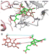
Common pharmacophores of colchicine and PAB, based on docking studies. A. Docked pose of PAB in the colchicine site, superimposed onto that of colchicine derived from the 1SA0 crystal structure [5]. Tubulin is rendered in ribbon, with amino acids within 4 Å shown as thin sticks. Hydrogen bonding amino acids are depicted as thick sticks, with carbon atoms from the α- and β-subunits colored purple and grey, respectively. Hydrogen bonds are highlighted by yellow dashed lines. PAB and colchicine are shown in stick with carbon atoms colored green and brown for PAB and colchicine, respectively. The α-tubulin backbone is tinted light purple, while the β-tubulin backbone is grey. Helices are shown as thick ribbons, and β-sheets as thin strands. Nitrogen atoms are blue, oxygen red, and sulfur yellow. Tubulin hydrogen atoms are white, while those of colchicine and PAB are not shown. B. Binding conformations of colchicine and PAB extracted from the models. Colors as in Panel A. Corresponding pharmacophoric features, as discussed in the text, are indicated. Hydrogen bond acceptors and hydrophobic features are shown as red and blue dashed circles, respectively.

References

    1. Hamel E. An overview of compounds that interact with tubulin and their effects on microtubule assembly. In: Fojo AT, editor. Microtubule targets in cancer therapy. Totowa, New Jersey: Humana Press; 2008. pp. 1–20.pp. 2008
    1. Wong VKW, Chiu P, Chung SSM, Chow LMC, Zhao YZ, Yang BB, Ko BCB. Pseudolaric acid B, a novel microtubule-destabilizing agent that circumvents multidrug resistance phenotype and exhibits antitumor activity in vivo. Clin Cancer Res. 2005;11:6002–11. - PubMed
    1. Tong YG, Zhang XW, Geng MY, Yue JM, Xin XL, Tian F, Shen X, Tong LJ, Zhang C, Li WH, Lin LP, Ding LP. Pseudolarix Acid B, a new tubulin-binding agent, inhibits angiogenesis by interacting with a novel binding site on tubulin. Mol Pharmacol. 2006;69:1226–33. - PubMed
    1. Bhattacharyya B, Wolff J. Promotion of fluorescence upon binding of colchicine to tubulin. Proc Natl Acad Sci USA. 1974;71:2627–31. - PMC - PubMed
    1. Ravelli RBG, Gigant B, Curmi PA, Jourdain I, Lachkar S, Sobel A, Knossow M. Insight into tubulin regulation from a complex with colchicine and a stathmin-like domain. Nature. 2004;428:198–202. - PubMed

Publication types

MeSH terms