

doi: 10.1038/nn.3128.
A color-coding amacrine cell may provide a blue-off signal in a mammalian retina
Affiliations
- PMID: 22634731
- PMCID: PMC3386466
- DOI: 10.1038/nn.3128
Item in Clipboard
A color-coding amacrine cell may provide a blue-off signal in a mammalian retina
Nat Neurosci.
.
Abstract
Retinal amacrine cells are thought to lack chromatic or color-selective light responses and have only a minor role in color processing. We found that a type of mammalian (Ictidomys tridecemlineatus) amacrine cell selectively carries a blue-On signal, which is received from a blue or short wavelength-sensitive (S) cone On bipolar cell. This glycinergic inhibitory S-cone amacrine cell is ideally placed for driving blue-Off responses in downstream ganglion cells.
Figures
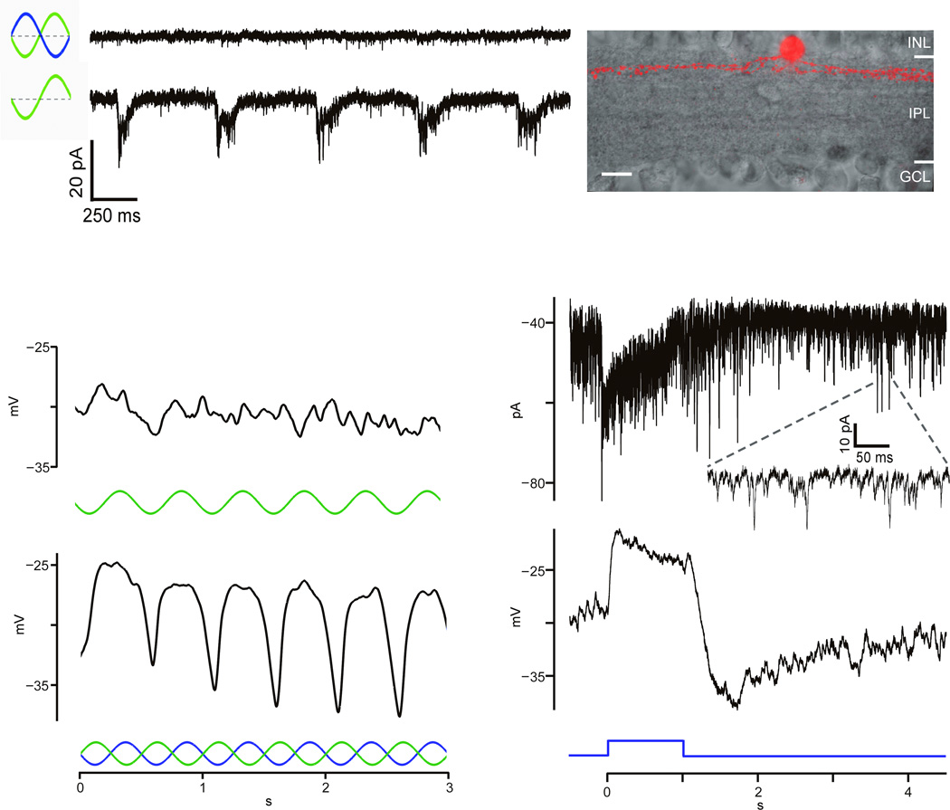
a) An amacrine cell is unresponsive to SCIS (upper) but responsive to green LED (lower); one–tier ramification (right). b) Sample SCA unresponsive to green LED stimulus (upper) but responsive to SCIS (lower). c) Sample SCA current (upper) and voltage (lower) responses to a 1s blue light pulse. Inset:spontaneous EPSCs.
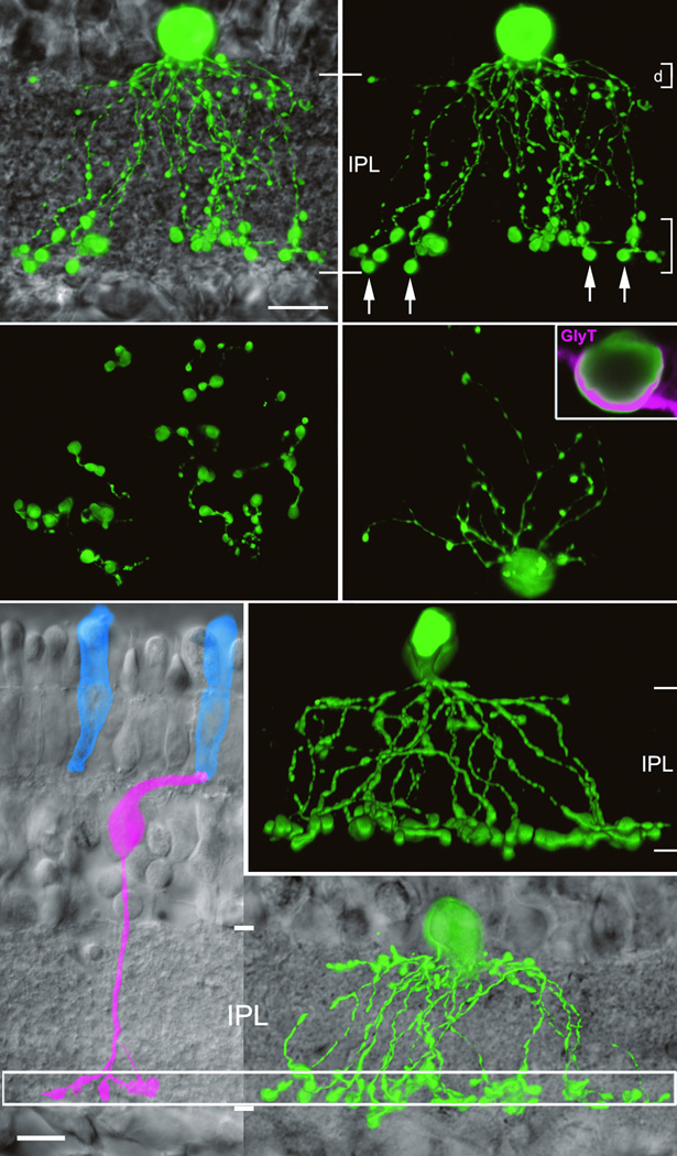
a) Tracer–filled SCA (green) against DIC background. b) Dual dendritic arbors shown as flatmounts in c) and d). Arrows indicate sample varicosities likely to be synaptic sites. Inset d) SCAs immunolabeled by anti– glycine transporter (magenta). f,g)Additional examples of SCAs. e) An SCB (magenta) contacts a single S–cone (blue). Aligning IPLs of e and f, SCA varicosities co–stratify with SCB axon terminals (box).
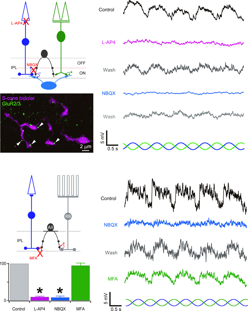
a) Proposed SCA pathway; below: SCB axon terminals (magenta) opposite puncta immunolabeled for GluR2/3 (green). Right: abolition of SCA responses to the SCIS ) by L–AP4 and NBQX. b) AII pathway. Graphics summarize abolition of SCA responses to SCIS by L–AP4 and NBQX, but not MFA . Mean ± S.D.; *p<0.01.
Comment in
-
Sensory systems: Inverting the blues.Nat Rev Neurosci. 2012 Jun 20;13(7):450. doi: 10.1038/nrn3288. Nat Rev Neurosci. 2012. PMID: 22714013 No abstract available.
-
Another blue neuron in the retina.Nat Neurosci. 2012 Jun 26;15(7):930-1. doi: 10.1038/nn.3146. Nat Neurosci. 2012. PMID: 22735511 No abstract available.
References
Publication types
MeSH terms
Grants and funding
LinkOut - more resources
Full Text Sources
