. 2012 Aug;40(8):2390-9.
doi: 10.1097/CCM.0b013e3182535aeb.

Toll-like receptor 3 plays a central role in cardiac dysfunction during polymicrobial sepsis

Affiliations

Toll-like receptor 3 plays a central role in cardiac dysfunction during polymicrobial sepsis

Ming Gao et al. Crit Care Med. 2012 Aug.

Abstract

Objective: To determine the role of Toll-like receptor 3 in cardiac dysfunction during polymicrobial sepsis.

Design: Controlled animal study.

Setting: University research laboratory.

Subjects: Male C57BL/6, wild-type, Toll-like receptor 3-/-.

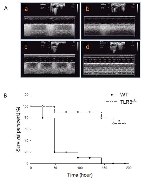
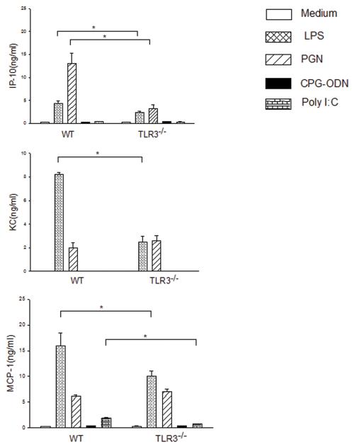
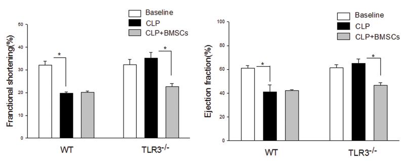
Intervention: Myocardial dysfunction is a major consequence of septic shock and contributes to the high mortality of sepsis. Toll-like receptors (TLRs) play a critical role in the pathophysiology of sepsis/septic shock. TLR3 is located in intracellular endosomes, and recognizes double-stranded RNA. This study examined the role of TLR3 in cardiac dysfunction following cecal ligation and puncture (CLP)-induced sepsis. TLR3 knockout (TLR3-/-, n=12) and age-matched wild-type (n=12) mice were subjected to CLP. Cardiac function was measured by echocardiography before and 6 hrs after CLP.

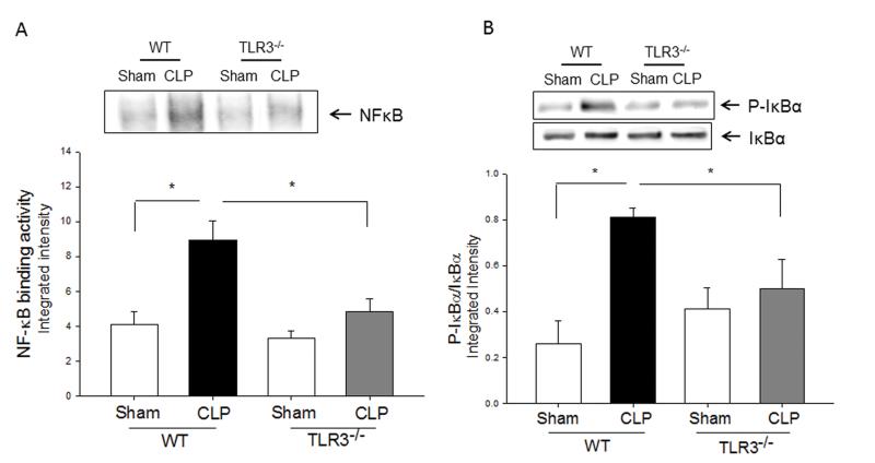
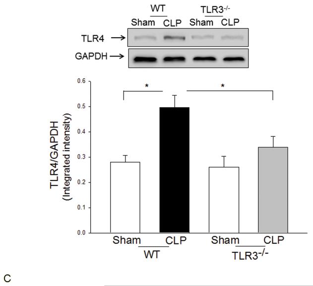
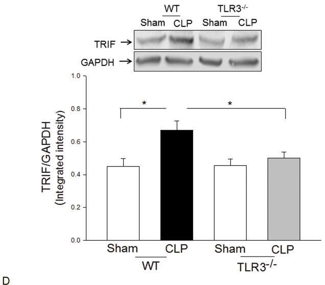
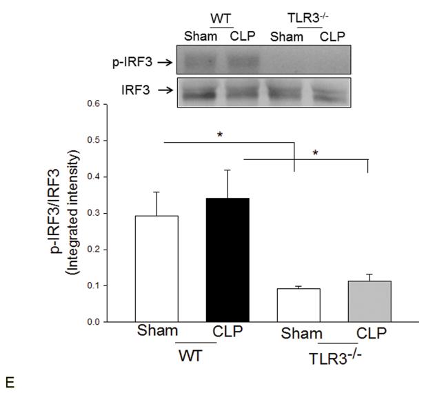
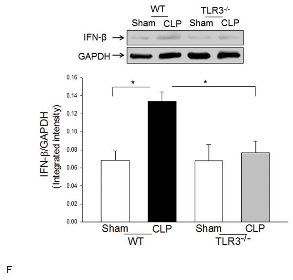
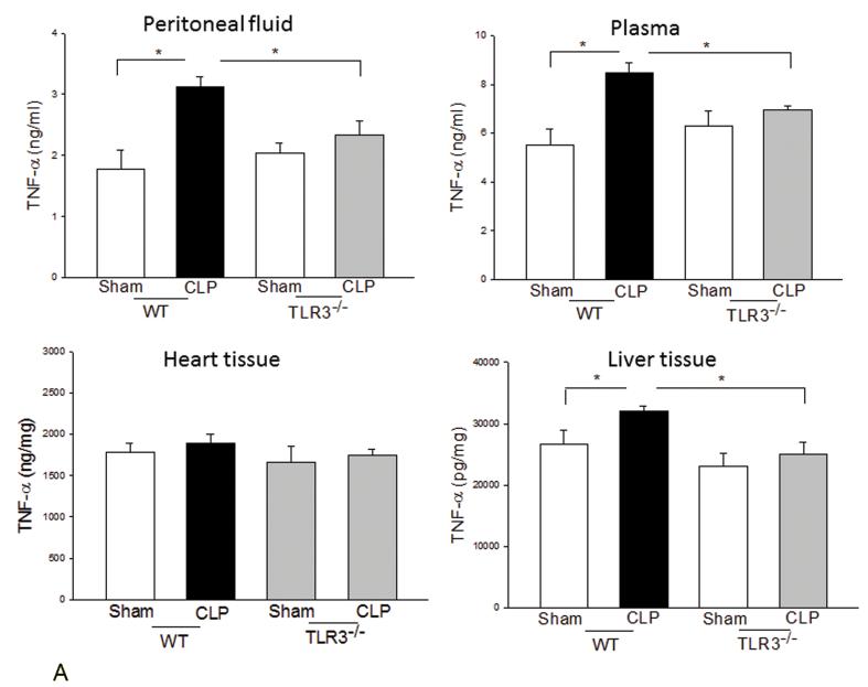
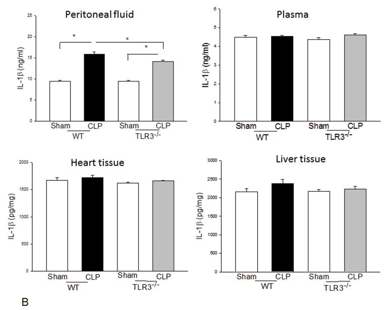
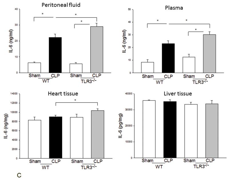
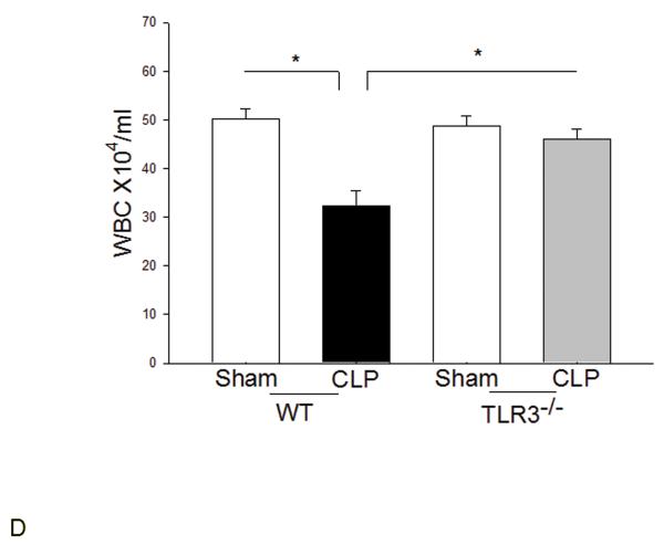
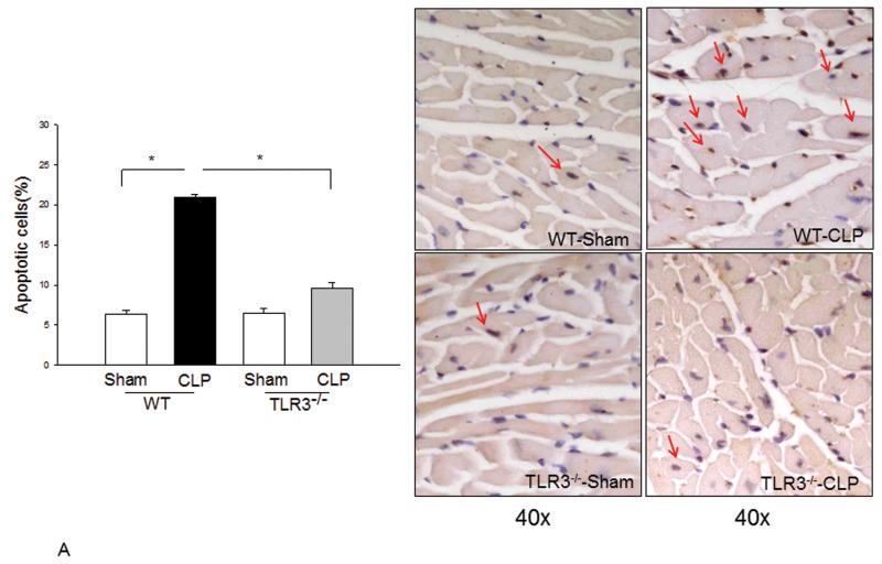
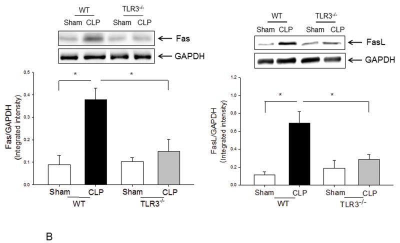
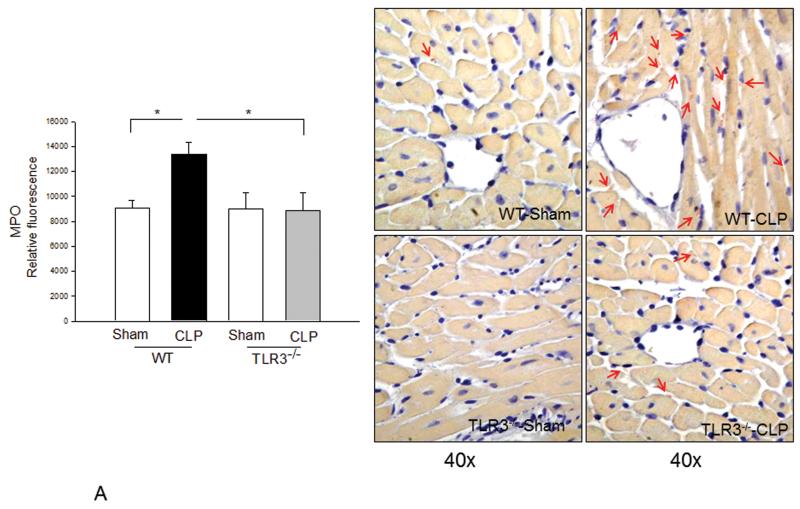
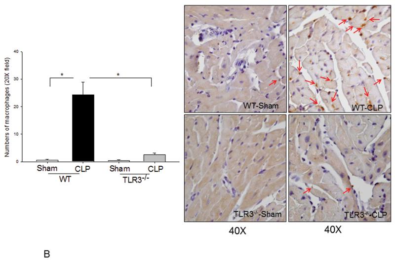
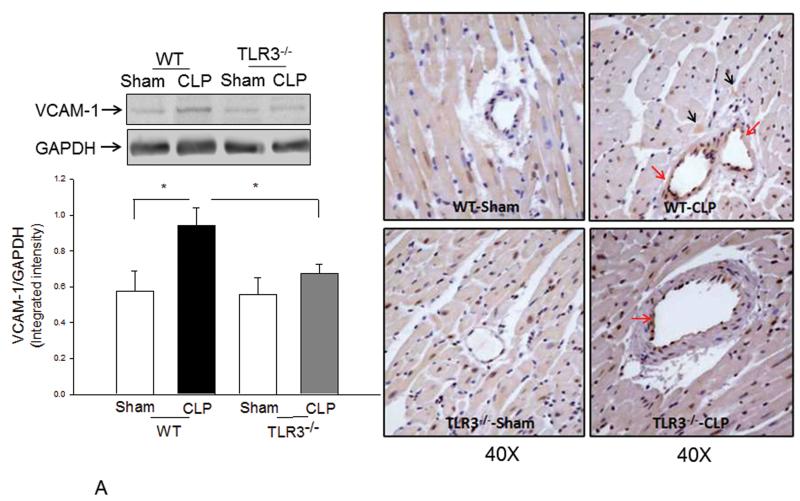
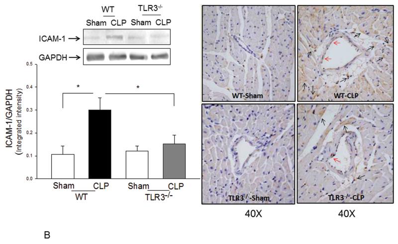
Measurements and main results: CLP resulted in significant cardiac dysfunction as evidenced by decreased ejection fraction by 25.7% and fractional shortening by 29.8%, respectively. However, TLR3-/- mice showed a maintenance of cardiac function at pre-CLP levels. Wild-type mice showed 50% mortality at 58 hrs and 100% mortality at 154 hrs after CLP. In striking contrast, 70% of TLR3-/- mice survived indefinitely, that is, >200 hrs. TLR3 deficiency significantly decreased CLP-induced cardiac-myocyte apoptosis and attenuated CLP-induced Fas and Fas ligand expression in the myocardium. CLP-activation of TLR4-mediated nuclear factor-κB and Toll/IL-1 receptor-domain-containing adapter-inducing interferon-β-dependant interferon signaling pathways was prevented by TLR3 deficiency. In addition, CLP-increased vascular cell adhesion molecule-1 and intercellular adhesion molecule-1 expression, and neutrophil and macrophage sequestration in the myocardium were also attenuated in septic TLR3-/- mice. More significantly, adoptive transfer of wild-type bone-marrow stromal cells to TLR3-/- mice abolished the cardioprotective effect in sepsis.

Conclusions: These data indicate that TLR3 plays a deleterious role in mediating cardiac dysfunction in sepsis. Thus, modulation of the TLR3 activity may be useful in preventing cardiac dysfunction in sepsis.


Figures

Figure 1
Figure 1. TLR3 deficiency attenuated cardiac dysfunction and increased survival outcome following CLP-induced sepsis in mice
TLR3−/− and age-matched WT mice (n=12/group) were subjected to CLP and cardiac function was measured before and 6 hrs after CLP. (A) Representative images generated by echocardiography. a: WT mice base line (before CLP); b: WT mice after CLP; c: TLR3−/− mice base line (before CLP); d: TLR3−/− mice after CLP. (B) TLR3 deficiency increases survival outcome in CLP-induced septic mice. TLR3−/− and age-matched WT mice (10/group) were subjected to CLP and the survival was carefully monitored daily. * P<0.05 compared with indicated groups.
Figure 2
Figure 2. TLR3 deficiency prevented sepsis-induced activation of TLR4-mediated NF-κB activation and TRIF/IRF-dependent IFN signaling pathways
TLR3−/− and WT mice were subjected to CLP (5-6/group). Sham operation served as sham control (5-6/group). Hearts were harvested six hrs after CLP and the nuclear and cytoplasmic proteins were prepared. (A) NFκB binding activity was determined by EMSA with nuclear proteins. The levels of p-IκBα/IκBα (B), TLR4 (C), TRIF (D), p-IRF3/IRF3 (E), and IFNβ (F) were examined by Western blot with specific antibodies, respectively. * p<0.05 compared with indicated groups.
Figure 3
Figure 3. The effect of TLR3 deficiency on systemic inflammatory responses following CLP
TLR3−/− and WT mice were subjected to CLP (4-6/group). Sham operation served as sham control (4-6/group). Peritoneal fluid, plasma, and heart and liver tissues were harvested six hrs after CLP. Inflammatory cytokines TNFα (A), IL-1β (B), and IL-6 (C) were measured using commercially available ELISA kits, respectively. (D) White blood cells (WBC) in the plasma were accounted. * p<0.05 compared with indicated groups.
Figure 4
Figure 4. TLR3 deficiency prevented CLP-apoptosis and Fas/FasL-mediated apoptotic signaling in the myocardium
TLR3−/− and WT mice were subjected to CLP (6/group). Sham operation served as sham control (6/group). Hearts were harvested for tissue section and cellular protein preparations. (A) Cardiac myocyte apoptosis was examined by TUNEL assay. Red arrows indicate cardiac myocyte apoptosis. Magnification is 40X. Quantitated data are shown on the left. (B) The levels of Fas and FasL were examined by Western blot with specific antibodies. * p<0.05 compared with indicated groups.
Figure 5
Figure 5. Neutrophil and macrophage myocardial infiltration was attenuated in TLR3 deficiency
TLR3−/− and WT mice were subjected to CLP (6/group). Sham operation served as sham control (6/group). Hearts were harvested six hrs after CLP and sectioned. (A) MPO activity was measured in cellular protein preparations by a kit (left). Neutrophil accumulation (pink color) was marked by red arrows (n=3/group). (B) Macrophages in the myocardium were examined by immunohistochemistry (right) with a specific antibody (n=3/group). The dark brown color indicates macrophage infiltration marked by red arrows. Quantitated data are shown on the left. * p<0.05 compared with indicated groups.
Figure 6
Figure 6. Sepsis-increased expression of myocardial VCAM-1 and ICAM-1 was attenuated by TLR3 deficiency
TLR3−/− and WT mice were subjected to CLP (6/group). Sham operation served as sham control (6/group). Hearts were harvested six hrs after CLP and sectioned. Expression of (A) VCAM-1 and (B) ICAM-1 was examined by Western blot (left) and immunohistochemistry (right). The dark brown color indicates expression of VCAM-1 and ICAM-1 marked by red and black arrows, respectively. * p<0.05 compared with indicated groups.
Figure 7
Figure 7. Attenuated chemokine secretion by TLR3−/− macrophages following TLR ligand stimulation
Peritoneal macrophages were isolated from TLR3−/− and WT mice and stimulated with LPS, PGN, CpG-ODN and Poly I:C, respectively. Secretion of chemokines into the supernatants was measured using commercial kits. There were four replicates in each group (4/group). * p<0.05 compared with indicated groups.
Figure 8
Figure 8. Adoptive transfer of WT bone marrow stromal cells abolished the cardioprotective effect of TLR3 deficiency
Bone marrow stromal cells were isolated from WT mice and transplanted to TLR3−/− mice and WT mice, immediately before the mice were subjected to CLP (5/group). Cardiac function was measured by echocardiography before and 6 hrs after CLP. * p<0.05 compared with indicated groups.

Comment in

Similar articles

Cited by

References

    1. Krishnagopalan S, Kumar A, Parrillo JE, Kumar A. Myocardial dysfunction in the patient with sepsis. Curr Opin Crit Care. 2002;8:376–388. - PubMed
    1. Oberholzer A, Oberholzer C, Moldawer LL. Sepsis Syndromes: Understanding the Role of Innate and Acquired Immunity. Shock. 2001;16:83–96. - PubMed
    1. Aderem A, Ulevitch RJ. Toll-like receptors in the induction of the innate immune response. Nature. 2000;406:782–787. - PubMed
    1. Akira S, Takeda K. Toll-Like Receptor Signalling. Nature Reviews. 2004;4:499–511. - PubMed
    1. Frantz S, Ertl G, Bauersachs J. Mechanisms of disease: Toll-like receptors in cardiovascular disease. Nat Clin Pract Cardiovasc Med. 2007;4:444–454. - PubMed

Publication types