. 2012 Jun 12;109(24):9372-7.
doi: 10.1073/pnas.1201130109. Epub 2012 May 25.

RNA 3'-end mismatch excision by the severe acute respiratory syndrome coronavirus nonstructural protein nsp10/nsp14 exoribonuclease complex

Affiliations

RNA 3'-end mismatch excision by the severe acute respiratory syndrome coronavirus nonstructural protein nsp10/nsp14 exoribonuclease complex

Mickaël Bouvet et al. Proc Natl Acad Sci U S A. .

Abstract

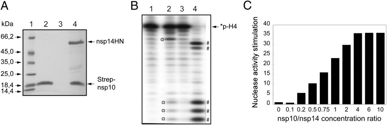
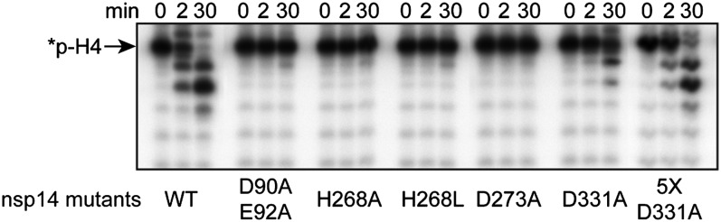
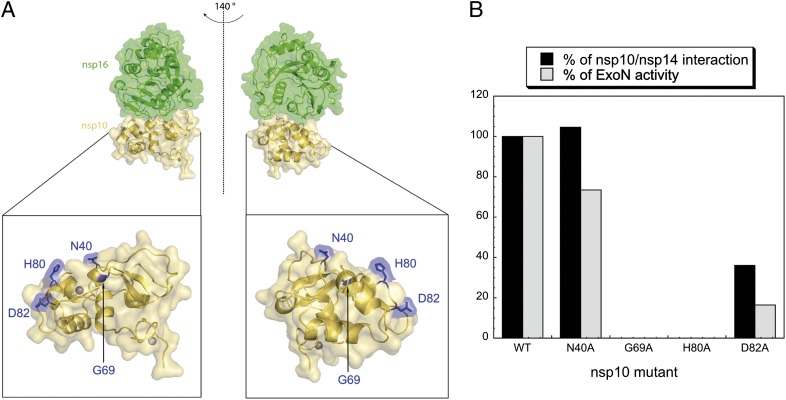
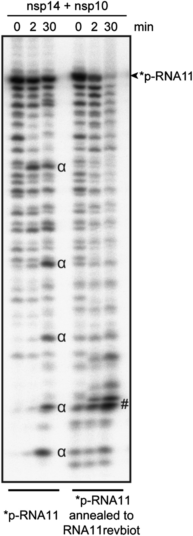
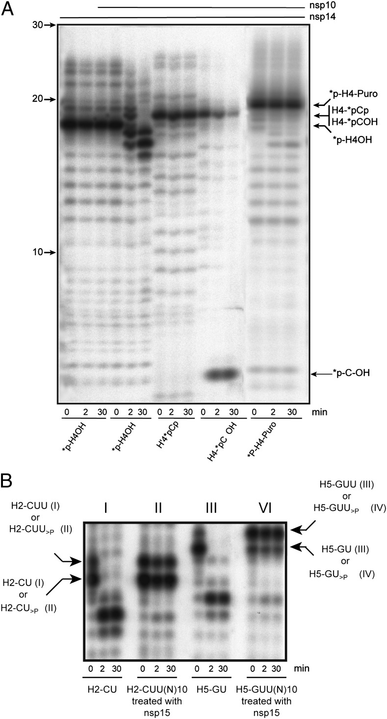
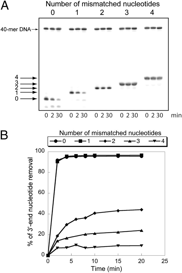
The replication/transcription complex of severe acute respiratory syndrome coronavirus is composed of at least 16 nonstructural proteins (nsp1-16) encoded by the ORF-1a/1b. This complex includes replication enzymes commonly found in positive-strand RNA viruses, but also a set of RNA-processing activities unique to some nidoviruses. The nsp14 protein carries both exoribonuclease (ExoN) and (guanine-N7)-methyltransferase (N7-MTase) activities. The nsp14 ExoN activity ensures a yet-uncharacterized function in the virus life cycle and must be regulated to avoid nonspecific RNA degradation. In this work, we show that the association of nsp10 with nsp14 stimulates >35-fold the ExoN activity of the latter while playing no effect on N7-MTase activity. Nsp10 mutants unable to interact with nsp14 are not proficient for ExoN activation. The nsp10/nsp14 complex hydrolyzes double-stranded RNA in a 3' to 5' direction as well as a single mismatched nucleotide at the 3'-end mimicking an erroneous replication product. In contrast, di-, tri-, and longer unpaired ribonucleotide stretches, as well as 3'-modified RNAs, resist nsp10/nsp14-mediated excision. In addition to the activation of nsp16-mediated 2'-O-MTase activity, nsp10 also activates nsp14 in an RNA processing function potentially connected to a replicative mismatch repair mechanism.


Conflict of interest statement

The authors declare no conflict of interest.

Figures

Fig. 1.
Fig. 1.
Nsp10 interacts with nsp14, and nsp10/nsp14 shows enhanced ExoN activity. (A) SARS-CoV nsp14HN and Strep-nsp10 proteins coexpressed or expressed alone were incubated with Strep-Tactin Sepharose. Strep-Tactin–bound proteins were eluted with d-desthiobiotin and analyzed by SDS/PAGE and Coomassie blue staining. Lane 1 corresponds to the molecular size markers; lane 2 to Strep-nsp10 expressed alone; lane 3 to nsp14HN expressed alone; and lane 4 to Strep-nsp10 coexpressed with nsp14HN. (B) Autoradiogram of RNA cleavage products. Synthetic *p-H4 RNA was radiolabeled at its 5′-end using PNK in the presence of [γ32P]ATP (the asterisk indicates the 32P-labeling position). *p-H4 RNA was incubated at 37 °C in Tris⋅HCl buffer 40 mM (pH 8), DTT 5 mM with no protein (lane 1), 0.7 μM of nsp14 (lane 2), nsp10 (lane 3), or both proteins (lane 4) during a 90-min period. The reaction products were then separated on a 20% (wt/vol) denaturing Urea-PAGE and revealed using photostimulated plates and a FujiImager (Fuji). (C) *p-H4 RNA was hydrolyzed with fixed nsp14 concentration (50 nM) in the presence of increasing concentration of nsp10 ranging from 0 to 1,600 nM (nsp10/nsp14 is indicated below the bar graph). ExoN activity was quantified using denaturing Urea-PAGE followed by measuring the hydrolysis of *p-H4 corresponding band using a FujiImager and Image Gauge software analysis.
Fig. 2.
Fig. 2.
Mutagenesis analysis of nsp14 ExoN activity. Residues from the nsp14 ExoN catalytic site and from the SAM-binding site of the nsp14 MTase domain were mutated into alanine. Equal amounts of each nsp14 mutant were incubated with nsp10 and *p-H4 RNA for 0, 2, and 30 min. In lane 5XD331A, the concentration of the mutant was fivefold higher. The panel shows the time-dependent hydrolysis of *p-H4 RNA after Urea-PAGE separation and autoradiography.
Fig. 3.
Fig. 3.
Nsp10/nsp14 interaction is required for nsp14 ExoN stimulation. (A) Selection of nsp10 surface mutants and their position in the nsp10/nsp16 dimer. The nsp10/nsp16 complex image (Upper) was represented using PyMOL, and an enlargement of nsp10 is shown (Lower) with the position of mutated amino acids (highlighted in blue). nsp16 is represented in green, nsp10 in gold, and nsp10 zinc structural ions as gray spheres. (B) Quantification of nsp10/nsp14 interaction and corresponding ExoN activity. Nsp10 WT or mutants proteins carrying a Strep-TagII were coexpressed with nsp14HN. After purification on Strep-Tactin beads, eluted proteins were separated using LabChip (Caliper), and the intensities of peaks corresponding to nsp10 and nsp14 proteins were quantified. Molecular ratio obtained for nsp10 WT/nsp14 complex was taken as 100%. Results are presented in percentage of interaction compared with the nsp10 WT/nsp14 complex (black bars). For ExoN quantification, equal amounts of nsp10 mutants were incubated with nsp14 and *p-H4 RNA. After 30 min of incubation, reaction products were separated using Urea-PAGE, revealed using a FujiImager, and quantified using Image Gauge software. Results are presented in percentage of ExoN activity (nsp10 WT/nsp14 was taken as 100% of activity; gray bars).
Fig. 4.
Fig. 4.
The nsp10/nsp14 ExoN activity is a dsRNA-dependent exonuclease. Autoradiogram of an ExoN assay. HPLC-purified ssRNA (RNA11) was labeled at its 5′-end using PNK and [γ32P]ATP, and hybridized to its complementary strand modified at its 3′-end with biotin (RNA11revbiot). Single- or double-stranded labeled-RNAs were subjected to nsp10/nsp14 ExoN digestion for 0, 2, and 30 min. Digestion products were separated on Urea-PAGE and revealed by autoradiography.
Fig. 5.
Fig. 5.
Nsp10/nsp14 requires a free 3′-hydroxyl end. (A) Autoradiogram of an ExoN assay performed with RNA carrying 3′-phosphate, 3′-puromycin, or 3′-hydroxyl end. The 3′ modified RNAs (*p-H4 as control, *p-H4-puromycin, H4-*pCp, and H4-*pCOH) were incubated with nsp14 or nsp10/nsp14 for 0, 2, and 30 min (asterisk indicates the 32P-labeling position). Digestion products were separated on 20% (wt/vol) Urea-PAGE and revealed by autoradiography. (B) Autoradiogram of nsp15-generated 2′-3′-cyclic phosphate RNAs cleavage assay. RNA substrates H2-CUU(N)10 and H5-GUU(N)10 were synthesized using T7 RNA polymerase and subsequently dephosphorylated and radiolabeled. These RNAs were then incubated with nsp15 to generate *p-H2-CU>P and *p-H2-CUU>P and, to a lesser extent, *p-H5-GUU>P and *p-H5-GU>P. Control RNAs *p-H2-CUOH and *p-H5-GUOH were synthesized using T7 RNA polymerase and led to the production of the side products *p-H2-CUUOH and *p-H5-GUUOH, respectively. After purification, these substrates were incubated with nsp10/nsp14 for 0, 2, and 30 min. Digestion products were separated on 20% (wt/vol) Urea-PAGE and revealed by autoradiography.
Fig. 6.
Fig. 6.
Comparison of nsp10/nsp14 ExoN activity on paired and mismatched 3′-end nucleotide base pairs. (A) A 40-nt RNA (LS1) was annealed with 5′-radiolabeled oligoribonucleotides carrying zero, one, two, three, or four noncomplementary bases at its 3′-end (respectively, LS2, LS3, LS4, LS5, or LS6 (Table S1 and Fig. S5). To avoid nsp10/nsp14-mediated LS1 degradation, this RNA carries a biotin group at its 3′-end. Duplex RNAs were then incubated (0, 2, and 30 min) with purified nsp10/nsp14 (200 nM/50 nM). Reaction products were separated on a 20% (wt/vol) denaturing Urea-PAGE and revealed by autoradiography. A radiolabeled 40-mer DNA was introduced in the reaction mixture as a quantification reference. (B) Kinetics of mismatch excision. The assay was performed as in A, using 100 nM of nsp14 and 400 nM of nsp10. RNA cleavage was quantified at 0, 2, 4, 6, 8, 10, 15, and 20 min. Data are presented as percent of 3′-nucleotide removal.

References

    1. Gorbalenya AE, Enjuanes L, Ziebuhr J, Snijder EJ. Nidovirales: Evolving the largest RNA virus genome. Virus Res. 2006;117:17–37. - PMC - PubMed
    1. Knoops K, et al. SARS-coronavirus replication is supported by a reticulovesicular network of modified endoplasmic reticulum. PLoS Biol. 2008;6:e226. - PMC - PubMed
    1. van Hemert MJ, et al. SARS-coronavirus replication/transcription complexes are membrane-protected and need a host factor for activity in vitro. PLoS Pathog. 2008;4:e1000054. - PMC - PubMed
    1. Snijder EJ, et al. Unique and conserved features of genome and proteome of SARS-coronavirus, an early split-off from the coronavirus group 2 lineage. J Mol Biol. 2003;331:991–1004. - PMC - PubMed
    1. Minskaia E, et al. Discovery of an RNA virus 3′→5′ exoribonuclease that is critically involved in coronavirus RNA synthesis. Proc Natl Acad Sci USA. 2006;103:5108–5113. - PMC - PubMed

Publication types

MeSH terms