. 2012 Aug;153(8):3780-91.
doi: 10.1210/en.2012-1313. Epub 2012 May 25.

Spinal motor and sensory neurons are androgen targets in an acrobatic bird

Affiliations

Spinal motor and sensory neurons are androgen targets in an acrobatic bird

Matthew J Fuxjager et al. Endocrinology. 2012 Aug.

Abstract

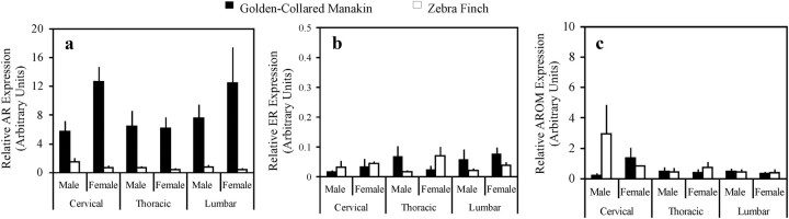
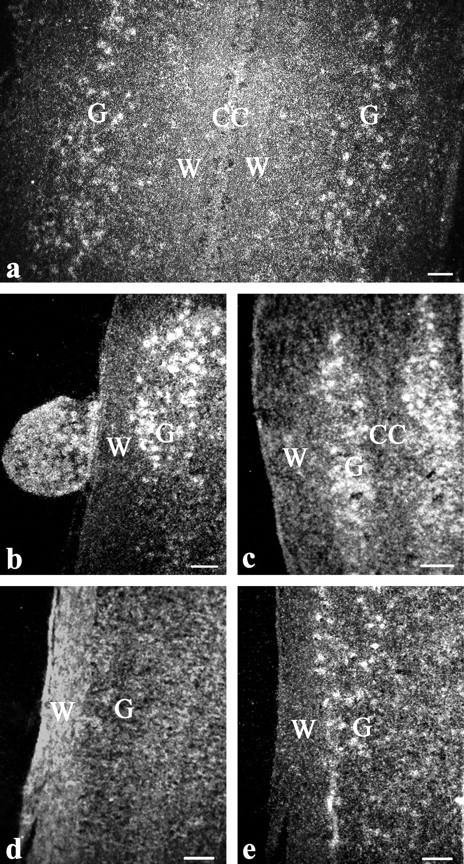
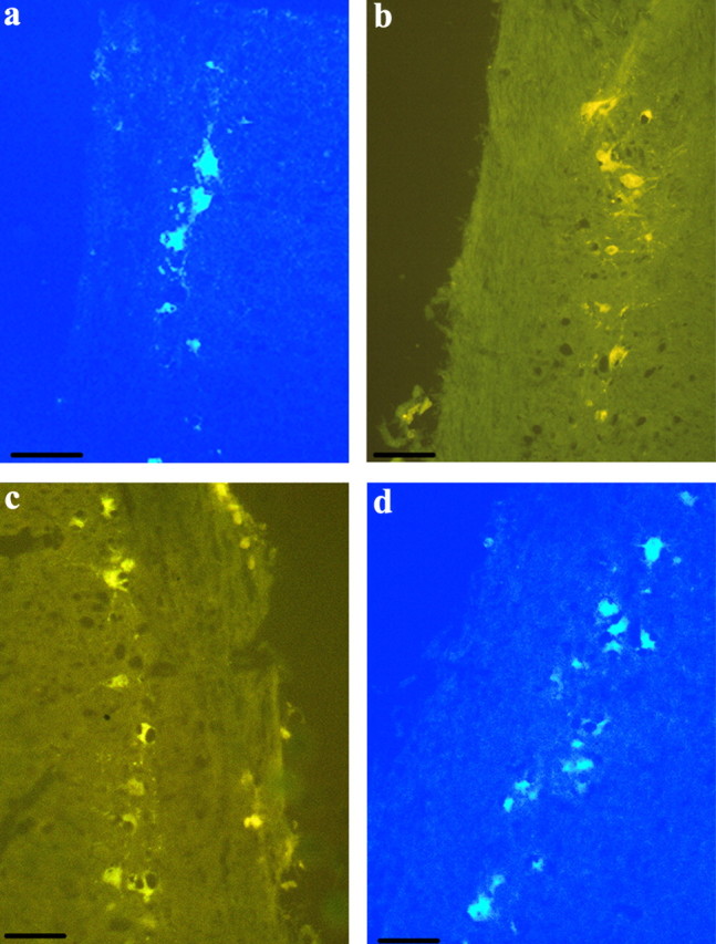
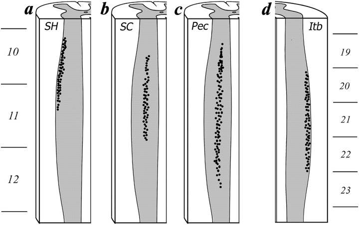
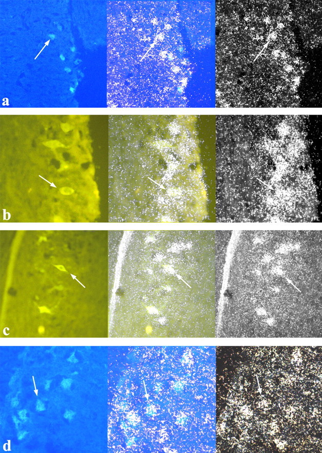
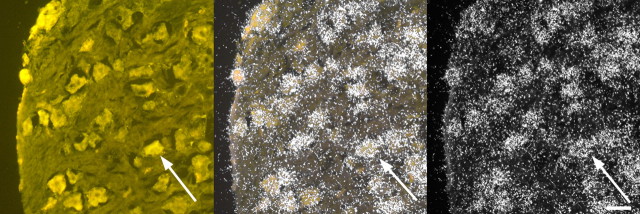
Sex steroids affect the motivation to court mates, but less is known about how they influence motor movements associated with courtship behavior. Steroidal control of motor function may be especially important for species in which courtship requires superior strength, stamina, and neuromuscular coordination. Here we use the golden-collared manakin (Manacus vitellinus) to examine whether the neuromuscular circuitry that controls motoric aspects of courtship activity is sensitive to androgens. Males of this tropical species attract mates by rapidly jumping among branches in a courtship arena and using their wings to produce loud wing snaps. Testosterone activates this display via the androgen receptor (AR), and past work reveals that manakins injected with radio-labeled T ((3)H-T) accumulate radioactivity in the spinal cord. Thus, we used quantitative PCR to measure AR, estrogen receptor-α (ER-α) subtype, and aromatase (AROM) mRNA in spinal cords of male and female manakins and zebra finches. Expression of AR, but not ER-α or aromatase, was higher throughout the manakin spinal cord compared with the zebra finch. Next, we tested whether AR-expressing skeletal muscles are innervated by motor and sensory neurons that also express AR. To do this, we backfilled spinal neurons by injecting fluorescent tracers into select AR-sensitive wing and leg muscles of wild caught male and female manakins. We then removed these spinal cords and measured AR expression with in situ hybridization. Both sexes showed abundant AR mRNA in the cervical and lumbosacral spinal enlargements as well as in dorsal root ganglia attached to these enlargements. Together our findings suggest that androgens act widely on peripheral motor and sensory circuits in golden-collared manakins to influence wing snapping displays.


Figures

Fig. 1.
Fig. 1.
Relative expression (ΔCt levels) of AR (a), ER-α (b), and AROM (c) mRNA in the cervical, thoracic, and lumbar regions of the spinal cord in male and female golden-collared manakins and zebra finches. Spinal AR expression was higher in manakins than in zebra finches. Within manakins, spinal AR expression was higher in females than in males. Data represent mean ± sem.
Fig. 2.
Fig. 2.
Darkfield photomicrographs of cells in the spinal cord that express AR mRNA. a, Cervical enlargement of a male implanted with T. b, Rostral cervical enlargement and attached DRG of a nonimplanted female. c, Thoracic region well outside the cervical and lumbosacral enlargements in a male implanted with T. d and e, Consecutive sections of the lumbosacral enlargement showing hybridization using the 33P-labeled AR sense probe (d) and the 33P-labeled antisense probe (e). CC, Position of the central canal; W, region of white matter; G, position of gray matter. Reference bar, 40 μm.
Fig. 3.
Fig. 3.
Photomicrographs of labeled motoneurons in the spinal cord after the im injection of the retrograde transport fluorescent tracer TB or HM. a, Motoneurons after TB was injected into the SH of a T-implanted male. b, HM was injected into the SC of a nonimplanted female. c, HM was injected into the PEC of a nonimplanted female. d, TB was injected into the ITB of a T-implanted female. Reference bar, 40 μm.
Fig. 4.
Fig. 4.
Schematic showing the rostrocaudal distribution of the motoneurons within spinal column that innervate the SH (a), SC (b), PEC (c), and ITB (d). The ventral third of the spinal cord is illustrated.
Fig. 5.
Fig. 5.
Photomicrographs of spinal motoneurons that innervate the SH (a), SC (b), PEC (c), and ITB (d). Photomicrographs in the left column show cells labeled with fluorescent tracers under UV illumination. Photomicrographs in the right column show the same tissue but viewed under dark-field illumination to illustrate the AR probe hybridization. Photomicrographs in the center column show an overlay of the left and right photomicrographs, demonstrating that fluorescent cells readily express AR mRNA. Arrows point to a representative cell that shows both florescence and silver grain accumulation.
Fig. 6.
Fig. 6.
Photomicrographs of a DRG containing fluorescently labeled neurons as well as hybridization with the AR probe. The photomicrograph to the left shows cells labeled with fluorescent traces under UV illumination. The photomicrograph to the right shows the same tissue viewed under dark-field illumination to illustrate the AR probe hybridization. The photomicrograph in the center shows an overlay of the left and right photomicrographs to demonstrate that fluorescent cells express AR mRNA. Arrows point to a representative cell that shows both florescence and silver grain accumulation. Reference bar, 40 μm.

References

    1. Andersson M. 1994. Sexual selection. Princeton, NJ: Princeton University Press
    1. Adkins-Regan E. 2005. Hormones and animal social behavior. Princeton, NJ: Princeton University Press
    1. Sipos ML , Nyby JG. 1996. Concurent androgenic stimulation of the ventral tegmental area and medial preoptic area: synergistic effects on male-typical reproductive behaviors in house mice. Brain Res 729:29–44 - PubMed
    1. Goodson JL , Evans AK , Lindberg L , Allen CD. 2005. Neuro-evolutionary patterning of sociality. Proc Biol Sci 272:227–235 - PMC - PubMed
    1. DiMeo AN , Wood RI. 2006. ICV testosterone induces fos in male Syrian hamster brain. Psychoneuroendocrinol 31:237–249 - PubMed

Publication types