. 2012 Aug;11(8):978-88.
doi: 10.1128/EC.00007-12. Epub 2012 May 25.

Role of Fig1, a component of the low-affinity calcium uptake system, in growth and sexual development of filamentous fungi

Affiliations

Role of Fig1, a component of the low-affinity calcium uptake system, in growth and sexual development of filamentous fungi

Brad Cavinder et al. Eukaryot Cell. 2012 Aug.

Abstract

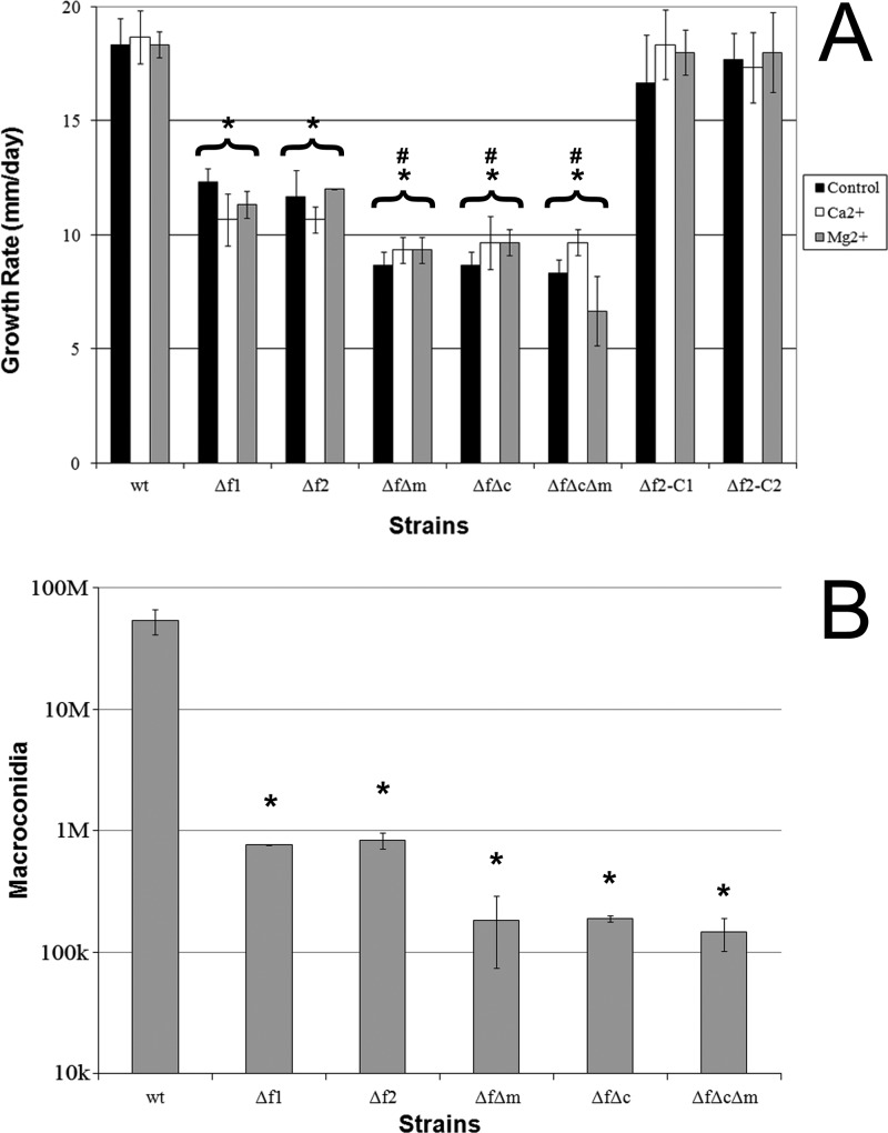
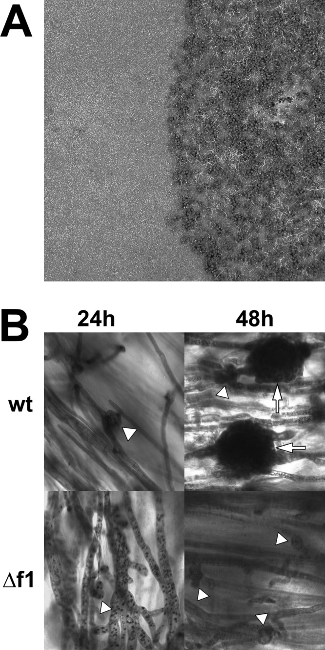
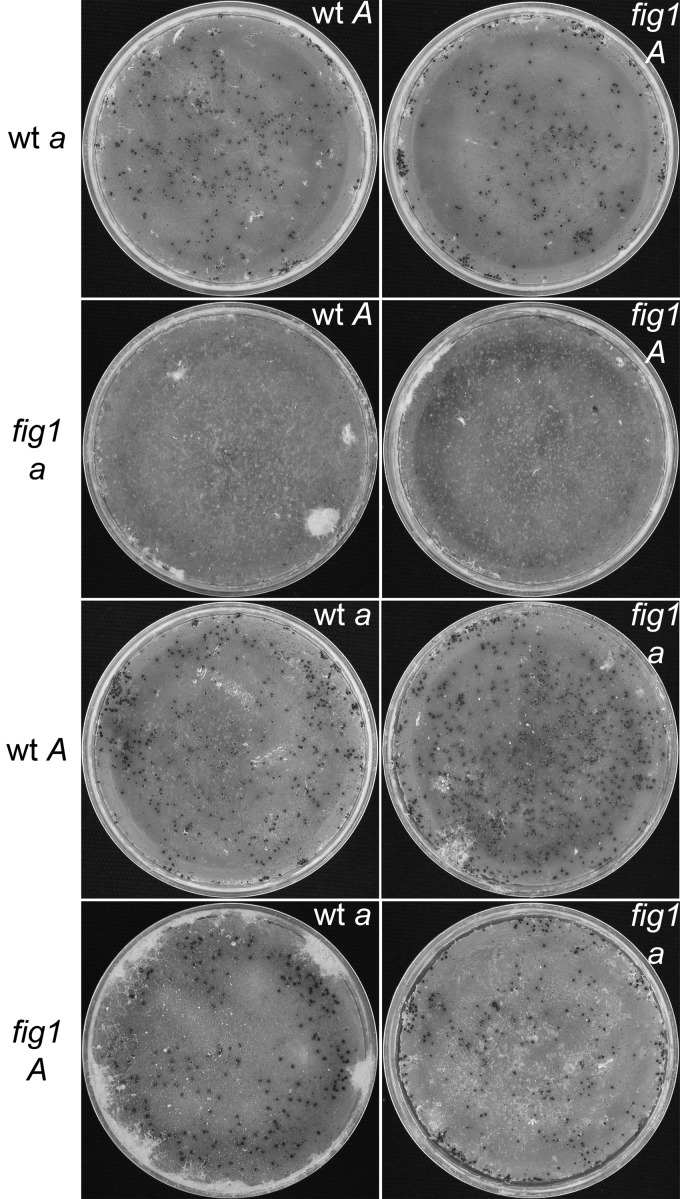
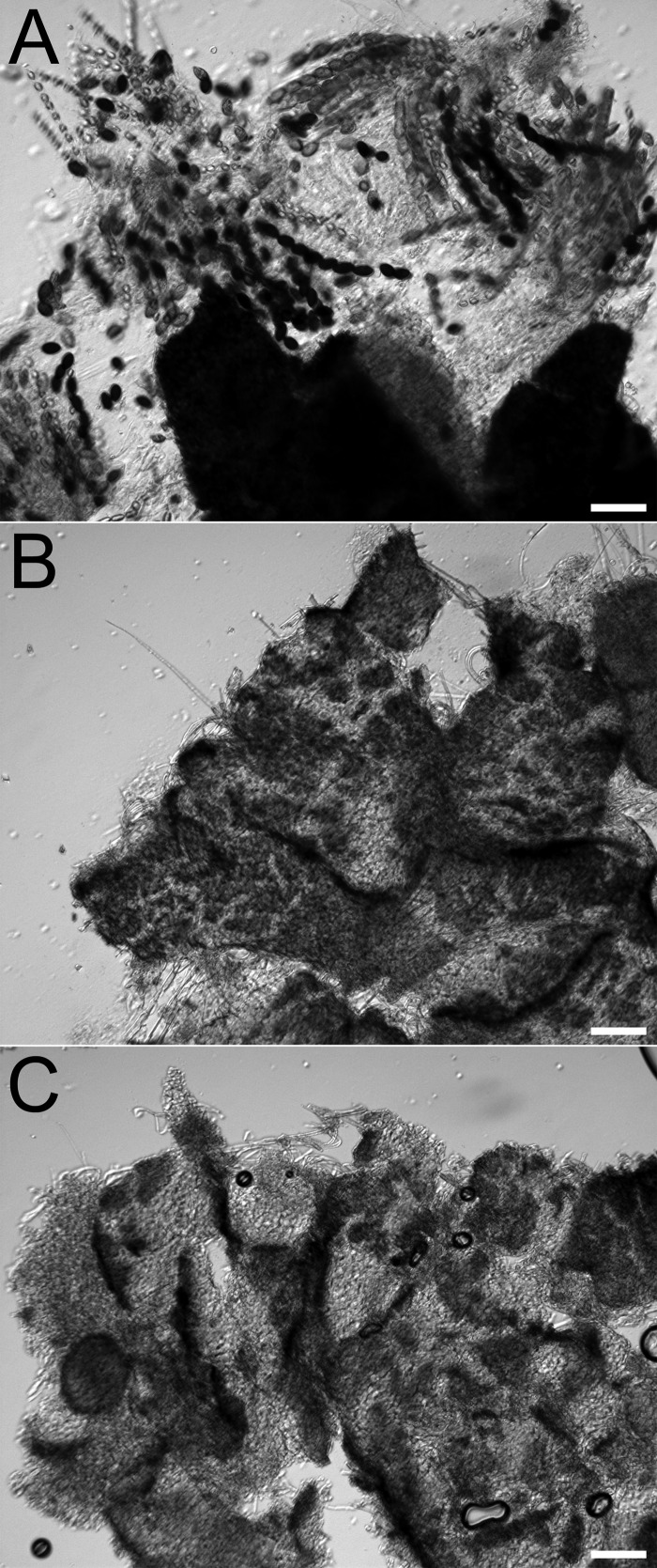
The function of Fig1, a transmembrane protein of the low-affinity calcium uptake system (LACS) in fungi, was examined for its role in the growth and development of the plant pathogen Fusarium graminearum. The Δfig1 mutants failed to produce mature perithecia, and sexual development was halted prior to the formation of perithecium initials. The loss of Fig1 function also resulted in a reduced vegetative growth rate. Macroconidium production was reduced 70-fold in the Δfig1 mutants compared to the wild type. The function of the high-affinity calcium uptake system (HACS), comprised of the Ca(2+) channels Mid1 and Cch1, was previously characterized for F. graminearum. To better understand the roles of the LACS and the HACS, Δfig1 Δmid1, Δfig1 Δcch1, and Δfig1 Δmid1 Δcch1 double and triple mutants were generated, and the phenotypes of these mutants were more severe than those of the Δfig1 mutants. Pathogenicity on wheat was unaffected for the Δfig1 mutants, but the Δfig1 Δmid1, Δfig1 Δcch1, and Δfig1 Δmid1 Δcch1 mutants, lacking both LACS and HACS functions, had reduced pathogenicity. Additionally, Δfig1 mutants of Neurospora crassa were examined and did not affect filamentous growth or female fertility in a Δfig1 mating type A strain, but the Δfig1 mating type a strain failed to produce fertile fruiting bodies. These results are the first report of Fig1 function in filamentous ascomycetes and expand its role to include complex fruiting body and ascus development.


Figures

Fig 1
Fig 1
F. graminearum FIG1 gene models. (A) Current gene model from MIPS. (B) Predicted gene model from a Velvet assembly of insertion mutant 729g-112. Gray bars above the model represent aligned 5′- and 3′-RACE clone sequences. The graph below the model shows the coverage of aligned wt RNAseq reads. (C) Predicted spliced transcript from wt RACE and RNAseq alignments in panel B. Only a portion of the 5′-untranslated region is shown; the locations of splice sites are indicated (arrowheads). An alignment of the wt RNAseq reads to the model sequence is graphed below the model. Scale bar, 200 bp.
Fig 2
Fig 2
Multiple-amino-acid sequence alignment of a region of Fig1 homologs. Highly conserved glycine and cysteine residues of a conserved Gly-Cys motif, at or near the end of the first transmembrane domain of the proteins (*), and a conserved claudin motif are highlighted with a black background. A. clavatus, Aspergillus clavatus; N. fischeri, Neosartorya fischeri; A. terreus, Aspergillus terreus; A. flavus, Aspergillus flavus; A. oryzae, Aspergillus oryzae; P. chrysogenum, Penicillium chrysogenum; S. sclerotiorum, Sclerotina sclerotiorum; M. oryzae, Magnaporthe oryzae; A. gossypii, Ashbya gossypii; S. kluyveri, Saccharomyces kluyveri.
Fig 3
Fig 3
Vegetative growth of F. graminearum strains. (A, top) Growth on carrot agar. While the wt, Δfig1-1, and Δfig1-2 fully colonized the surface of the medium in 4 days, the growth rates of strains fm-5, fc-6, and fcm-2 were reduced. (Bottom) Growth (14 days) on carrot agar supplemented with1 mM BAPTA. The wt, Δfig1-1, and Δfig1-2 fully colonized the surface of the medium, but strains fm-5, fc-6, and fcm-2 ceased growth after some initial colonization. (B, top) Growth on V8 agar with EtOH (control). While the wt fully colonized the surface of the medium in 4 days, strains Δfig1-1 and Δfig1-2 had reduced growth and fewer aerial hyphae than the wt. Strains fm-5, fc-6, and fcm-2 had more severe phenotypes than did strains with single mutations. (Bottom) Growth on V8 in the presence of the Ca2+ ionophore A23187 (the application point is indicated by a black dot). The wt fully colonized the ionophore-treated medium (lower portion of the colonies), but in all mutant strains, the presence of the ionophore halted growth. Strains and genotypes are abbreviated as follows: wt (FIG1 MID1 CCH1); Δf1, Δfig1-1 (Δfig1 MID1 CCH1); Δf2, Δfig1-2 (Δfig1 MID1 CCH1); ΔfΔm, fm-5 (Δfig1 Δmid1 CCH1); ΔfΔc, fc-6 (Δfig1 MID1 Δcch1); ΔfΔmΔc, fcm-2 (Δfig1 Δmid1 Δcch1).
Fig 4
Fig 4
Characterization of F. graminearum growth and asexual development. (A) Growth rates on carrot agar with and without Ca2+ or Mg2+ supplementation (change of diameter in mm/day). Asterisks above brackets indicate a significant difference between all growth conditions of the bracketed strain and the wild type on carrot agar. Number signs above brackets indicate a significant difference between the bracketed strain on carrot agar and strain Δfig1-1, but not Δfig1-2, on carrot agar. (B) Macroconidium production. Error bars represent the standard deviations of the means from 3 biological replicates. *, P < 0.005; #, P < 0.05. Abbreviations for strains and genotypes are listed in the legend of Fig. 3, except that Δf2-C1 indicates f12-C1 (Δfig1::FIG1 MID1 CCH1) and Δf2-C2 indicates f12-C2 (Δfig1::FIG1 MID1 CCH1).
Fig 5
Fig 5
Sexual development of F. graminearum wt and Δfig1-1. (A) Mature wt culture (right) with normal sexual development and Δfig1-1 culture (left) with no perithecia. (B) Sexually induced cultures at 24 and 48 h stained with toluidine blue. Perithecium initials (arrowheads), which developed at 24 h in the wt, were barely initiated in Δfig1-1 cells. By 48 h, there were immature walled perithecia (arrow) in wt cells; in Δfig1-1 cells, perithecium initials have not matured and do not develop further. Scale bars, 20 μm. Abbreviations for strains and genotypes are listed in the legend of Fig. 3.
Fig 6
Fig 6
Reciprocal crosses of N. crassa wt and Δfig1 strains. Strains listed to the left served as females, developing protoperithecia that were fertilized by conidia of the strain listed in the upper right of each image. All crosses produced wt perithecia, except for crosses with Δfig1 mating type a strains as the female, which produced small infertile fruiting bodies. Abbreviations for strains are as follows: wt a, FGSC_4200; wt A, FGSC_2489; Δfig1 a, FGSC_17273; Δfig1 A, fig1A-18.
Fig 7
Fig 7
N. crassa perithecium squash mounts from wt and Δfig1 crosses. (A) Perithecium from a wt a × Δfig1 A cross showing normal development, with asci and ascospores. (B) Infertile fruiting body from a Δfig1 a × wt A cross. (C) Infertile fruiting body from a Δfig1 a × Δfig1 A cross. Scale bars, 75 μm. Abbreviations for strains are described in the legend of Fig. 6.

References

    1. Abràmoff MD, Magalhães PJ, Ram SJ. 2004. Image processing with ImageJ. Biophotonics Int. 11: 36–42
    1. Alby K, Schaefer D, Sherwood RK, Jones SK, Jr, Bennett RJ. 2010. Identification of a cell death pathway in Candida albicans during the response to pheromone. Eukaryot. Cell 9: 1690–1701 - PMC - PubMed
    1. Beckett A, Crawford RM. 1973. The development and fine structure of the ascus apex and its role during spore discharge in Xylaria longipes. New Phytol. 72: 357–369
    1. Berridge MJ, Bootman MD, Roderick HL. 2003. Calcium signalling: dynamics, homeostasis and remodelling. Nat. Rev. Mol. Cell Biol. 4: 517–529 - PubMed
    1. Booth C. 1971. The genus Fusarium. Commonwealth Mycological Institute, Kew, Surrey, England

Publication types

MeSH terms

LinkOut - more resources