. 2012 Jul;135(Pt 7):2155-68.
doi: 10.1093/brain/aws127. Epub 2012 May 26.

Apolipoprotein E4 effects in Alzheimer's disease are mediated by synaptotoxic oligomeric amyloid-β

Affiliations

Apolipoprotein E4 effects in Alzheimer's disease are mediated by synaptotoxic oligomeric amyloid-β

Robert M Koffie et al. Brain. 2012 Jul.

Abstract

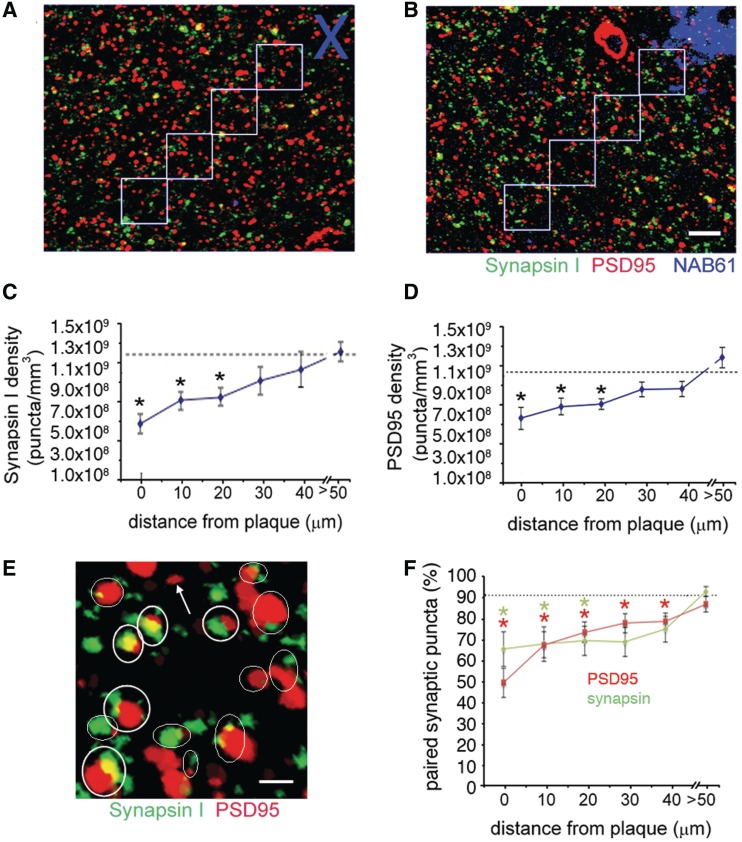
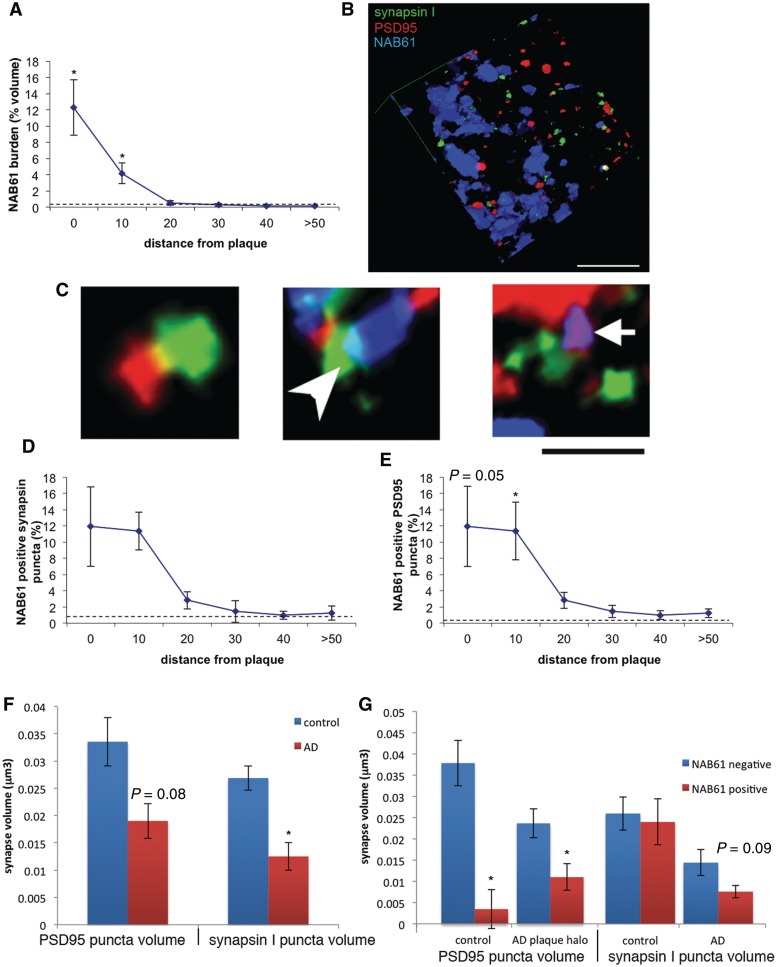
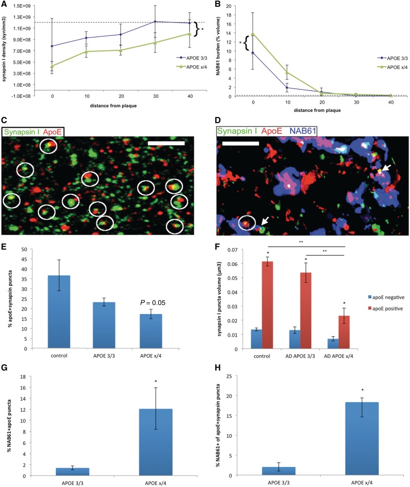
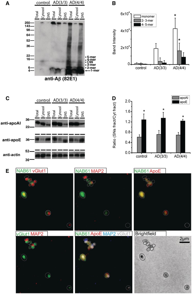
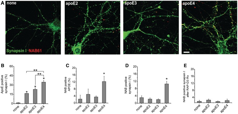
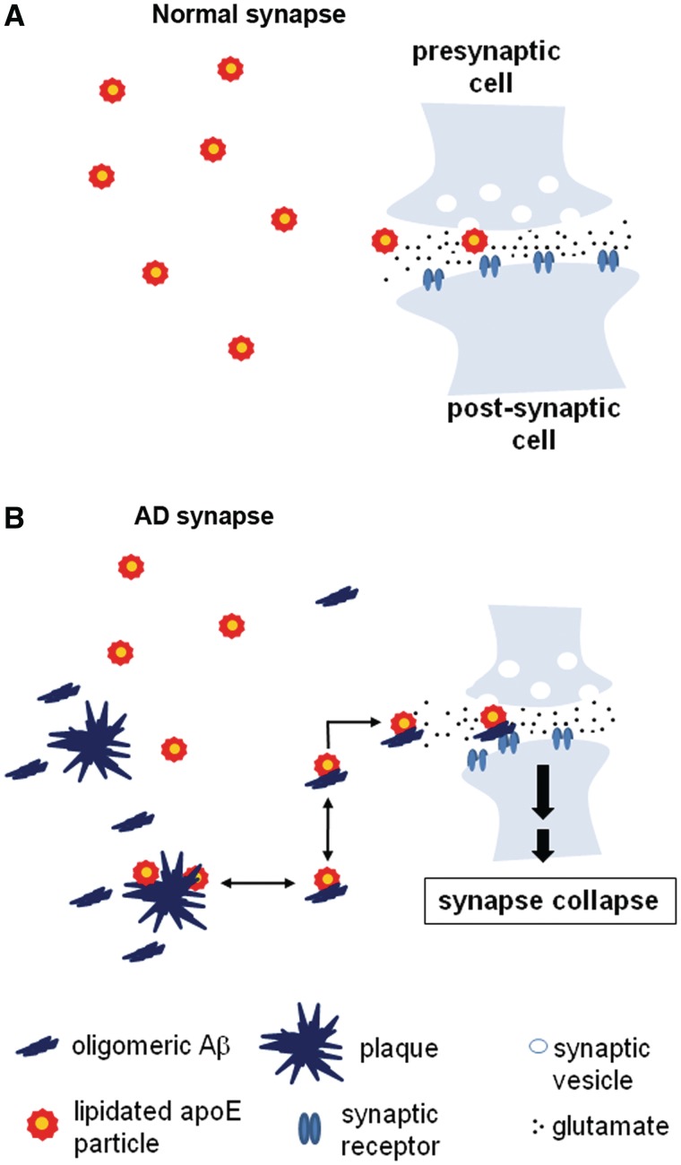
The apolipoprotein E ε4 gene is the most important genetic risk factor for sporadic Alzheimer's disease, but the link between this gene and neurodegeneration remains unclear. Using array tomography, we analysed >50000 synapses in brains of 11 patients with Alzheimer's disease and five non-demented control subjects and found that synapse loss around senile plaques in Alzheimer's disease correlates with the burden of oligomeric amyloid-β in the neuropil and that this synaptotoxic oligomerized peptide is present at a subset of synapses. Further analysis reveals apolipoprotein E ε4 patients with Alzheimer's disease have significantly higher oligomeric amyloid-β burden and exacerbated synapse loss around plaques compared with apolipoprotein E ε3 patients. Apolipoprotein E4 protein colocalizes with oligomeric amyloid-β and enhances synaptic localization of oligomeric amyloid-β by >5-fold. Biochemical characterization shows that the amyloid-β enriched at synapses by apolipoprotein E4 includes sodium dodecyl sulphate-stable dimers and trimers. In mouse primary neuronal culture, lipidated apolipoprotein E4 enhances oligomeric amyloid-β association with synapses via a mechanism involving apolipoprotein E receptors. Together, these data suggest that apolipoprotein E4 is a co-factor that enhances the toxicity of oligomeric amyloid-β both by increasing its levels and directing it to synapses, providing a link between apolipoprotein E ε4 genotype and synapse loss, a major correlate of cognitive decline in Alzheimer's disease.


Figures

Figure 1
Figure 1
Synapse density decreases approaching plaques in Alzheimer’s disease brain. Array tomography was used to obtain image stacks of immunostained pre- (synapsin I) and postsynaptic (PSD95) elements in control (A) and Alzheimer’s disease (B) brains at 10-µm increments from a plaque edge (phantom plaques in control represented by blue cross in A were chosen randomly) or at distances further than 50 µm from the nearest plaque. Quantification reveals a significant loss of both (C) pre- and (D) postsynaptic elements approaching plaques in Alzheimer’s disease brains compared with control levels (dotted lines show control averages; Kruskal–Wallis test, synapse density independent variable, plaque distance dependent variable, P < 0.05, post hoc Wilcoxon all pairs *P < 0.05). Pairing analysis was done to determine which synaptic puncta had a partner within 0.5 µm (examples circled in E). Quantification (F) shows that ∼90% of both postsynaptic density and synapsin puncta have a partner in control brain and >50 µm from plaques in Alzheimer’s disease brain, but that the pairing is significantly decreased near plaques in Alzheimer’s disease leaving ‘orphaned’ puncta (arrow) (Kruskal–Wallis test, pairing independent variable, plaque distance dependent variable, P < 0.05, post hoc Wilcoxon all pairs *P < 0.05). Images are maximum intensity z-projections of sections from array tomography ribbons (10 sections projected in A and B, 17 sections projected in E). Scale bars: B = 5 µm, E = 1 µm.
Figure 2
Figure 2
Synapse loss correlates with oligomeric amyloid-β at synapses. The burden of oligomeric amyloid-β in Alzheimer’s disease brain is highest around plaques and decreases exponentially to control levels (represented by dotted line) with increasing distance from the plaque (A). Three dimensional reconstructions of array tomograms (12 sections reconstructed in B) reveal that oligomeric amyloid-β associates with a subset of synapses in Alzheimer’s disease brain. (C) Enlargements of the reconstruction showing examples of opposed pre- and postsynapses with no oligomeric amyloid-β (left), an oligomeric amyloid-β positive presynaptic element (arrowhead) and an oligomeric amyloid-β positive postsynaptic element (arrow). The level of co-localization between NAB61-positive puncta and presynaptic (D) or postsynaptic elements (E) is highest near plaques in Alzheimer’s disease and decreases exponentially to reach normal levels at distances >50 µm away from plaques (Kruskal–Wallis test with distance from plaque independent variable, P < 0.05, *P < 0.05 Wilcoxon all pairs post hoc test compared with control level). In Alzheimer’s disease brains, presysnaptic elements are shrunken compared with control brains (F, *P < 0.05 Wilcoxon test) and there is a trend towards shrinkage of postsynaptic densities (P = 0.08) that becomes significant when only the volumes within 10 µm of a plaque edge are considered. The presence of NAB61 positive oligomeric amyloid-β puncta at synapses is associated with smaller postsynaptic densities in this same region near plaques in Alzheimer’s disease brains and in control brains (G). Synapsin I puncta volumes are not significantly affected by the presence of oligomeric amyloid-β although there is a trend towards presynapse shrinkage in Alzheimer’s disease brains. Scale bars: B = 5 µm; C = 2 µm. AD = Alzheimer’s disease.
Figure 3
Figure 3
ApoE4 increases accumulation of oligomeric amyloid-β at synaptic sites. Array tomograms of brains of Alzheimer’s disease cases reveal that synapse density near plaques (<50 µm away from nearest plaque) is lower in cases with one or two copies of ε4 (APOE ε3/4 or APOE ε4/4, combined referred to as APOE εx/4) compared with APOE ε3/3 cases (A). In this same region around plaques, oligomeric amyloid-β burden (represented by neuropil volume occupied by NAB61 staining) is higher in APOE εx/4 cases (B). Using array tomograms stained with apoE, NAB61 and synapsin I, we found that apoE is localized to a subset of synapses. A z-projection of four consecutive array tomogram sections stained with apoE and synapsin I in a control brain (C) shows the localization of apoE at presynaptic sites (circles). In Alzheimer’s disease brains near plaques (within 50 µm), we observe that apoE and oligomeric amyloid-β can be present in the same synapses (arrows show examples of synapses containing both apoE and oligomeric amyloid-β in a 3D reconstruction of 14 sections in D). There is a genotype-dependent effect in apoE association with synapses, with apoE3 being more likely to co-localize with synaptic elements than apoE4 (E, P = 0.05 Wilcoxon test). ApoE positive synapsin I puncta are significantly larger than apoE negative puncta in each group (F, *P < 0.05 Wilcoxon test apoE positive versus apoE negative synapsin volume). In APOE εx/4 cases, this apoE associated increase in synapse volume is not as pronounced as in control and APOE ε3/3 cases (post hoc Wilcoxon each pair **P < 0.05 for control and APOE ε3/3 versus APOE εx/4 apoE positive synapsin volume). ApoE co-localizes with oligomeric amyloid-β in an isoform dependent fashion, with apoE4 being more likely to co-localize with oligomeric amyloid-β than apoE3 (G). Furthermore, apoE4-positive synaptic elements are more likely to be positive for oligomeric amyloid-β than apoE3-positive synapses (*P < 0.05 APOE3/3 vs APOEx/4 Wilcoxon test) (H). Scale bars = 3 µm.
Figure 4
Figure 4
Biochemical fractionation confirms synaptic localization of oligomeric amyloid-β and ApoE. Biochemical fractionation experiments of brain homogenates from patients with Alzheimer’s disease with APOE ε3/3 (n = 6) or APOE ε4/4 (n = 6) matched for plaque burden, and controls (n = 6) show that APOE ε4 is associated with increased amyloid-β oligomers and monomers in synaptoneurosomes (A and B). Since subjects with Alzheimer’s disease were carefully matched to have similar plaque burdens through stereologic histopathological analysis, these results were not due to higher amyloid-β burden in APOE ε4/4 brains. ApoE is significantly enriched in synaptoneuromes in both Alzheimer’s disease and control brains, whereas apoAI is not (actin served as loading control) (C). There is no isoform specific difference in the level of enrichment of apoE in synaptoneurosomes (D). To confirm that apoE and oligomeric amyloid-β are present at a subset of both pre- and postsynaptic elements as observed with array tomography, synaptoneurosome preparations were spread thinly on a slide and immunostained for oligomeric amyloid-β (NAB61), apoE, presynaptic marker vGlut1 and postsynaptic dendritic marker MAP2 (E). These preparations (pseudocoloured in the two channel images so that co-localized puncta appear yellow) confirm the presence of both oligomeric amyloid-β and apoE at a subset of isolated pre- (closed circles) and postsynaptic elements (dashed circles). For (B) *P < 0.05 one-way ANOVA, Bonferroni test; for (D) *P < 0.05 compared with apoAI in each group. AD = Alzheimer’s disease.
Figure 5
Figure 5
ApoE4 enhances colocalization of oligomeric amyloid-β with synaptic elements in vitro. For in vitro experiments, cultured mouse neurons were treated with oligomeric amyloid-β-containing media and lipidated apoE particles for 48 h, fixed, permeabilized and immunostained to determine the level of colocalization between oligomeric amyloid-β, apoE and presynaptic elements. ApoE4 treated cultured neurons showed enhanced localization of oligomeric amyloid-β at synaptic sites (A). While all isoforms of apoE colocalized with synaptic elements to a marked extent (B), only apoE4 colocalized significantly with oligomeric amyloid-β in vitro (C). Further, apoE4, but not apoE2 or apoE3, enhances colocalization of oligomeric amyloid-β with synaptic elements (D) (n = 4 replicates per group, 4767 synapses). Treating cultured neurons with RAP-D3 to block low-density lipoprotein-receptor family members for 24 h under the same conditions as above abolished the apoE4 isoform specific accumulation of oligomeric amyloid-β at synaptic sites (E). *P < 0.01 Mann–Whitney test compared with no treatment, **P < 0.05 compared with indicated treatment group. Data shown as mean ± SEM. Scale bar = 5 µm.
Figure 6
Figure 6
Model of synaptic effects of oligomeric amyloid-β and apoE. (A) Normal synapses receive lipid support from lipidated apoE particles, which interact with synaptic apoE receptors and deliver lipids to neuronal synapses. (B) In Alzheimer’s disease (AD), however, these apoE particles, particularly lipidated apoE4 particles, tend to stabilize synaptotoxic oligomeric amyloid-β species and inadvertently enhance their accumulation at synapses, leading to synapse shrinkage and loss.

Comment in

Similar articles

Cited by

References

    1. Bellosta S, Nathan BP, Orth M, Dong LM, Mahley RW, Pitas RE. Stable expression and secretion of apolipoproteins E3 and E4 in mouse neuroblastoma cells produces differential effects on neurite outgrowth. J Biol Chem. 1995;270:27063–71. - PubMed
    1. Blacklow SC. Versatility in ligand recognition by LDL receptor family proteins: advances and frontiers. Curr Opin Struct Biol. 2007;17:419–26. - PMC - PubMed
    1. Castellano JM, Kim J, Stewart FR, Jiang H, DeMattos RB, Patterson BW, et al. Human apoE isoforms differentially regulate brain amyloid-beta peptide clearance. Sci Transl Med. 2011;3:89ra57. - PMC - PubMed
    1. Chartier-Harlin MC, Parfitt M, Legrain S, Perez-Tur J, Brousseau T, Evans A, et al. Apolipoprotein E, epsilon 4 allele as a major risk factor for sporadic early and late-onset forms of Alzheimer's disease: analysis of the 19q13.2 chromosomal region. Hum Mol Genet. 1994;3:569–74. - PubMed
    1. Cleary JP, Walsh DM, Hofmeister JJ, Shankar GM, Kuskowski MA, Selkoe DJ, et al. Natural oligomers of the amyloid-beta protein specifically disrupt cognitive function. Nat Neurosci. 2005;8:79–84. - PubMed

Publication types