. 2012 Sep;342(3):642-53.
doi: 10.1124/jpet.112.191890. Epub 2012 May 25.

Durable pharmacological responses from the peptide ShK-186, a specific Kv1.3 channel inhibitor that suppresses T cell mediators of autoimmune disease

Affiliations

Durable pharmacological responses from the peptide ShK-186, a specific Kv1.3 channel inhibitor that suppresses T cell mediators of autoimmune disease

Eric J Tarcha et al. J Pharmacol Exp Ther. 2012 Sep.

Abstract

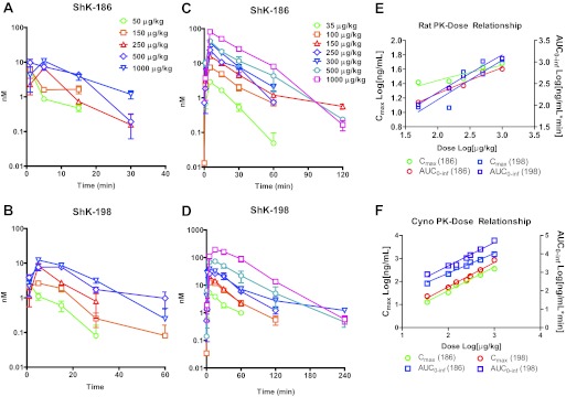
The Kv1.3 channel is a recognized target for pharmaceutical development to treat autoimmune diseases and organ rejection. ShK-186, a specific peptide inhibitor of Kv1.3, has shown promise in animal models of multiple sclerosis and rheumatoid arthritis. Here, we describe the pharmacokinetic-pharmacodynamic relationship for ShK-186 in rats and monkeys. The pharmacokinetic profile of ShK-186 was evaluated with a validated high-performance liquid chromatography-tandem mass spectrometry method to measure the peptide's concentration in plasma. These results were compared with single-photon emission computed tomography/computed tomography data collected with an ¹¹¹In-1,4,7,10-tetraazacyclododecane-1,4,7,10-tetraacetic acid-conjugate of ShK-186 to assess whole-blood pharmacokinetic parameters as well as the peptide's absorption, distribution, and excretion. Analysis of these data support a model wherein ShK-186 is absorbed slowly from the injection site, resulting in blood concentrations above the Kv1.3 channel-blocking IC₅₀ value for up to 7 days in monkeys. Pharmacodynamic studies on human peripheral blood mononuclear cells showed that brief exposure to ShK-186 resulted in sustained suppression of cytokine responses and may contribute to prolonged drug effects. In delayed-type hypersensitivity, chronic relapsing-remitting experimental autoimmune encephalomyelitis, and pristane-induced arthritis rat models, a single dose of ShK-186 every 2 to 5 days was as effective as daily administration. ShK-186's slow distribution from the injection site and its long residence time on the Kv1.3 channel contribute to the prolonged therapeutic effect of ShK-186 in animal models of autoimmune disease.


Figures

Fig. 1.
Fig. 1.
Biophysical properties of ShK-186, ShK-186 dephosphorylated form (ShK-198), and ShK-186 analogs. A, representative whole-cell Kv1.3 currents in the absence and presence of dephosphorylated ShK-186 (ShK-198). B, representative whole-cell Kv1.3 currents in the absence and presence of In-ShK-221 (a ShK-186 analog). In-ShK-221 was in 100% serum. C, comparison of dose-response curves for ShK-186, ShK-198, Gd-ShK-221, and In-ShK-221 on Kv1.3. The IC50 values were: ShK-186, 68.99 ± 4.01 pM (n = 5); ShK-198, 41.4 ± 7.25 pM (n = 5); Gd-ShK-221, 58.23 ± 1.38 pM (n = 5), and In-ShK-221, 63.8 ± 2.25 pM (n = 3). D, dose-response curves of ShK-186 on Kv1.3 in the presence of varying concentrations of serum. E, dose-response curves of In-ShK-221 in the presence or absence of serum. Electrophysical recordings were carried out in the whole-cell configuration of the patch-clamp technique as described previously (Beeton et al., 2005; Wulff et al., 2000). The external solution was sodium Ringer's, and the pipette solution was potassium fluoride (300 mOsm). Kv1.3 currents were elucidated by 200-ms depolarizing pulses from a holding potential of −80 to 40 mV. All analogs were each tested at several concentrations. The reduction in peak current at 40 mV for each concentration was used to generate a dose-response curve by using Origin software (OriginLab Corp., Northhampton, MA).
Fig. 2.
Fig. 2.
Pharmacokinetic profiles of ShK-186 and ShK-198 in plasma after a single dose to rats and monkeys. A to D, ShK-186 was administered by subcutaneous injection to SD rats (A and B; n = 3–9 animals/dose/time point) and cynomolgus monkeys (C and D; n = 2–16 animals/dose/time point), and plasma samples were collected at time intervals ranging from 1 min to 24 h. Samples were analyzed by using a validated HPLC-MS/MS method that can resolve both the parent compound (ShK-186; A and C) and its metabolite (ShK-198; B and D). E and F, Cmax and AUC0-inf were computed for rat (E) and monkey (F) at each dose level by using noncompartmental analysis and the linear trapezoidal method.
Fig. 3.
Fig. 3.
Biodistribution studies with radiolabeled ShK in rat and squirrel monkey. 111In-labeled ShK-221 was administered to SD rats (100 μg/kg; 1.0mCi) and squirrel monkeys (35 μg/kg; 0.84mCi) as a single subcutaneous injection to the scapular region of each animal. SPECT and CT scans were collected continuously during the first hour (4 × 15-m intervals) and at 4, 8, 24, 48, 72, 120, and 160 h postdose. A and C, flattened 2D images of the 3D reconstructions are shown for the 4-, 24-, 72-, and 160-h time points for monkeys (A) and the 1-, 8-, and 24-h time points for rats (C). Both animals showed slow absorption of drug from the injection site and significant early and sustained distribution to kidney and to a lesser extent liver. B and D, kidney-associated radioactivity in monkeys (B) and rats (D) was principally identified in the cortex. E, quantification of 111In-ShK-221 at the injection site in monkeys (top) and rats (bottom) revealed a biphasic decay with an initial half-life of approximately 0.5 to 1.5 h and a terminal half-life of >48 h. F, drug concentrations in monkey (top) and rat (bottom) whole blood followed a similar biphasic decay with an initial half-life of approximately 1.5 h and a terminal half-life of >64 h. Blood concentrations remained above the IC50 for Kv1.3 throughout the entire study period and above the 80% saturation concentration (233 pM) through the first 120 h, which is consistent with a slow, continuous distribution from the injection site throughout the study period.
Fig. 4.
Fig. 4.
Durable effect of drug treatment on human PBMCs. Time of treatment studies were conducted in human PBMCs in which the effect of drug treatment timing and duration on thapsigargin-stimulated IL-2 responses was measured. Continuous drug treatment during the 48-h stimulation period (A; black bars) was compared with drug pretreatment for 1 h followed by a brief washout and stimulation for 48 h (B; open bars) or drug pretreatment (1 h) followed by washout, media incubation (16 h), and thapsigargin stimulation (48 h) (C; hatched bars). Drug treatment for a 1-h period up to 16 h before stimulation was statistically indistinguishable from continuous drug treatment. None of the treatment times and durations were significantly different (one-way ANOVA; F3,12 = 0.40; p = 0.76).
Fig. 5.
Fig. 5.
Time of administration and dose-dependent suppression of the DTH response. Lewis rats were immunized with ovalbumin in CFA and challenged 7 to 9 days later by injection of ovalbumin to the ear pinna. The day of challenge is represented as day 0. The DTH reaction was measured 24 h postchallenge (day 1). A, ShK-186 administered at 10 or 100 μg/kg in two doses on days 0 and 1 or as a single 100 μg/kg dose on days −1, −2, −3, and −4 resulted in a significant reduction in ear swelling relative to placebo (PL)-treated animals when measured on day 1 postchallenge (two-tailed Mann-Whitney). These data indicate that a single ShK-186 dose can suppress the DTH response for up to 5 days. B, using the DTH model, a dose-response study was conducted with a single dose of ShK-186 administered on day −2. Doses between 0.1 and 100 μg/kg showed a clear dose-response relationship, with all but 0.1 μg/kg achieving statistical significance relative to placebo. *, p < 0.05; **, p < 0.01.
Fig. 6.
Fig. 6.
Alternate dosing schedules are effective in the chronic EAE model. A, an initial study evaluated lead-in dosing followed by less than daily drug administration. Male DA rats were immunized with spinal cord homogenate in CFA and administered ShK-186 (100 μg/kg/day; n = 25) or vehicle (n = 12) during the prodromal period and for 7 days after the onset of EAE, after which drug-treated animals were randomized to receive a maintenance dose of 100 μg/kg ShK-186 every 2 days (n = 12) or 3 days (n = 13). ShK-186 significantly reduced (p < 0.001) the severity of the initial episode of disease. Maintenance therapy administered at 48- or 72-h intervals significantly (p < 0.001; one-way repeated-measures ANOVA) reduced the severity of CR-EAE. B, a second study measured the effect of infrequent drug administration beginning at disease onset. Female DA rats (n = 13/group) were immunized with spinal cord homogenate in CFA to elicit a chronic, relapsing EAE. After the onset of symptoms (clinical score >1), animals were randomized to receive daily placebo, daily ShK-186, or ShK-186 every 2 or 3 days. Drug treatment resulted in significantly lower clinical scores and a lower frequency of disease incidence (inset) during the chronic phase in the groups treated daily and groups treated every third day compared with placebo (repeated-measures ANOVA; F2,60 = 39.4; p < 0.0001). Mean clinical scores were not lower relative to placebo in the group treated on alternate days in this study (data not shown). C, a final study measured the durability of response of a period of drug therapy. Female DA rats (n = 23) were immunized and allowed to proceed to CR-EAE. Ten days after the onset of symptoms, animals were randomized to receive daily placebo (n = 10) or 100 μg/kg ShK-186 (n = 13) for 14 days. Therapy was discontinued on day 24, and the time required for treated animals to return to baseline disease was evaluated. The previously treated group had mean clinical scores significantly lower than placebo controls on days 24, 25, 26, and 28 (Supplemental Table 2), which is consistent with a 5-day window for disease recurrence.
Fig. 7.
Fig. 7.
Pristane-induced arthritis treated with alternate day administration of ShK-186. Arthritis was induced in DA rats by injection of 75 μl of pristane at the base of the tail. Disease was allowed to evolve to a clinical score of 1 when animals were randomized to receive placebo or 100 μg/kg ShK-186 every other day (EOD). Clinical score is computed as according to Gillett et al. (2010). Overall disease severity in the study was high. Animals receiving ShK-186 every other day had a statistically significant reduction in mean score (placebo, 20.4 ± 5.9; drug, 14.6 ± 4.1; paired t test; p < 0.0001).

References

    1. Audebert F, Urtizberea M, Sabouraud A, Scherrmann JM, Bon C. (1994) Pharmacokinetics of Vipera aspis venom after experimental envenomation in rabbits. J Pharmacol Exp Ther 268:1512–1517 - PubMed
    1. Azam P, Sankaranarayanan A, Homerick D, Griffey S, Wulff H. (2007) Targeting effector memory T cells with the small molecule Kv1.3 blocker PAP-1 suppresses allergic contact dermatitis. J Invest Dermatol 127:1419–1429 - PMC - PubMed
    1. Barral-Netto M, Schriefer A, Vinhas V, Almeida AR. (1990) Enzyme-linked immunosorbent assay for the detection of Bothrops jararaca venom. Toxicon 28:1053–1061 - PubMed
    1. Beeton C, Chandy KG. (2007) Induction and monitoring of active delayed type hypersensitivity (DTH) in rats. J Vis Exp 6:237. - PMC - PubMed
    1. Beeton C, Pennington MW, Wulff H, Singh S, Nugent D, Crossley G, Khaytin I, Calabresi PA, Chen CY, Gutman GA, et al. (2005) Targeting effector memory T cells with a selective peptide inhibitor of Kv1.3 channels for therapy of autoimmune diseases. Mol Pharmacol 67:1369–1381 - PMC - PubMed

Publication types

MeSH terms