. 2012 Aug;40(15):7303-18.
doi: 10.1093/nar/gks466. Epub 2012 May 25.

Myh7b/miR-499 gene expression is transcriptionally regulated by MRFs and Eos

Affiliations

Myh7b/miR-499 gene expression is transcriptionally regulated by MRFs and Eos

Fan Yeung et al. Nucleic Acids Res. 2012 Aug.

Abstract

The sarcomeric myosin gene, Myh7b, encodes an intronic microRNA, miR-499, which regulates cardiac and skeletal muscle biology, yet little is known about its transcriptional regulation. To identify the transcription factors involved in regulating Myh7b/miR-499 gene expression, we have mapped the transcriptional start sites and identified an upstream 6.2 kb region of the mouse Myh7b gene whose activity mimics the expression pattern of the endogenous Myh7b gene both in vitro and in vivo. Through promoter deletion analysis, we have mapped a distal E-box element and a proximal Ikaros site that are essential for Myh7b promoter activity in muscle cells. We show that the myogenic regulatory factors, MyoD, Myf5 and Myogenin, bind to the E-box, while a lymphoid transcription factor, Ikaros 4 (Eos), binds to the Ikaros motif. Further, we show that through physical interaction, MyoD and Eos form an active transcriptional complex on the chromatin to regulate the expression of the endogenous Myh7b/miR-499 gene in muscle cells. We also provide the first evidence that Eos can regulate expression of additional myosin genes (Myosin 1 and β-Myosin) via the miR-499/Sox6 pathway. Therefore, our results indicate a novel role for Eos in the regulation of the myofiber gene program.


Figures

Figure 1.
Figure 1.
Myh7b/miR-499 has different transcription start sites in heart and skeletal muscle. Top: Schematic of the location of the 6.2 kb Myh7b promoter region relative to the transcription start site (+1) and the translation start site (ATG). Middle: UCSC Genome Browser graphical map of the mouse 6.2 kb Myh7b promoter indicates multiple high homology regions (denoted by peaks) across species. Bottom: 5′-RACE analysis of the mouse Myh7b transcripts revealed striking differences in position of the TSSs in skeletal and cardiac muscle. While multiple TSSs were identified for Myh7b transcripts in cardiac muscle as denoted by bolded nucleotides, a single TSS was identified for the mouse soleus (as indicated by +1). All TSSs identified from the striated muscle are located in exon 1 upstream of the putative translational start site (underlined). Exon boundaries are indicated by lowercase letters.
Figure 2.
Figure 2.
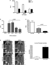
A 6.2 kb upstream promoter region of the Myh7b gene mimics endogenous Myh7b gene expression patterns. (A) The 6.2 kb Myh7b promoter shows cell type-specific activity in cardiac and skeletal muscle cells. C2C12, NRVM and NIH3T3 cells were transiently transfected with m7b-6.2/luc, CMV/β-gal and vector control (VC) or pcDNA-Sox6. *Significantly different from VC, P < 0.05; **significantly different from C2C12, P < 0.05. (B) For differentiation experiments, C2C12 myoblasts were transiently transfected with m7b-6.2/luc and CMV/β-gal, transferred to differentiation medium 24 h post-transfection and harvested at the indicated time points for luciferase and β-gal assays. (C) NRVMs were transfected as in (B). Cells were either left untreated (No add), treated overnight with PE (20 uM) or thyroid hormone (T3, 100 nM). (D) In vivo imaging of the m7b-6.2/luc construct activity in skeletal muscle. Bioluminescent images of a representative individual mouse co-injected with either m7b-6.2/luc or the minimal TATA promoter luciferase control (minTATA) and the internal control (CMV-mKate). Quantification of photons emitted is displayed as Luc activity normalized to mKate activity in the graph. Data presented are means of two mice ± SD. *P < 0.01.
Figure 3.
Figure 3.
Mapping of the critical cis-elements in the Myh7b promoter. (A) Deletion studies indicated a distal and a proximal region within the Myh7b promoter that are important for promoter activity. Indicated deletion or truncation constructs of m7b-6.2/luc were co-transfected with CMV/β-gal into C2C12 cells. Activities of the constructs were shown as luciferase activity normalized to β-gal activity (RLU). (B) Fine deletion of the 2.6 kb distal region of m7b-6.2/luc identified a 450 bp DNA fragment that is responsible for promoter activity. Transient transfections were performed as in (A). (C) D450 and P500 regions contain critical cis-elements for Myh7b promoter activity. Top: The positions of D450 and P500 are shown in the context of the 6.2 kb promoter while the graphical map from the UCSC Genome Browser is shown below with the corresponding regions circled, indicating D450 and P500 share high homology across multiple species. D450 was cloned upstream of a minimal TATA box (D450-TATA) and P500 was inserted upstream in the pGL3 basic vector (P500/luc).
Figure 4.
Figure 4.
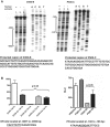
Identification of critical nucleotides in D450 and P500. (A) DNase I footprinting assays were performed with the 150 bp fragments containing either Site B of D450 (D450-B, -4710/-4560) or Site C of P500 (P500-C, -191/-32). Lanes 2, 3, 5 and 6 were naked DNA controls with different concentrations of DNase I in the absence of C2C12 myoblast nuclear extracts indicating even DNA ladders. The footprints were observed (brackets) in D450-B and P500-C only in the presence of C2C12 nuclear extracts (Lanes 1 and 4). The corresponding sequencing reactions are shown to the right of the footprinting assays. The sequences of the protected regions are shown beneath the autoradiogram. (B) Linker-scanning mutagenesis identified DR and PR as the important nucleotides for D450-TATA and P500/luc activities, respectively. A 17 bp fragment in each of the protected regions was replaced systematically with a GAL4 binding site to generate a panel of mutants. The sequences of the PR site and DR site within the respective G2 mutants are shown beneath the graphs. The potential E-box and Ikaros motifs are underlined.
Figure 5.
Figure 5.
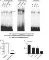
The E-box motif is crucial for D450 transcriptional activity. A,B: EMSA and site-directed mutagenesis indicated that the E-box is essential for D450-TATA activity. C2C12 nuclear extracts (Lanes 1–9 and 12) and the DR probes were used in EMSA. The E-box motif is underlined in the DR probe sequence. DR-mut-Ebox competitor contains nucleotide mutations as indicated in capital letters. D450-mEbox-TATA contains the same mutations as the DR-mut-Ebox probe. C: Overexpression of MRFs activates the distal E-box in the Myh7b promoter. One microgram of D450-TATA or D450-G2-TATA was co-transfected with 0.1 µg of MyoD, Myf5, Myogenin or empty vector (VC) into C2C12 cells.
Figure 6.
Figure 6.
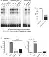
Eos associates with the Ikaros motif within P500 to regulate Myh7b promoter activity. (A and B) EMSA and site-directed mutagenesis indicated the Ikaros site is important for P500 activity. C2C12 nuclear extracts (Lanes 1–7, 9–12) and the PR probes were used in the EMSA experiments. The Ikaros motif is underlined in the PR probe sequence. The PR-mut-Ik competitor contained the indicated point mutations (capital letters). Multiple point mutations were introduced into the Ikaros site within P500/luc as indicated in PR-mut-Ik to generate P500-mIkaros. C2C12 were transiently transfected with either P500/luc or P500-mIkaros. (C): Wild-type m7b-6.2/luc construct or m7b-6.2/luc constructs containing point mutations in the distal E-box (mut-Ebox), proximal Ikaros site (mut-Ikaros) or both sites (mut-Ebox + Ikaros) were transiently transfected into C2C12 cells. *Significantly different from the wild-type (WT) promoter, P < 0.05; #significantly different from WT, mut-Ebox and mut-Ikaros, P < 0.05. (D) Ikaros family members were overexpressed in 293 T cells together with the m7b-6.2/luc and CMV/β-gal. The RLU of the vector control (VC) group was set to 1 and the activities of the various overexpression groups are presented relative to the VC. (E) Mutation of the Ikaros site within P500/luc abolished the effects of Eos overexpression. One microgram of P500-mIkaros was co-transfected with either 0.1 µg of VC or Flag-Eos plasmids into C2C12 cells. (F) EMSA super-shift experiment using Eos antibody and control antibody p65. *Indicates the disappearance of Complex A.
Figure 6.
Figure 6.
Eos associates with the Ikaros motif within P500 to regulate Myh7b promoter activity. (A and B) EMSA and site-directed mutagenesis indicated the Ikaros site is important for P500 activity. C2C12 nuclear extracts (Lanes 1–7, 9–12) and the PR probes were used in the EMSA experiments. The Ikaros motif is underlined in the PR probe sequence. The PR-mut-Ik competitor contained the indicated point mutations (capital letters). Multiple point mutations were introduced into the Ikaros site within P500/luc as indicated in PR-mut-Ik to generate P500-mIkaros. C2C12 were transiently transfected with either P500/luc or P500-mIkaros. (C): Wild-type m7b-6.2/luc construct or m7b-6.2/luc constructs containing point mutations in the distal E-box (mut-Ebox), proximal Ikaros site (mut-Ikaros) or both sites (mut-Ebox + Ikaros) were transiently transfected into C2C12 cells. *Significantly different from the wild-type (WT) promoter, P < 0.05; #significantly different from WT, mut-Ebox and mut-Ikaros, P < 0.05. (D) Ikaros family members were overexpressed in 293 T cells together with the m7b-6.2/luc and CMV/β-gal. The RLU of the vector control (VC) group was set to 1 and the activities of the various overexpression groups are presented relative to the VC. (E) Mutation of the Ikaros site within P500/luc abolished the effects of Eos overexpression. One microgram of P500-mIkaros was co-transfected with either 0.1 µg of VC or Flag-Eos plasmids into C2C12 cells. (F) EMSA super-shift experiment using Eos antibody and control antibody p65. *Indicates the disappearance of Complex A.
Figure 7.
Figure 7.
MRFs and Eos regulate Myh7b/miR-499 expression. (A) Myh7b promoter requires MRFs and Eos for activity. Indicated siRNA was transfected into C2C12 cells for 24 h followed by m7b-6.2/luc plasmid transfection. The RLU of m7b-6.2/luc in the presence of respective siRNAs is depicted relative to the non-targeting siRNA control, which was set as 100. Western blot analysis of C2C12 cells demonstrated efficient knockdown of the target proteins by the siRNAs. Endogenous α-actin protein levels are shown as controls. (B) Eos and MRFs are essential for the endogenous expression of Myh7b/miR-499 in muscle cells. Indicated siRNA knockdowns were performed in C2C12 cells. After RNA isolation, Myh7b and miR-499 levels were determined by SYBR green qPCR and Taqman microRNA assays, respectively. α-Actin was used as an internal control for Myh7b PCR and Sno202 was used as the internal control for miR-499 expression. (C) MRFs and Eos are chromatin-bound on D450 and P500 regions of the Myh7b promoter. ChIP assays were performed with the indicated antibodies on the Myh7b promoter. IgG served as the no antibody control, no DNA served as the PCR negative control, Myh7b −3660 bp and Gapdh served as a non-specific gene control.
Figure 8.
Figure 8.
MyoD interacts with Eos through its bHLH and AD domains. (A) MM14 muscle cells were transfected with expression constructs encoding Eos or MyoD, either individually or together. *Significantly different from vector control (VC), P < 0.05; #significantly different from VC, Eos and MyoD, P < 0.05, Δ indicates fold increase compared to VC. (B) 35S-labeled Eos and recombinant GST-MyoD as well as different domains of MyoD (AD = activation domain, bHLH = basic helix-loop-helix, CT = C-terminal) were used for in vitro GST pull-down assays and analyzed by autoradiography. Inputs were determined by immunoblotting with a GST antibody to show that the same amounts of recombinant proteins were used in the reactions. (C) IP-western with 293 T cells overexpressing Flag-Eos and HA-MyoD as indicated. Cell lysates were immunoprecipitated (IP) with a Flag antibody, followed by immunoblotting (WB) with a HA antibody. Inputs were analyzed by WB using indicated antibodies.
Figure 9.
Figure 9.
The role of Eos in myosin gene regulation. Indicated siRNA knockdowns were performed in C2C12 cells. mRNA levels of Myh1, Myh7 and Sox6 were determined by qPCR. 18S was used as an internal control for q-PCR. Protein levels of Sox6 and Eos were analyzed by western blot with α-actin as internal control. *Significantly different from the siRNA control (siControl), P < 0.05.

Similar articles

Cited by

References

    1. van Rooij E, Quiat D, Johnson BA, Sutherland LB, Qi X, Richardson JA, Kelm RJ, Jr, Olson EN. A family of microRNAs encoded by myosin genes governs myosin expression and muscle performance. Dev. Cell. 2009;17:662–673. - PMC - PubMed
    1. Rubio MD, Johnson R, Miller CA, Huganir RL, Rumbaugh G. Regulation of synapse structure and function by distinct myosin II motors. J. Neurosci. 2011;31:1448–1460. - PMC - PubMed
    1. Bell ML, Buvoli M, Leinwand LA. Uncoupling of expression of an intronic microRNA and its myosin host gene by exon skipping. Mol. Cell. Biol. 2010;30:1937–1945. - PMC - PubMed
    1. Rossi AC, Mammucari C, Argentini C, Reggiani C, Schiaffino S. Two novel/ancient myosins in mammalian skeletal muscles: MYH14/7b and MYH15 are expressed in extraocular muscles and muscle spindles. J. Physiol. 2010;588:353–364. - PMC - PubMed
    1. Wilson KD, Hu S, Venkatasubrahmanyam S, Fu JD, Sun N, Abilez OJ, Baugh JJ, Jia F, Ghosh Z, Li RA, et al. Dynamic microRNA expression programs during cardiac differentiation of human embryonic stem cells: role for miR-499. Circ. Cardiovasc. Genet. 2011;3:426–435. - PMC - PubMed

Publication types

MeSH terms