. 2012 Aug;40(15):7016-45.
doi: 10.1093/nar/gks382. Epub 2012 May 25.

Sequence, structure and functional diversity of PD-(D/E)XK phosphodiesterase superfamily

Affiliations

Sequence, structure and functional diversity of PD-(D/E)XK phosphodiesterase superfamily

Kamil Steczkiewicz et al. Nucleic Acids Res. 2012 Aug.

Abstract

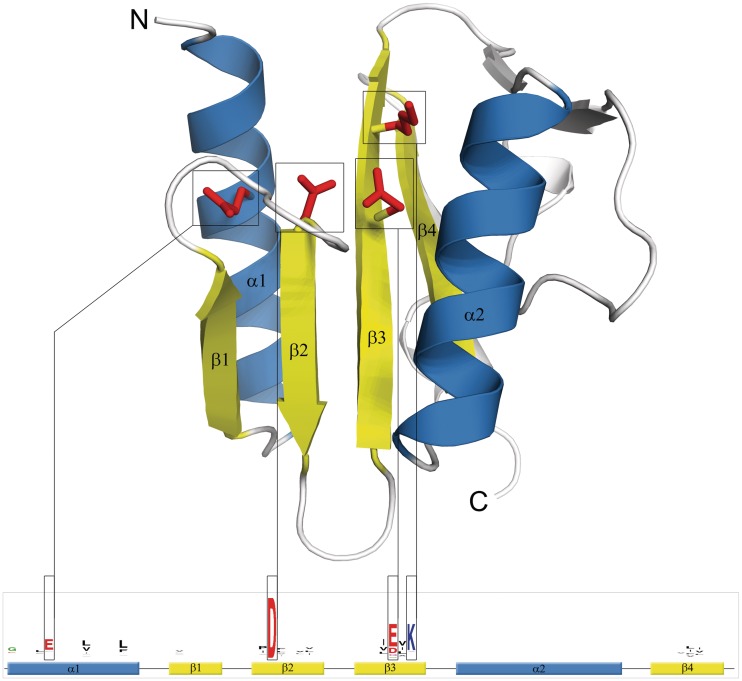
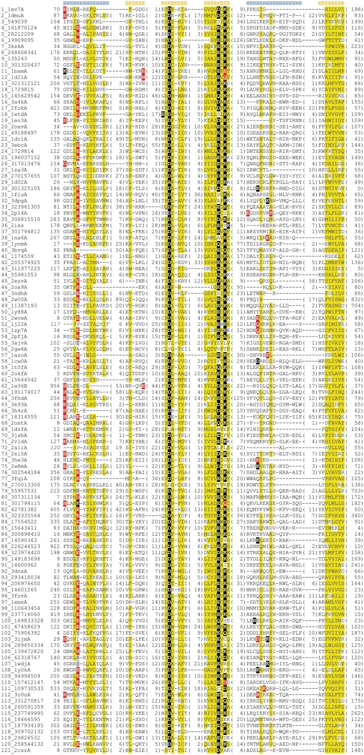
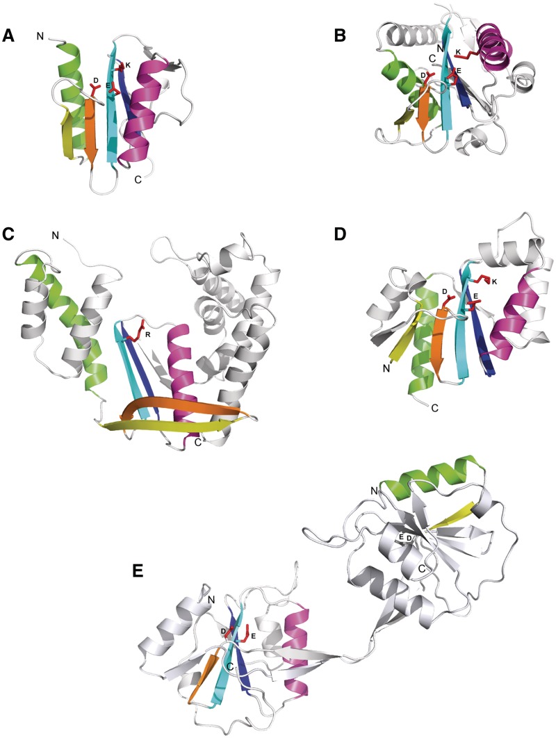
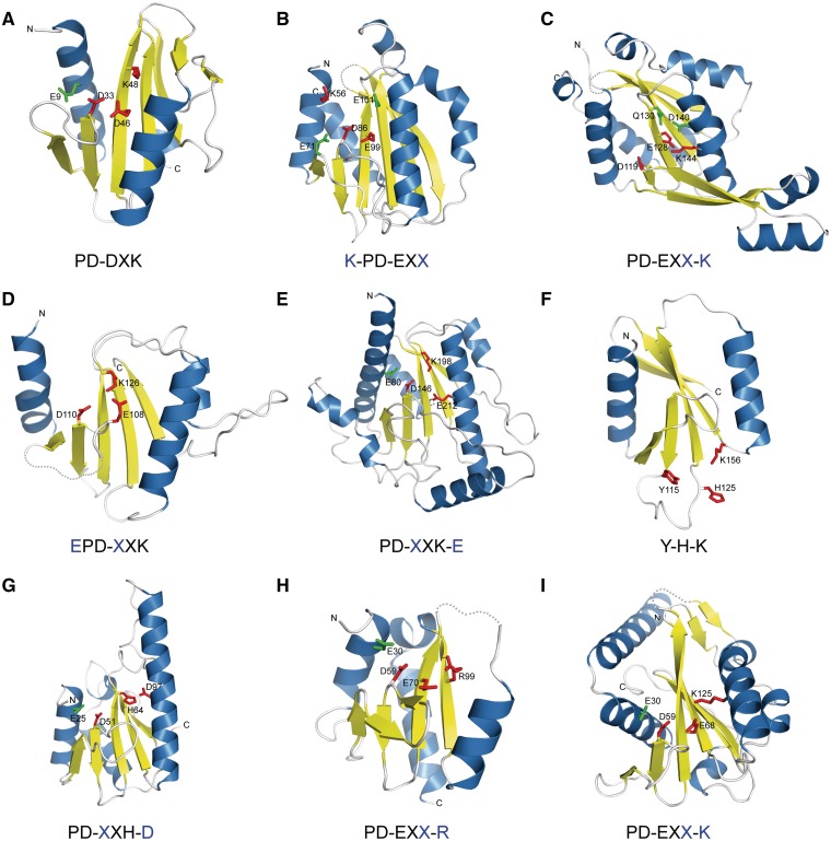
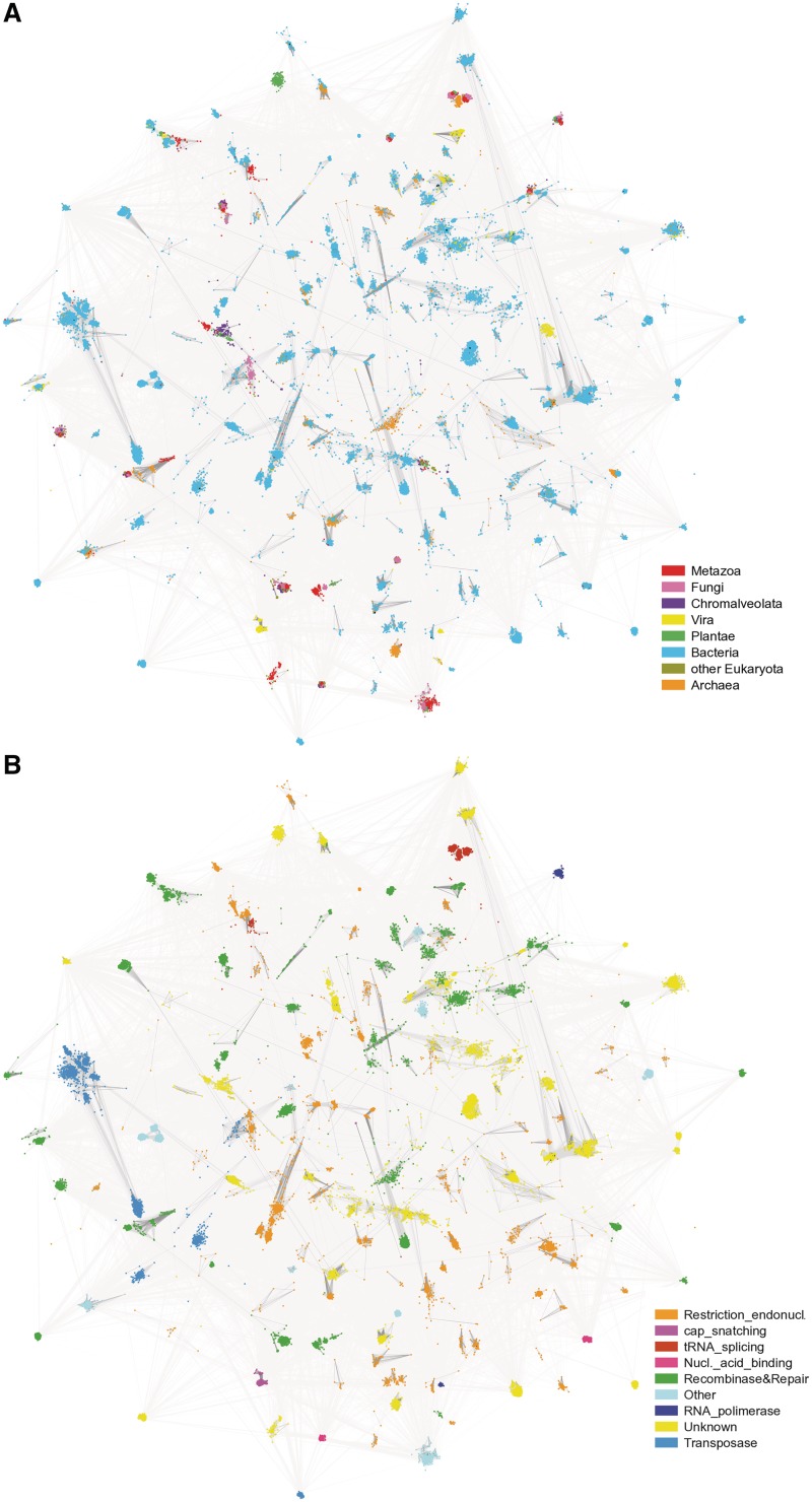
Proteins belonging to PD-(D/E)XK phosphodiesterases constitute a functionally diverse superfamily with representatives involved in replication, restriction, DNA repair and tRNA-intron splicing. Their malfunction in humans triggers severe diseases, such as Fanconi anemia and Xeroderma pigmentosum. To date there have been several attempts to identify and classify new PD-(D/E)KK phosphodiesterases using remote homology detection methods. Such efforts are complicated, because the superfamily exhibits extreme sequence and structural divergence. Using advanced homology detection methods supported with superfamily-wide domain architecture and horizontal gene transfer analyses, we provide a comprehensive reclassification of proteins containing a PD-(D/E)XK domain. The PD-(D/E)XK phosphodiesterases span over 21,900 proteins, which can be classified into 121 groups of various families. Eleven of them, including DUF4420, DUF3883, DUF4263, COG5482, COG1395, Tsp45I, HaeII, Eco47II, ScaI, HpaII and Replic_Relax, are newly assigned to the PD-(D/E)XK superfamily. Some groups of PD-(D/E)XK proteins are present in all domains of life, whereas others occur within small numbers of organisms. We observed multiple horizontal gene transfers even between human pathogenic bacteria or from Prokaryota to Eukaryota. Uncommon domain arrangements greatly elaborate the PD-(D/E)XK world. These include domain architectures suggesting regulatory roles in Eukaryotes, like stress sensing and cell-cycle regulation. Our results may inspire further experimental studies aimed at identification of exact biological functions, specific substrates and molecular mechanisms of reactions performed by these highly diverse proteins.


Figures

Figure 1.
Figure 1.
The commonly conserved core of PD-(D/E)XK nuclease fold. Critical active site residues are shown as red sticks and marked in corresponding sequence logo. Sequence logo was derived from multiple sequence alignment for PD-(D/E)XK phosphodiesterase superfamily using WebLogo (20).
Figure 2.
Figure 2.
Multiple sequence alignment for the conserved core regions of the PD-(D/E)XK superfamily. Each group of closely related Pfam, COG, KOG families and PDB90 structures (detectable with PSI-BLAST) is represented by available PDB90 sequence or selected representative if the cluster does not contain solved structure. Sequences are labeled according to the group number followed by NCBI gene identification number or PDB code. The first residue numbers are indicated before each sequence, while the numbers of excluded residues are specified in parentheses. Sequence given in italic corresponds to circularly permuted α-helix. Residue conservation is denoted with the following scheme: uncharged, highlighted in yellow; polar, highlighted in grey; active site PD-(D/E)XK signature residues, highlighted in black; other conserved polar/charged residues augmenting the active site, highlighted in red. Locations of secondary structure elements are shown above the corresponding alignment blocks.
Figure 3.
Figure 3.
Examples of structural diversity in the PD-(D/E)XK phosphodiesterase superfamily. (A) typical PD-(D/E)XK enzyme (Holiday junction resolvase, Pyrococcus furiosus, pdb|1gef); (B) highly diverged structure with short first β-strand and perpendicular orientation of core α-helices (Pa4535 protein, P. aeruginosa, pdb|1y0k); (C) structure deterioration and the loss of active site (RecC, E. coli, pdb|1w36C); (D) circular permutation of the first core α-helix (Hef endonuclease, Pyrococcus furiosus, pdb|1j22); (E) domain swapping (endonuclease I, Enterobacteria phage T7, pdb|2pfj). Active site PD-(D/E)XK signature residues are shown as red sticks.
Figure 4.
Figure 4.
Active site variations observed in the PD-(D/E)XK phosphodiesterase superfamily structures. Observed variant of ‘PD-(D/E)XK’ signature motif is given below each structure with residue migration denoted in blue. (A) archaeal HJC resolvase (P. furiosus, pdb|1gef); (B) BamHI restriction endonuclease (Oceanobacter kriegii, pdb|3odh); (C) BstYI restriction endonuclease (Geobacillus stearothermophilus, pdb|1sdo); (D) EcoO109I restriction endonuclease (E. coli, pdb|1wtd); (E) Bse634I restriction endonuclease (Geobacillus stearothermophilus, pdb|1knv); (F) tRNA splicing endonuclease (Methanocaldococcus jannaschii, pdb|1a79); (G) Vsr repair endonuclease (E. coli, pdb|1cw0); (H) a putative endonuclease-like protein (Neisseria gonorrhoeae, pdb|3hrl); (I) Pa4535 protein (P. aeruginosa, pdb|1y0k).
Figure 5.
Figure 5.
CLANS clustering of 21 911 sequences belonging to 121 clades of the PD-(D/E)XK superfamily. The image was drawn with an in-house script based on CLANS run files. (A) illustrates the taxonomic distribution of analyzed sequences and (B) summarizes their functional annotation.

References

    1. Orlowski J, Bujnicki JM. Structural and evolutionary classification of Type II restriction enzymes based on theoretical and experimental analyses. Nucleic Acids Res. 2008;36:3552–3569. - PMC - PubMed
    1. Belfort M, Weiner A. Another bridge between kingdoms: tRNA splicing in archaea and eukaryotes. Cell. 1997;89:1003–1006. - PubMed
    1. Hickman AB, Li Y, Mathew SV, May EW, Craig NL, Dyda F. Unexpected structural diversity in DNA recombination: the restriction endonuclease connection. Mol. Cell. 2000;5:1025–1034. - PubMed
    1. Dahlroth SL, Gurmu D, Schmitzberger F, Engman H, Haas J, Erlandsen H, Nordlund P. Crystal structure of the shutoff and exonuclease protein from the oncogenic Kaposi's sarcoma-associated herpesvirus. FEBS J. 2009;276:6636–6645. - PubMed
    1. Aravind L, Makarova KS, Koonin EV. SURVEY AND SUMMARY: holliday junction resolvases and related nucleases: identification of new families, phyletic distribution and evolutionary trajectories. Nucleic Acids Res. 2000;28:3417–3432. - PMC - PubMed

Publication types

MeSH terms

Substances