Comparative Study
. 2012 Sep;36(9):1915-21.
doi: 10.1007/s00264-012-1574-x. Epub 2012 May 27.

The locking attachment plate for proximal fixation of periprosthetic femur fractures--a biomechanical comparison of two techniques

Affiliations
Comparative Study

The locking attachment plate for proximal fixation of periprosthetic femur fractures--a biomechanical comparison of two techniques

Mark Lenz et al. Int Orthop. 2012 Sep.

Abstract

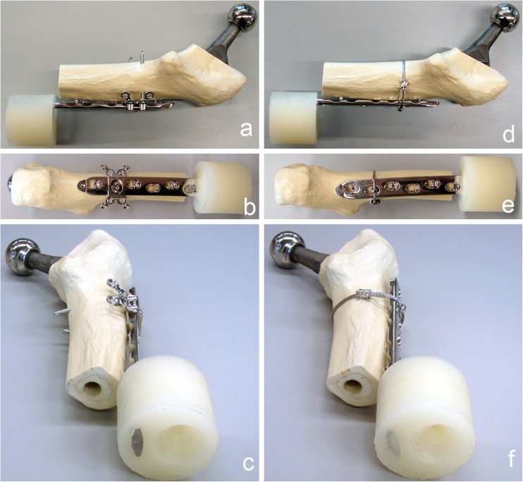
Purpose: Mechanical properties of a locking attachment plate construct (LAP-LCP), allowing bicortical screw placement laterally to the prosthesis stem, are compared to a cerclage-LCP construct.

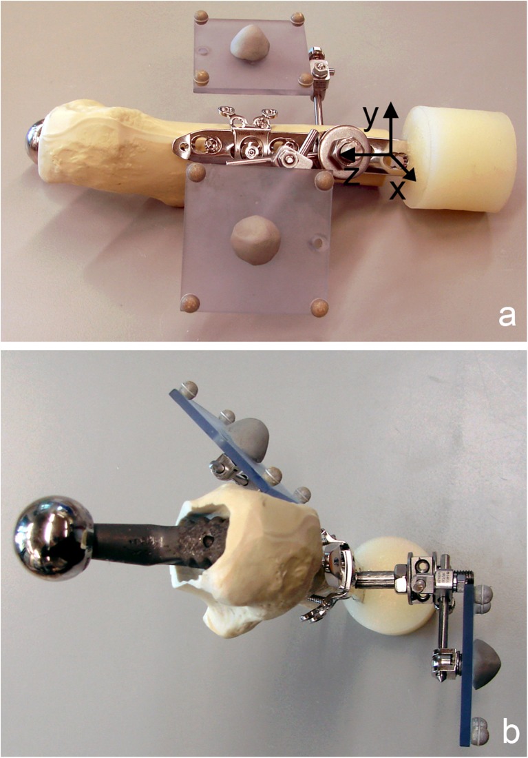
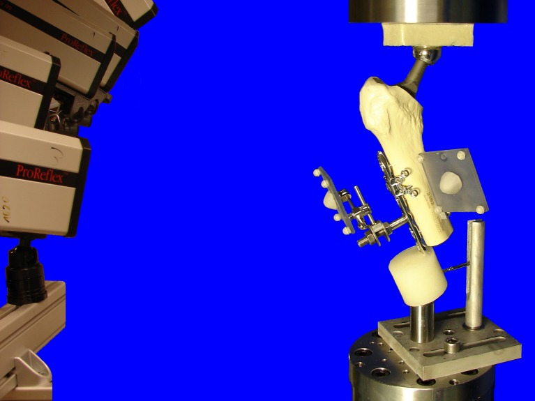
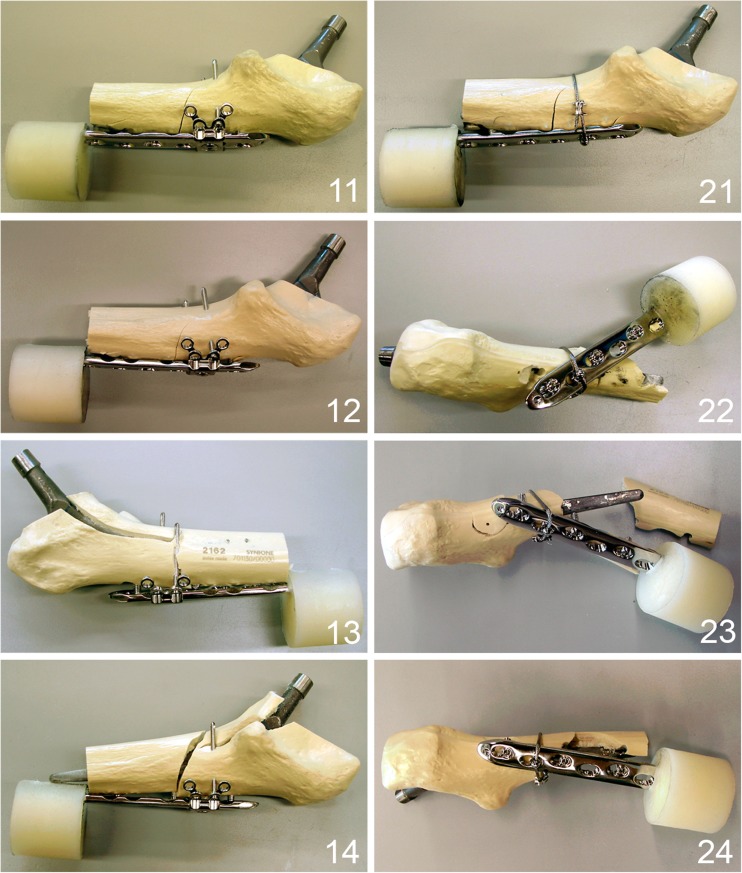
Methods: Eight right synthetic femora with implanted uncemented hip endoprosthesis were cut distally and fixed with LCP, monocortical locking screws and either LAP (n = 4) or cerclage (n = 4). Cyclic testing was performed with monotonically increasing sinusoidal load until failure. Relative movements at the plate-femur interface were registered by motion tracking. Statistical differences were detected by unpaired t-test and general linear model repeated measures.

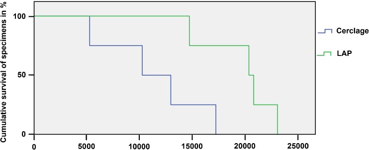
Results: Stiffness of the LAP-LCP was significantly higher at the beginning (875.4 N/mm ± 29.8) and after 5000 cycles (1213.0 N/mm ± 101.1) compared to the cerclage-LCP (644.96 N/mm ± 50.1 and 851.9 N/mm ± 81.9), with p = 0.013. Relative movements for AP-bending (B) and axial translation (T) of the LAP-LCP at the beginning (0.07° ± 0.02, 0.20 mm ± 0.08), after 500 cycles (0.16° ± 0.10, 0.26 mm ± 0.07) and after 5000 cycles (0.26° ± 0.11, 0.31 mm ± 0.07) differed significantly from the cerclage-LCP (beg.: 0.26° ± 0.04, 0.28 mm ± 0.05; 500 cyc: 0.47° ± 0.03, 0.53 mm ± 0.07; 5000 cyc.: 0.63° ± 0.18, 0.79 mm ± 0.13), with B: p = 0.02, T: p = 0.04. Relative movements for medial bending were not significantly different between the two constructs. Cycles to failure (criterion 1 mm axial translation) differed significantly between LAP-LCP (19,519 ± 1,758) and cerclage-LCP (11,265 ± 2,472), with p = 0.035.

Conclusions: Biomechanically, the LAP-LCP construct improves proximal fixation of periprosthetic fractures compared to the cerclage-LCP construct.


Figures

Fig. 1
Fig. 1
Instrumented synthetic femur prepared for testing, cut distally to the tip of the prosthesis stem. The distal end of the locking compression plate (LCP) was potted into PMMA to test only the proximal fixation. ac Fixation with locking attachment plate (LAP) and unicortical screws. df Fixation with cerclage and unicortical screws
Fig. 2
Fig. 2
Specimen prepared for mechanical testing with two retro-reflective marker sets, consisting of four markers each, attached to the locking compression plate (LCP) (reference marker set) and to the synthetic proximal femur (a lateral view with schematic coordinate system for motion tracking; b cranial view). The reference marker set with markers aligned parallel to the LCP axis and the sagittal plane defines a Cartesian coordinate system (a) with centre on the lateral aspect of the femur on the level of the fourth LCP hole, and the z-axis pointing in the proximal direction along the central axis of the plate and the y-axis anteriorly in the plane of the plate
Fig. 3
Fig. 3
Test setup with a specimen, mounted on the testing machine in a 20° valgus position ensuring physiological weight bearing and avoiding distal bending moment of the locking compression plate (LCP). The head of the prosthesis was used as a ball-and-socket joint, connecting the specimen to the actuator. Distally the specimen was connected to the load cell via a ball-and-socket-joint. A Schanz-screw in the PMMA block prevented rotational instability around the load axis of the specimens. Cameras on the left recorded the movements of the retroflective marker sets attached to the constructs
Fig. 4
Fig. 4
Cumulative survival in both groups upon cycle number. Cumulative survival rate (y-axis) is scaled from 100 % of the specimens intact down to 0 % of the specimens intact where all specimens failed. Failure criterion was defined as a relative movement of 1 mm in axial translation at the plate–femur interface
Fig. 5
Fig. 5
Different failure types of the tested specimens. 11–14 Specimens fixed with locking attachment plate (LAP) plate showing an unique failure mode with cracks around all screws. 21–24 Specimens fixed with cerclage and screws showing a fracturing of the bone around the screws as a result of anteroposterior bending as preponderant failure mode except specimen 21, showing the same failure mode as in the LAP group with cracks around all screws

References

    1. Duncan CP, Masri BA. Fractures of the femur after hip replacement. Instr Course Lect. 1995;44:293–304. - PubMed
    1. Lindahl H, Garellick G, Regner H, Herberts P, Malchau H. Three hundred and twenty-one periprosthetic femoral fractures. J Bone Joint Surg Am. 2006;88:1215–1222. doi: 10.2106/JBJS.E.00457. - DOI - PubMed
    1. Pike J, Davidson D, Garbuz D, Duncan CP, O’Brien PJ, Masri BA. Principles of treatment for periprosthetic femoral shaft fractures around well-fixed total hip arthroplasty. J Am Acad Orthop Surg. 2009;17:677–688. - PubMed
    1. Ricci WM, Bolhofner BR, Loftus T, Cox C, Mitchell S, Borrelli J., Jr Indirect reduction and plate fixation, without grafting, for periprosthetic femoral shaft fractures about a stable intramedullary implant. J Bone Joint Surg Am. 2005;87:2240–2245. doi: 10.2106/JBJS.D.01911. - DOI - PubMed
    1. Virolainen P, Mokka J, Seppanen M, Makela K. Up to 10 years follow up of the use of 71 cortical allografts (strut-grafts) for the treatment of periprosthetic fractures. Scand J Surg. 2010;99:240–243. - PubMed

Publication types