Odontogenic myxoma of the maxilla: A report of a rare case and review on histogenetic and diagnostic concepts
- PMID: 22639512
- PMCID: PMC3343396
- DOI: 10.4103/0975-5950.94480
Odontogenic myxoma of the maxilla: A report of a rare case and review on histogenetic and diagnostic concepts
Abstract
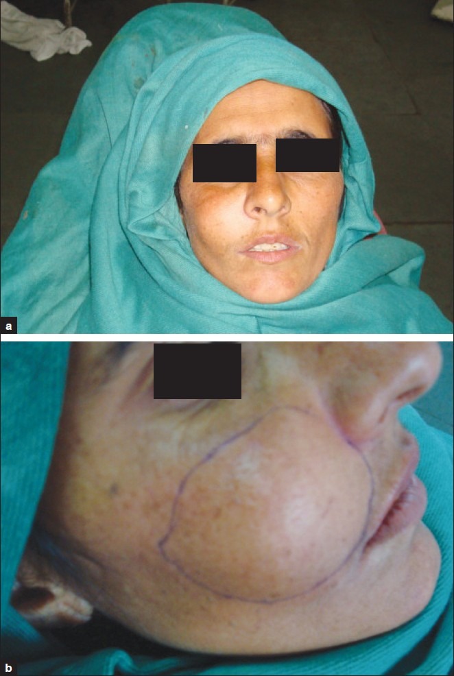
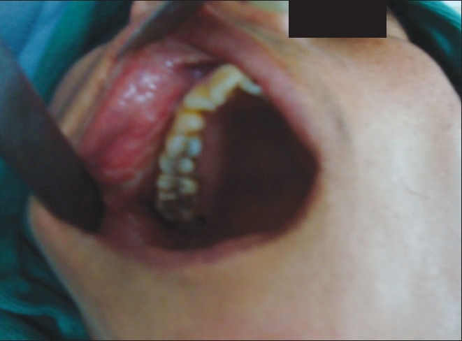
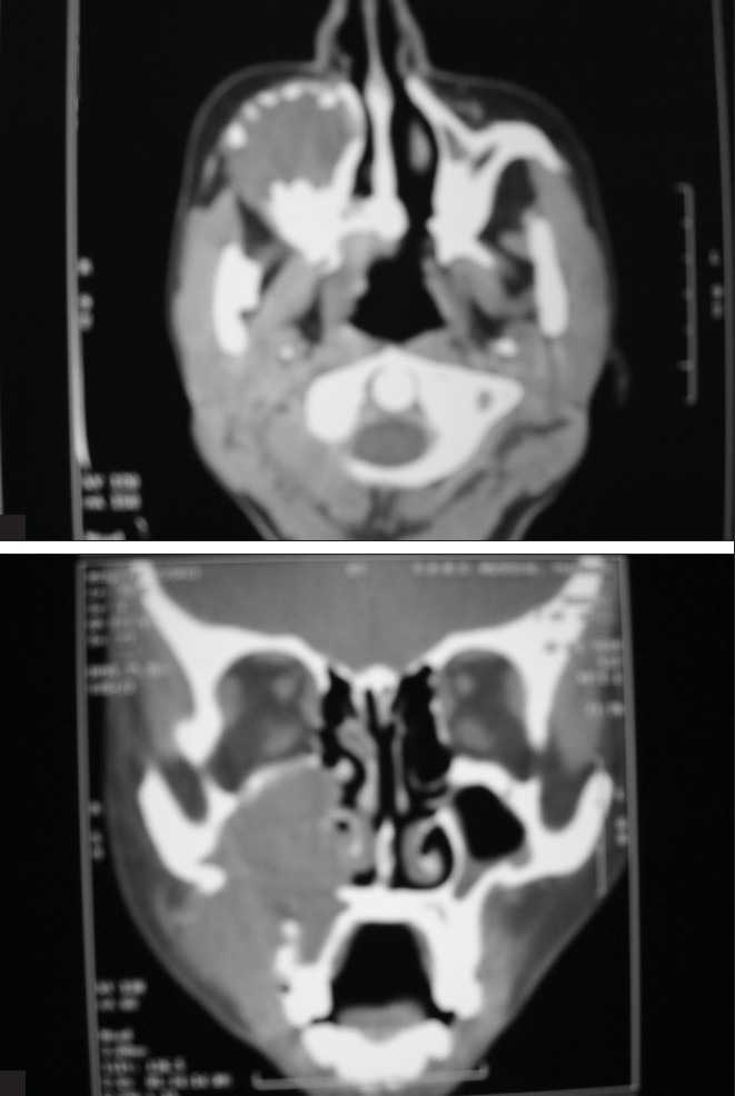
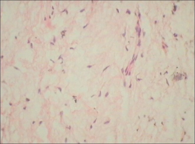
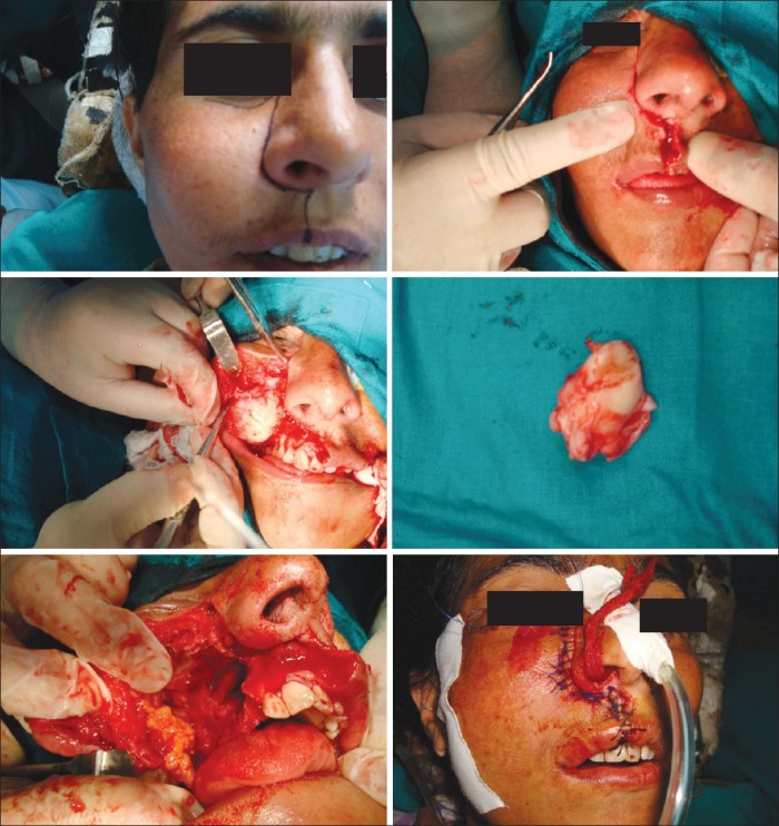
Odontogenic myxoma (OM) is a rare and locally invasive benign neoplasm (comprising of 3-6% of all odontogenic tumors) found exclusively in the jaws. OM commonly occurs in the second and third decades, and the mandible is involved more commonly than the maxilla. The lesion often grows without symptoms and presents as a painless swelling. The radiographic features are variable, and the diagnosis is therefore not easy. This article presents a rare case of OM occurring in the maxilla of a 37-year-old female patient with a brief review of the pathogenesis, clinical, radiological, histopathological, ultrastructural and immunohistochemical characteristics of OM.
Keywords: Fibromyxoma; odontogenic myxomas; odontogenic tumors.
Conflict of interest statement
Figures
References
-
- Abiose BO, Ajagbe HA, Thomas O. Fibromyxomas of the jaws: A study of 10 cases. Br J Oral Maxillofac Surg. 1987;25:415–21. - PubMed
-
- Lu Y, Xuan M, Takata T, Wang C, He Z, Zhou Z, et al. Odontogenictumors: A demographic study of 759 cases in a Chinese population. Oral Surg Oral Med Oral Pathol Oral Radiol Endod. 1998;86:707–14. - PubMed
-
- Pindborg JJ, Kramer IR. International Histological classification of tumors, No.5. Geneva: World Health Organization; 1971. Histological typing of Odontogenictumors. Jaw cysts and Allied lesions.
-
- Halfpenny W, Verey A, Bardsley V. Myxoma of mandibular condyle: A case report and review of literature. Oral Surg Oral Med Oral Pathol Oral Radiol Endod. 2000;90:348–53. - PubMed
-
- Stout AP. Myxoma the tumor of primitive mesenchyme. Ann Surg. 1948;127:706–19. - PubMed