. 2011 Oct 31:2:71.
doi: 10.3389/fpls.2011.00071. eCollection 2011.

Mutations in an Atypical TIR-NB-LRR-LIM Resistance Protein Confer Autoimmunity

Affiliations

Mutations in an Atypical TIR-NB-LRR-LIM Resistance Protein Confer Autoimmunity

Dongling Bi et al. Front Plant Sci. .

Abstract

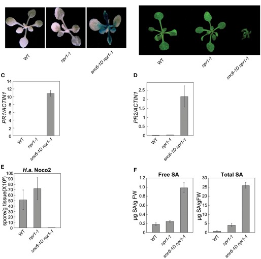
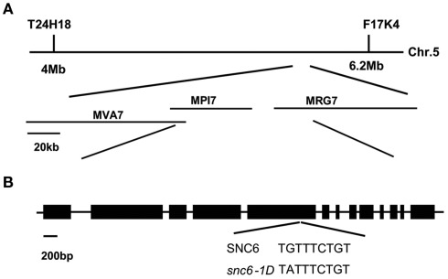
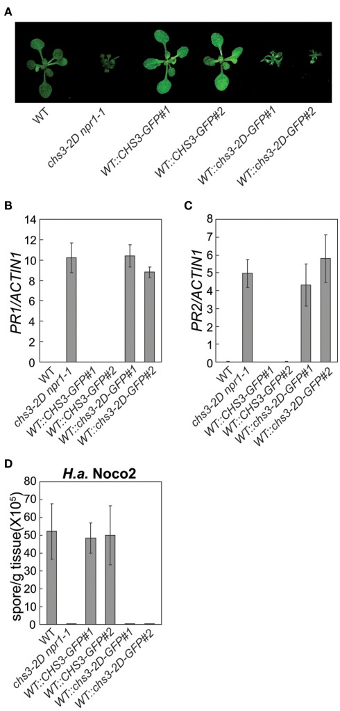
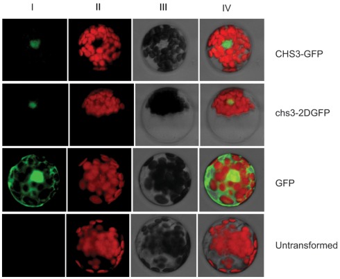
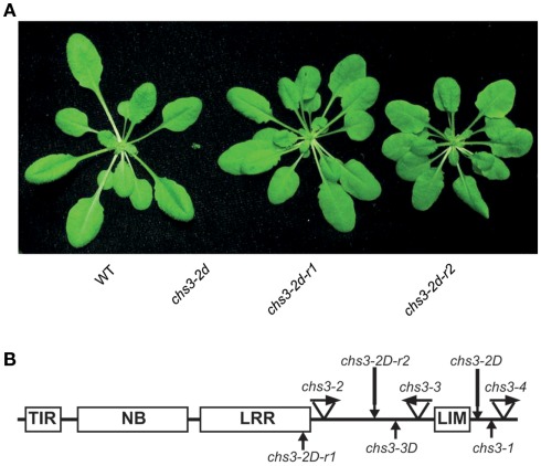
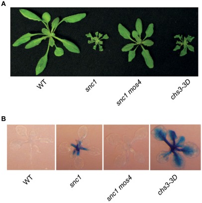
In order to defend against microbial infection, plants employ a complex immune system that relies partly on resistance (R) proteins that initiate intricate signaling cascades upon pathogen detection. The resistance signaling network utilized by plants is only partially characterized. A genetic screen conducted to identify novel defense regulators involved in this network resulted in the isolation of the snc6-1D mutant. Positional cloning revealed that this mutant contained a molecular lesion in the chilling sensitive 3 (CHS3) gene, thus the allele was renamed chs3-2D. CHS3 encodes a TIR-NB-LRR R protein that contains a C-terminal zinc-binding LIM (Lin-11, Isl-1, Mec-3) domain. Although this protein has been previously implicated in cold stress and defense response, the role of the LIM domain in modulating protein activity is unclear. The chs3-2D allele contains a G to A point mutation causing a C1340 to Y1340 substitution close to the LIM domain. It encodes a dominant gain-of-function mutation. The chs3-2D mutant is severely stunted and displays curled leaf morphology. Additionally, it constitutively expresses PATHOGENESIS-RELATED (PR) genes, accumulates salicylic acid, and shows enhanced resistance to the virulent oomycete isolate Hyaloperonospora arabidopsidis (H.a.) Noco2. Subcellular localization assays using GFP fusion constructs indicate that both CHS3 and chs3-2D localize to the nucleus. A third chs3 mutant allele, chs3-3D, was identified in an unrelated genetic screen in our lab. This allele contains a C to T point mutation resulting in an M1017 to V1017 substitution in the LRR-LIM linker region. Additionally, a chs3-2D suppressor screen identified two revertant alleles containing secondary mutations that abolish the mutant morphology. Analysis of the locations of these molecular lesions provides support for the hypothesis that the LIM domain represses CHS3 R-like protein activity. This repression may occur through either autoinhibition or binding of a negative defense regulator.

Keywords: Arabidopsis; CHS3; LIM domain; innate immunity; resistance protein.


Figures

Figure 1
Figure 1
Phenotypic characterization of snc6-1D npr1-1. (A) Expression of the pPR2-GUS reporter gene, present in wild type (WT) and npr1-1 mutant background, in WT, npr1-1, and snc6-1D npr1-1 plants. Seedlings were grown on MS media for 14 days prior to staining for GUS activity. The three photographs shown were taken at different magnifications in order to effectively capture GUS staining, thus seedling size should not be compared between panels. (B) Morphology of wild type (WT), npr1-1, and snc6-1D npr1-1 plants. Representative 4-week-old soil-grown plants were photographed. (C,D) Endogenous expression of (C) PR1 and (D) PR2 relative to Actin 1 in WT, npr1-1, and snc6-1D npr1-1. Values presented are averages of three replicates ± SD. (E) Growth of H.a. Noco2 on WT, npr1-1, and snc6-1D npr1-1 mutants 7 days post-infection. Values presented are averages of three replicates ± SD. (F) Endogenous free and total SA levels in WT, npr1-1, and snc6-1D npr1-1. Plants were grown on soil for 4 weeks and leaf tissue was collected for SA extraction. SA levels were analyzed using high-pressure liquid chromatography. Values presented are averages of four replicates ± SD.
Figure 2
Figure 2
Map-based cloning of the snc6 locus on chromosome 5. (A) A genetic map of the region of chromosome 5 containing the SNC6 locus, with markers used for mapping indicated. (B) Gene structure of SNC6 (At5g17890, previously named CHS3) and the position of the molecular lesion in snc6-1D/chs3-2D. Boxes and lines represent exons and introns, respectively.
Figure 3
Figure 3
Characterization of transgenic plants expressing CHS3-GFP or chs3-2D-GFP under the control of its endogenous promoter. (A) Morphology of wild type (WT), chs3-2D npr1-1, CHS3-GFPline 1, CHS3-GFPline 2, chs3-2D-GFPline 1, and chs3-2D-GFPline 2 soil-grown plants. CHS3-GFP and chs3-2D-GFP constructs were expressed in the WT Col-0 background. Representative plants were photographed 4 weeks after germination. (B,C) Endogenous expression of (B) PR1 and (C) PR2 relative to Actin 1 in the above-mentioned genotypes. Values presented are averages of three replicates ± SD. (D) Growth of H.a. isolate Noco2 on the above-mentioned genotypes 7 days post-infection. Values presented are averages of three replicates ± SD.
Figure 4
Figure 4
CHS3-GFP and chs3-2D-GFP localize to the nucleus. CHS3-GFP and chs3-2D-GFP fluorescence as observed by confocal microscopy in Arabidopsis mesophyll protoplast cells (I: GFP, II: Autofluorescence, III: Bright field, IV: Merged). Experiments were repeated with multiple cells for each construct.
Figure 5
Figure 5
Identification of chs3-2D revertants and location of a number of chs3 mutations. (A) Morphology of WT, chs3-2D npr1-1, chs3-2D-r1, and chs3-2D-r2 soil-grown plants. Representative plants were photographed 4 weeks after germination. (B) Predicted CHS3 protein structure and locations of chs3-1 (Yang et al., 2010), chs3-2D, chs3-3D, chs3-2D-r1, and chs3-2D-r2 mutations. TIR, Toll/Interleukin-1-receptor-like; NB, nucleotide-binding site; LRR, leucine-rich repeat; LIM, Lin-11, Isl-1, and Mec-3 domain. Arrows indicate the locations of the point mutations corresponding to chs3 mutations.
Figure A1
Figure A1
Characterization of chs3-3D. (A) Morphology of WT, snc1, snc1 mos4, and chs3-3D soil-grown plants. Representative plants were photographed 4 weeks after germination. (B) Expression of the pPR2-GUS reporter gene, present in WT and snc1 mutant background, in the above-mentioned genotypes. Seedlings were grown on MS media for 14 days prior to staining for GUS activity. The three photographs shown were taken at different magnifications in order to effectively capture GUS staining, thus seedling size should not be compared between panels.

References

    1. Aarts N., Metz M., Holub E., Staskawicz B. J., Daniels M. J., Parker J. E. (1998). Different requirements for EDS1 and NDR1 by disease resistance genes define at least two R gene-mediated signaling pathways in Arabidopsis. Proc. Natl. Acad. Sci. U.S.A. 95, 10306–10311 10.1073/pnas.95.17.10306 - DOI - PMC - PubMed
    1. Arber S., Caroni P. (1996). Specificity of single LIM motifs in targeting and LIM/LIM interactions in situ. Genes Dev. 10, 289–300 10.1101/gad.10.3.289 - DOI - PubMed
    1. Baltz R., Evrard J. L., Bourdon V., Steinmetz A. (1996). The pollen-specific LIM protein PLIM-1 from sunflower binds nucleic acids in vitro. Sex. Plant Reprod. 9, 264–268 10.1007/BF02152700 - DOI
    1. Bendahmane A., Famham G., Moffett P., Baulcombe D. C. (2002). Consitutive gain-of-function mutants in a nucleotide binding site-leucine rich repeat protein encoded at the Rx locus of potato. Plant J. 32, 195–204 10.1046/j.1365-313X.2002.01413.x - DOI - PubMed
    1. Bernoux M., Ve T., Williams S., Warren C., Hatters D., Valkov E., Zhang X., Ellis J. G., Kobe B., Dodds P. N. (2011). Structural and functional analysis of a plant resistance protein TIR domain reveals interfaces for self-association, signaling, and autoregulation. Cell Host Microbe 9, 200–211 10.1016/j.chom.2011.02.009 - DOI - PMC - PubMed