. 2012 May 28:12:10.
doi: 10.1186/1472-6807-12-10.

Modeling holo-ACP:DH and holo-ACP:KR complexes of modular polyketide synthases: a docking and molecular dynamics study

Affiliations

Modeling holo-ACP:DH and holo-ACP:KR complexes of modular polyketide synthases: a docking and molecular dynamics study

Swadha Anand et al. BMC Struct Biol. .

Abstract

Background: Modular polyketide synthases are multifunctional megasynthases which biosynthesize a variety of secondary metabolites using various combinations of dehydratase (DH), ketoreductase (KR) and enoyl-reductase (ER) domains. During the catalysis of various reductive steps these domains act on a substrate moiety which is covalently attached to the phosphopantetheine (P-pant) group of the holo-Acyl Carrier Protein (holo-ACP) domain, thus necessitating the formation of holo-ACP:DH and holo-ACP:KR complexes. Even though three dimensional structures are available for DH, KR and ACP domains, no structures are available for DH or KR domains in complex with ACP or substrate moieties. Since Ser of holo-ACP is covalently attached to a large phosphopantetheine group, obtaining complexes involving holo-ACP by standard protein-protein docking has been a difficult task.

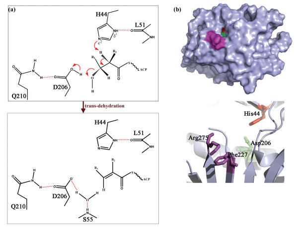
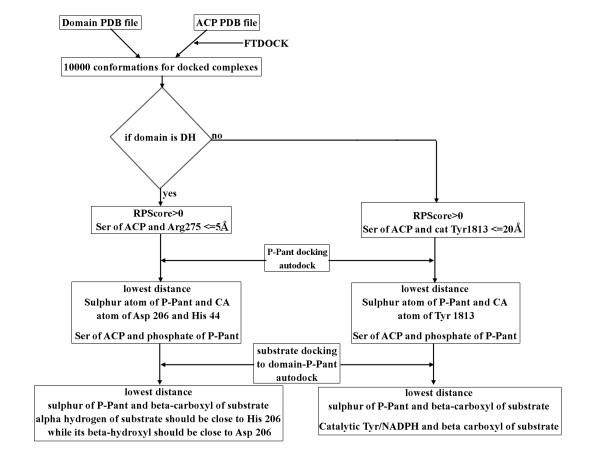
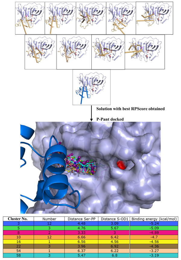
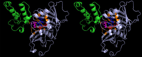
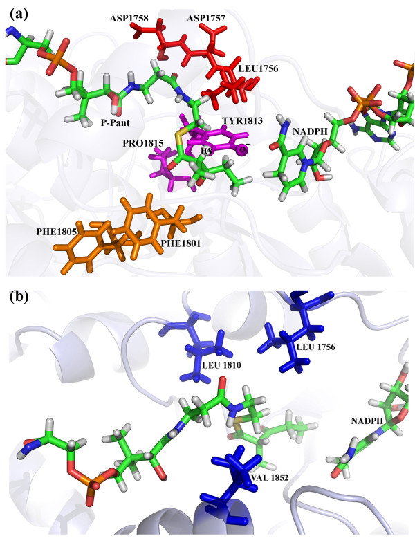
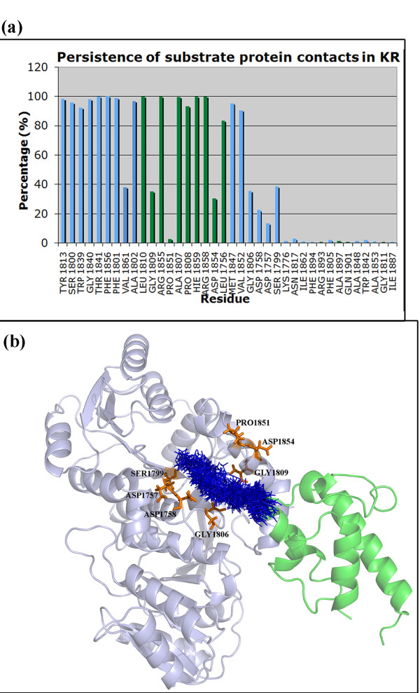
Results: We have modeled the holo-ACP:DH and holo-ACP:KR complexes for identifying specific residues on DH and KR domains which are involved in interaction with ACP, phosphopantetheine and substrate moiety. A novel combination of protein-protein and protein-ligand docking has been used to first model complexes involving apo-ACP and then dock the phosphopantetheine and substrate moieties using covalent connectivity between ACP, phosphopantetheine and substrate moiety as constraints. The holo-ACP:DH and holo-ACP:KR complexes obtained from docking have been further refined by restraint free explicit solvent MD simulations to incorporate effects of ligand and receptor flexibilities. The results from 50 ns MD simulations reveal that substrate enters into a deep tunnel in DH domain while in case of KR domain the substrate binds a shallow surface exposed cavity. Interestingly, in case of DH domain the predicted binding site overlapped with the binding site in the inhibitor bound crystal structure of FabZ, the DH domain from E.Coli FAS. In case of KR domain, the substrate binding site identified by our simulations was in proximity of the known stereo-specificity determining residues.

Conclusions: We have modeled the holo-ACP:DH and holo-ACP:KR complexes and identified the specific residues on DH and KR domains which are involved in interaction with ACP, phosphopantetheine and substrate moiety. Analysis of the conservation profile of binding pocket residues in homologous sequences of DH and KR domains indicated that, these results can also be extrapolated to reductive domains of other modular PKS clusters.


Figures

Figure 1
Figure 1
a) Schematic diagram depicting the mechanism of reaction catalyzed by DH domain.b) The Arg 275 and Phe 227 shown in magenta are hypothesized to be involved in ACP binding while His 44 (red) and Asp 206 (green) are catalytic residues.
Figure 2
Figure 2
Schematic diagram depicting the mechanism of reaction catalyzed by KR domain. The catalytic residues Tyr 1813 and Ser 1800 are marked in red in left panel along with the NADPH shown as stick representation. The LDD motif has been marked in cyan color in right panel.
Figure 3
Figure 3
Protocol forin silicomodeling of substrate bound holo ACP-DH and holo ACP-KR complexes.
Figure 4
Figure 4
The upper panel shows 10 solutions with positive RPScore obtained from protein-protein docking of apo-ACP onto DH domain. Complex 2, which was selected for P-pant docking is shown with helices of ACP colored as blue. The ACP helices are colored orange for other solutions. The middle panel shows representative P-pant docking solutions from each cluster that satisfy distance constraints while bottom panel shows a table with these distances (Å) as well as corresponding binding energies (kcal/mol) for each of these clusters. Minimized final solution of holo-ACP:DH chosen for further substrate docking is shown in deep purple color with sticks in large thickness in middle panel.
Figure 5
Figure 5
(a) RMSD (Å) vs. Time (ps) plots for the substrate bound holo-ACP DH complex over 50 ns trajectory. (b) Distance (Å) between Oδ atom of Asp 206 (DH) and the beta-hydroxyl group of the substrate (blue), alpha carbon of the substrate and Nϵ of His (red) over 50 ns MD trajectory.
Figure 6
Figure 6
The figure depicts the final solution obtained after 50 ns MD simulations in stereo view. The DH (lightblue) and ACP (green) domains are depicted as cartoon representation while the sticks represent final conformation of P-pant (magenta) and substrate (blue). The catalytic His and Asp are depicted in red sticks.
Figure 7
Figure 7
(a) The figure shows the percentage (y-axis) of time each residue shown on x-axis was involved in contact with DH domain. The residues forming contacts with P-pant have been shown as green bars while those in contact with substrate have been shown as blue bars.(b) The figure depicts (blue colored line representation) conformations of P-pant and substrate extracted every 1 ns from the MD trajectory of duration 50 ns for DH domain. The residues which show contacts with the P-pant or substrate moiety for less than 40% of the simulation time have been shown as orange sticks.
Figure 8
Figure 8
The upper panel of the figure shows 6 solutions with positive RPScore obtained from protein-protein docking apo-ACP onto KR domain. Complex 1, which was selected for P-pant docking is shown with helices of ACP colored as blue. The ACP helices are colored orange for other solutions.The middle panel shows representative P-pant docking solutions from each cluster that satisfy distance constraints while bottom panel shows a table with these distances (Å) as well as corresponding binding energies (kcal/mol) for each of these clusters.Minimized final solution of holo-ACP:KR chosen for further substrate docking is shown in deep purple color with sticks in large thickness in middle panel.
Figure 9
Figure 9
The figure depicts the residues involved in determination of stero-chemistry of reduction as well as the epimerization at alpha position. (a) The residues of LDD motif are depicted in red sticks, catalytic Tyr 1813 and Pro1815 are shown in magenta color while the Phe1805 and Phe1801 are shown in orange representation. The hydrogen atom of substrate and O- of Tyr1813 which are hypothesized to be involved in epimerization have been depicted as spheres. (b) The residues Val1852, Leu1810 and Leu1756 which form hydrophobic pocket around substrate are depicted in blue sticks.
Figure 10
Figure 10
RMSD (Å) vs. Time (ps) plots for the substrate bound holo-ACP KR complex over 20 ns MD trajectory. Distance (Å) between between keto group at beta position of the substrate and hydroxyl of Tyr 1813 as well as the side chain oxygen of Ser 1800 over 20 ns MD trajectory. The lower right panel shows the variation of the distance (Å) between NADPH and beta carbon of the substrate over the MD trajectory.
Figure 11
Figure 11
The figure depicts the final solution obtained after 50 ns MD simulations in stereo view. The KR (lightblue) and ACP (green) domains are depicted as cartoon representation while the sticks represent final conformation of P-pant (magenta) and substrate (blue). The catalytic Tyr and Ser are depicted in red sticks.
Figure 12
Figure 12
(a) The figure shows the percentage (y-axis) of time each residue shown on x-axis was involved in contact with KR domain. The residues forming contacts with P-pant have been shown as green bars while those in contact with substrate have been shown as blue bars.(b) The figure depicts (blue colored line representation) conformations of P-pant and substrate extracted every 1 ns from the MD trajectory of duration 50 ns for KR domain. The residues which show contacts with the P-pant or substrate moiety for less than 40% of the simulation time have been shown as orange sticks.
Figure 13
Figure 13
(a) The figure shows the DH-ACP complex transformed on to the structural model for module 4 of erythromycin PKS. There are no steric clashes of ACP with any other domain in the module. (b) The figure shows the KR-ACP complex transformed on to the structural model for module 4 of erythromycin PKS. There are no steric clashes of ACP with any other domain in the module.

References

    1. Fischbach MA, Walsh CT. Assembly-line enzymology for polyketide and nonribosomal peptide antibiotics: logic, machinery, and mechanisms. Chem Rev. 2006;106(8):3468–3496. doi: 10.1021/cr0503097. - DOI - PubMed
    1. Ansari MZ, Yadav G, Gokhale RS, Mohanty D. NRPS-PKS: a knowledge-based resource for analysis of NRPS/PKS megasynthases. Nucleic Acids Res. 2004;32:405–413. doi: 10.1093/nar/gkh359. - DOI - PMC - PubMed
    1. Dillon SC, Bateman A. The hotdog fold: wrapping up a superfamily of thioesterases and dehydratases. BMC Bioinformatics. 2004;5:109. doi: 10.1186/1471-2105-5-109. - DOI - PMC - PubMed
    1. Starcevic A, Zucko J, Simunkovic J, Long PF, Cullum J, Hranueli D. ClustScan: an integrated program package for the semi-automatic annotation of modular biosynthetic gene clusters and in silico prediction of novel chemical structures. Nucleic Acids Res. 2008;36(21):6882–6892. doi: 10.1093/nar/gkn685. - DOI - PMC - PubMed
    1. Keatinge-Clay AT. A tylosin ketoreductase reveals how chirality is determined in polyketides. Chem Biol. 2007;14(8):898–908. doi: 10.1016/j.chembiol.2007.07.009. - DOI - PubMed

Publication types