

Hailey-Hailey disease and tight junctions: Claudins 1 and 4 are regulated by ATP2C1 gene encoding Ca(2+) /Mn(2+) ATPase SPCA1 in cultured keratinocytes
- PMID: 22639968
- PMCID: PMC3879075
- DOI: 10.1111/j.1600-0625.2012.01520.x
Hailey-Hailey disease and tight junctions: Claudins 1 and 4 are regulated by ATP2C1 gene encoding Ca(2+) /Mn(2+) ATPase SPCA1 in cultured keratinocytes
Abstract
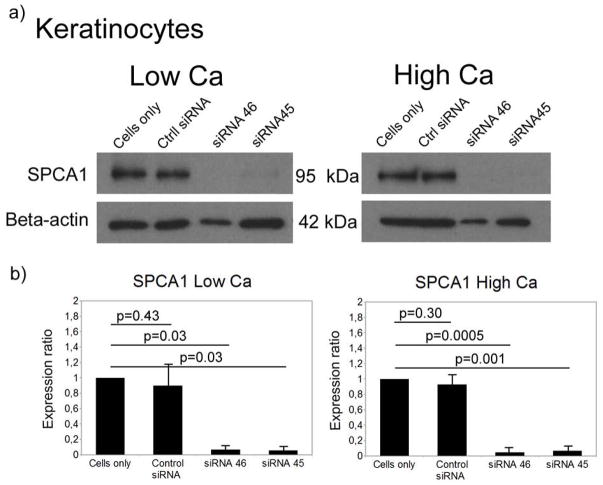
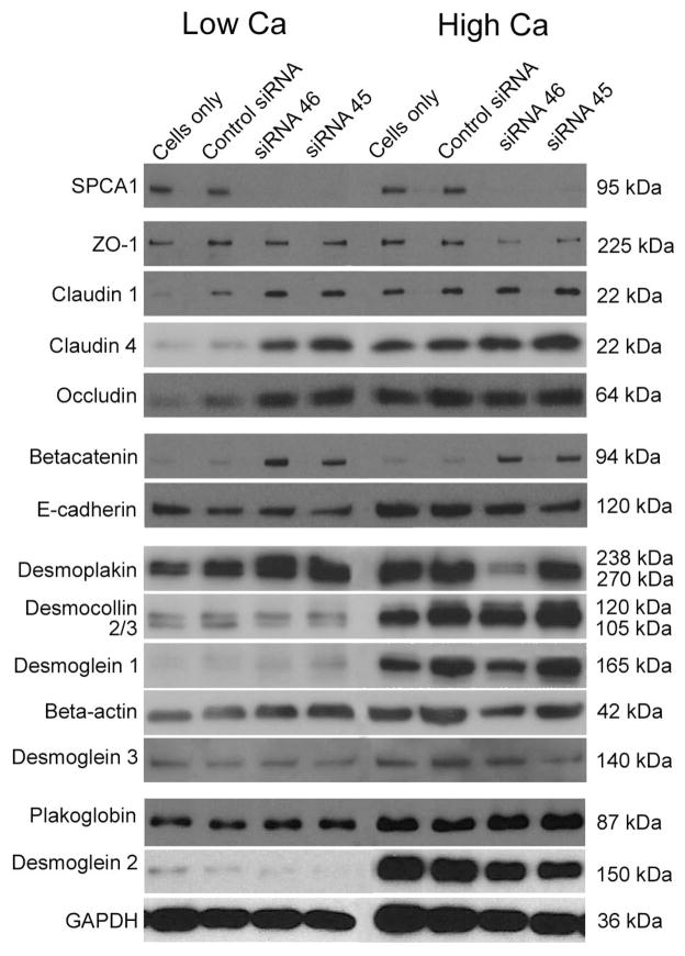
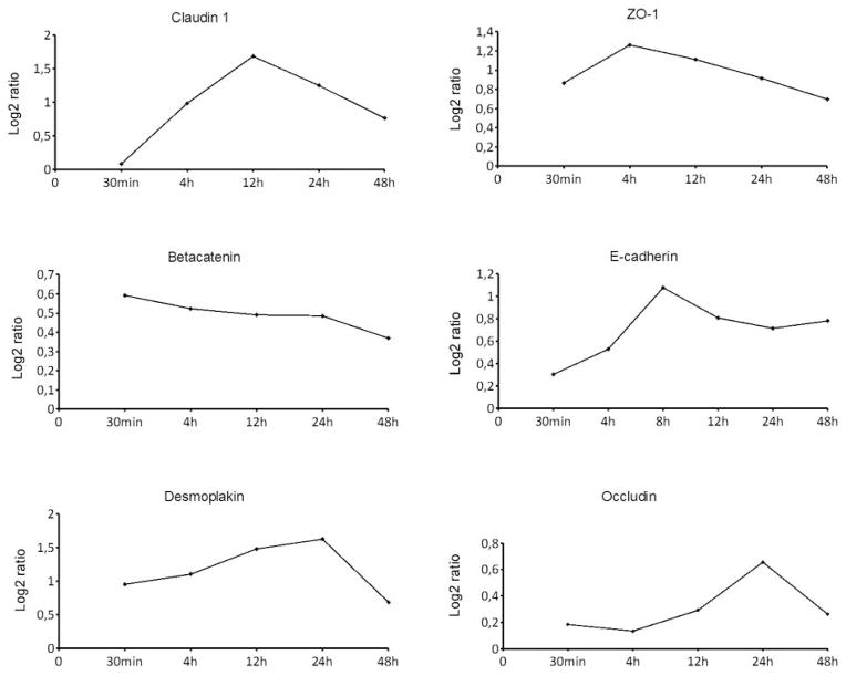
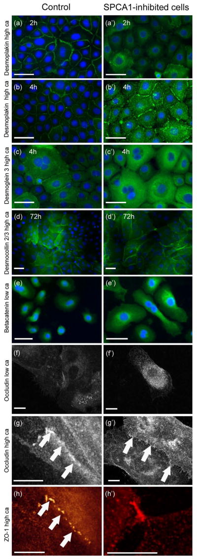
Mutations in the ATP2C1 gene encoding Ca(2+) /Mn(2+) ATPase SPCA1 cause Hailey-Hailey disease (HHD, OMIM 16960). HHD is characterized by epidermal acantholysis. We attempted to model HHD using normal keratinocytes, in which the SPCA1 mRNA was down-regulated with the small inhibitory RNA (siRNA) method. SiRNA inhibition significantly down-regulated the SPCA1 mRNA, as demonstrated by qPCR, and decreased the SPCA1 protein beyond detectable level, as shown by Western analysis. The expression of selected desmosomal, adherens and tight junction (TJ) proteins was then studied in the SPCA1-deficient and control keratinocytes cultured in low (0.06 mm) or high (1.2 mm) calcium concentration. The mRNA and protein levels of most TJ components were up-regulated in non-treated control keratinocyte cultures upon switch from low to high calcium concentration. In contrast, SPCA1-deficient keratinocytes displayed high levels of TJ proteins claudins 1 and 4 even in low calcium. ZO-1 did not, however, follow similar expression patterns. Protein levels of occludin, beta-catenin, E-cadherin, desmoplakin, desmogleins 1-3, desmocollin 2/desmocollin 3 and plakoglobin did not show marked changes in SPCA1-deficient keratinocytes. Indirect immunofluorescence labelling revealed delayed translocation of desmoplakin and desmoglein 3 in desmosomes and increased intracellular pools of TJ and desmosomal components in SPCA1-inhibited keratinocytes. The results show that SPCA1 regulates the levels of claudins 1 and 4, but does not affect desmosomal protein levels, indicating that TJ proteins are differently regulated. The results also suggest a potential role for claudins in HHD.
© 2012 John Wiley & Sons A/S.
Conflict of interest statement
The authors report no conflict of interest.
Figures
Similar articles
-
ATP2C1 knockdown induces abnormal expressions of cytoskeletal and tight junction proteins mimicking Hailey-Hailey disease.Indian J Dermatol Venereol Leprol. 2024 Nov-Dec;90(6):722-730. doi: 10.25259/IJDVL_853_2023. Indian J Dermatol Venereol Leprol. 2024. PMID: 38841932
-
Hailey-Hailey disease: investigation of a possible compensatory SERCA2 up-regulation and analysis of SPCA1, p63, and IRF6 expression.Arch Dermatol Res. 2015 Mar;307(2):143-9. doi: 10.1007/s00403-014-1506-2. Epub 2014 Sep 26. Arch Dermatol Res. 2015. PMID: 25256005 Clinical Trial.
-
Involucrin expression is decreased in Hailey-Hailey keratinocytes owing to increased involucrin mRNA degradation.J Invest Dermatol. 2007 Aug;127(8):1973-9. doi: 10.1038/sj.jid.5700785. Epub 2007 Mar 29. J Invest Dermatol. 2007. PMID: 17392835
-
Calcium pumps and keratinocytes: lessons from Darier's disease and Hailey-Hailey disease.Br J Dermatol. 2004 May;150(5):821-8. doi: 10.1111/j.1365-2133.2004.05904.x. Br J Dermatol. 2004. PMID: 15149492 Review.
-
ATP2C1 gene mutations in Hailey-Hailey disease and possible roles of SPCA1 isoforms in membrane trafficking.Cell Death Dis. 2016 Jun 9;7(6):e2259. doi: 10.1038/cddis.2016.147. Cell Death Dis. 2016. PMID: 27277681 Free PMC article. Review.
Cited by
-
A Ca2+-ATPase Regulates E-cadherin Biogenesis and Epithelial-Mesenchymal Transition in Breast Cancer Cells.Mol Cancer Res. 2019 Aug;17(8):1735-1747. doi: 10.1158/1541-7786.MCR-19-0070. Epub 2019 May 10. Mol Cancer Res. 2019. PMID: 31076498 Free PMC article.
-
The role of the ATP2C1 gene in Hailey-Hailey disease.Cell Mol Life Sci. 2017 Oct;74(20):3687-3696. doi: 10.1007/s00018-017-2544-7. Epub 2017 May 27. Cell Mol Life Sci. 2017. PMID: 28551824 Free PMC article. Review.
-
Cytokine-Mediated Inflammation in the Oral Cavity and Its Effect on Lipid Nanocarriers.Nanomaterials (Basel). 2021 May 18;11(5):1330. doi: 10.3390/nano11051330. Nanomaterials (Basel). 2021. PMID: 34070004 Free PMC article.
-
Cell-cell adhesions and cell contractility are upregulated upon desmosome disruption.PLoS One. 2014 Jul 9;9(7):e101824. doi: 10.1371/journal.pone.0101824. eCollection 2014. PLoS One. 2014. PMID: 25006807 Free PMC article.
-
Hypotonic, Acidic Oxidizing Solution Containing Hypochlorous Acid (HClO) as a Potential Treatment of Hailey-Hailey Disease.Molecules. 2019 Dec 4;24(24):4427. doi: 10.3390/molecules24244427. Molecules. 2019. PMID: 31817098 Free PMC article.
References
-
- Burge SM. Br J Dermatol. 1992;126:275–282. - PubMed
-
- Palmer D, Perry H. Arch Dermatol. 1962;86:493–502. - PubMed
-
- Sudbrak R, Brown J, Dobson-Stone C, et al. Hum Mol Genet. 2000;9:1131–1140. - PubMed
-
- Hu Z, Bonifas JM, Beech J, et al. Nat Genet. 2000;24:61–65. - PubMed
-
- Foggia L, Hovnanian A. Am J Med Genet C Semin Med Genet. 2004;131(C):20–31. - PubMed
Publication types
MeSH terms
Substances
Grants and funding
LinkOut - more resources
Full Text Sources
Medical
Miscellaneous
