Review
. 2012 May 24;7 Suppl 1(Suppl 1):S6.
doi: 10.1186/1750-1172-7-S1-S6. Epub 2012 May 24.

Cherubism: best clinical practice

Affiliations
Review

Cherubism: best clinical practice

Maria E Papadaki et al. Orphanet J Rare Dis. .

Abstract

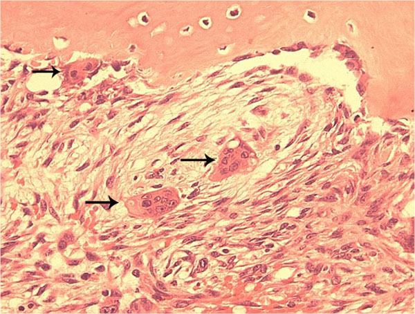
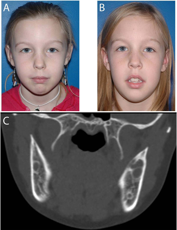
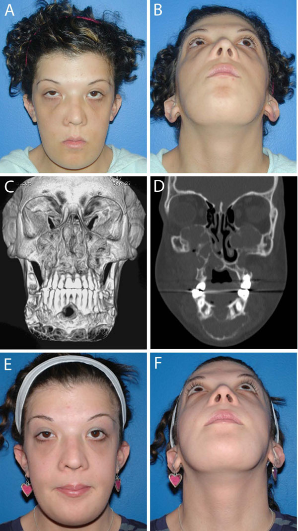
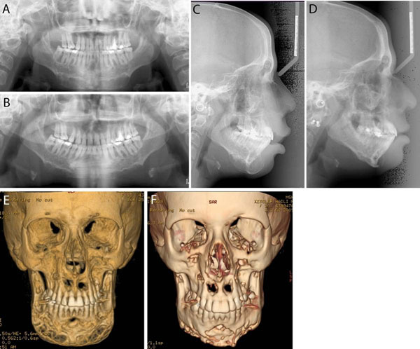
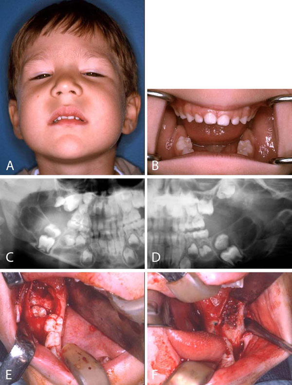
Cherubism is a skeletal dysplasia characterized by bilateral and symmetric fibro-osseous lesions limited to the mandible and maxilla. In most patients, cherubism is due to dominant mutations in the SH3BP2 gene on chromosome 4p16.3. Affected children appear normal at birth. Swelling of the jaws usually appears between 2 and 7 years of age, after which, lesions proliferate and increase in size until puberty. The lesions subsequently begin to regress, fill with bone and remodel until age 30, when they are frequently not detectable.Fibro-osseous lesions, including those in cherubism have been classified as quiescent, non-aggressive and aggressive on the basis of clinical behavior and radiographic findings. Quiescent cherubic lesions are usually seen in older patients and do not demonstrate progressive growth. Non-aggressive lesions are most frequently present in teenagers. Lesions in the aggressive form of cherubism occur in young children and are large, rapidly growing and may cause tooth displacement, root resorption, thinning and perforation of cortical bone.Because cherubism is usually self-limiting, operative treatment may not be necessary. Longitudinal observation and follow-up is the initial management in most cases. Surgical intervention with curettage, contouring or resection may be indicated for functional or aesthetic reasons. Surgical procedures are usually performed when the disease becomes quiescent. Aggressive lesions that cause severe functional problems such as airway obstruction justify early surgical intervention.


Figures

Figure 1
Figure 1
Typical histopathology of Cherubism. A histological section from a cherubism lesion demonstrates the typical finding of multinucleated osteoclast-like giant cells (arrows) near bone and within soft fibrous stroma.
Figure 2
Figure 2
A. Photograph of a 10 year old girl with bilateral cheek and jaw swelling. Cherubism was confirmed with genetic testing that was positive for the SH3BP2 gene. The patient had genetic counseling and was followed longitudinally. B. Patient one year later with no progression of facial swelling. C. CT scan at initial presentation showed typical bilateral lytic lesions in the mandible that remained unchanged at follow ups.
Figure 3
Figure 3
Noonan/multiple giant cell lesion syndrome. A 20-year old woman with Noonan/multiple giant cell lesion syndrome with bilateral involvement of the mandible and the maxilla. A and B. Frontal and submental photographs reveal prominent maxillary and mandibular contours, slight frontal bossing and increased intercanthal distance, downslanting palpebral features, epicanthal folds and posteriorly angulated ears. Lesions were painless to palpation. C and D. Coronal and 3D CT scan at initial presentation demonstrate involvement of the maxilla, including the maxillary sinuses and the nasal-maxillary region and the mandible from the antegonial notch to antegonial notch. The lesions are expansile and mixed radiolucent - radiopaque with cortical thinning and expansion into the orbits bilaterally. Patient was positive for a Noonan syndrome mutation in SOS-1. She underwent a contour resection of the maxillary and the mandibular giant cell lesions in two sessions. Postoperatively, she was treated with 4 courses of zoledronic acid, given at 6-week-intervals. E and F. Post-treatment frontal and submental photographs.
Figure 4
Figure 4
Imaging studies of the patient in Figure 3 pre-treatment (A, C, E) and post-treatment (B, D, F). A and B. Panoramic radiographs. C and D. Lateral cephalograms. E and F. 3D CT scans. Pre- and post-treatment images show improvement of the giant cell lesions and the contours of the mandible.
Figure 5
Figure 5
Cherubism. A 5 year old boy who after a mild trauma underwent a dental evaluation. A. Examination revealed marked fullness of the cheeks, expansion of the malar eminences and the mandibular angle regions. B. Intraoral examination was significant for expansion of the mandibular alveolus in the permanent molar regions. C and D. Panoramic radiograph revealed bilateral radiolucent lesions in the mandible and maxilla (arrows). The lesions were displacing the permanent second molar tooth buds superior to the first molar teeth. E and F. Intraoral biopsy and removal of the displaced second molar tooth buds leaving the first molar tooth buds in place to erupt. Pathologic diagnosis was fibro-osseous lesion consistent with cherubism.

References

    1. Jones WA. Familial multilocular cystic disease of the jaws. American Journal of Cancer. 1933;17:946–950.
    1. Jones WA. Further observations regarding familal multilocular cystic disease of the jaws. British Journal of Radiology. 1938;11:227–241. doi: 10.1259/0007-1285-11-124-227. - DOI
    1. Jones WA, Gerrie J, Pritchard J. Cherubism--familial fibrous dysplasia of the jaws. Journal of Bone and Joint Surgery. British Volume. 1950;32-B(3):334–47. - PubMed
    1. Arnott DG. Cherubism--an initial unilateral presentation. British Journal of Oral Surgery. 1978;16(1):38–46. doi: 10.1016/S0007-117X(78)80054-7. - DOI - PubMed
    1. Seward GR, Hankey GT. Cherubism. Oral Surg Oral Med Oral Pathol. 1957;10(9):952–74. doi: 10.1016/S0030-4220(57)80047-4. - DOI - PubMed

Publication types