. 2012 May 28:7:23.
doi: 10.1186/1750-1326-7-23.

Absence of amyloid β oligomers at the postsynapse and regulated synaptic Zn2+ in cognitively intact aged individuals with Alzheimer's disease neuropathology

Affiliations

Absence of amyloid β oligomers at the postsynapse and regulated synaptic Zn2+ in cognitively intact aged individuals with Alzheimer's disease neuropathology

Nicole L Bjorklund et al. Mol Neurodegener. .

Abstract

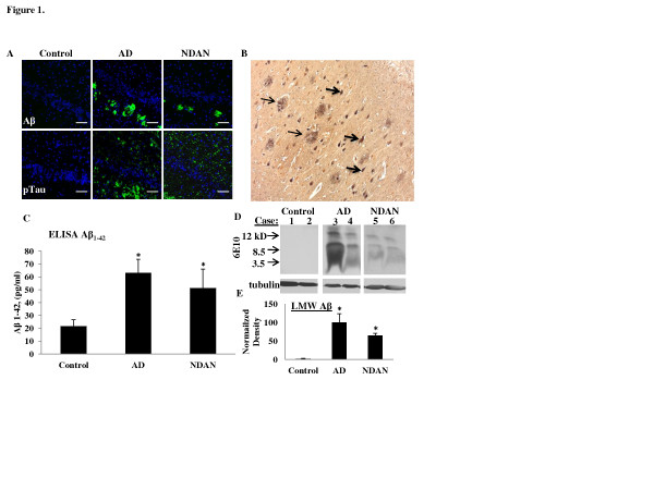
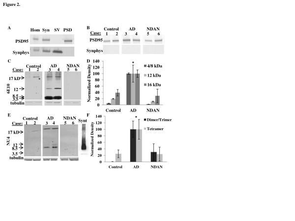
Background: Early cognitive impairment in Alzheimer Disease (AD) is thought to result from the dysfunctional effect of amyloid beta (Aβ) oligomers targeting the synapses. Some individuals, however, escape cognitive decline despite the presence of the neuropathologic features of AD (Aβ plaques and neurofibrillary tangles). We term this group Non-Demented with AD Neuropathology or NDAN. The present study illustrates one putative resistance mechanism involved in NDAN cases which may suggest targets for the effective treatment of AD.

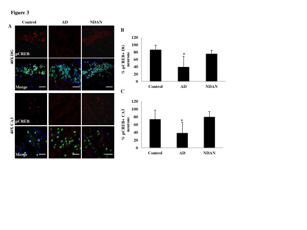
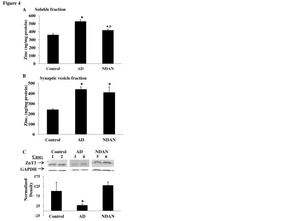
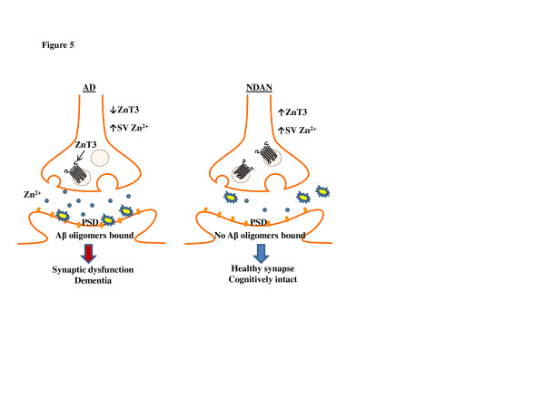
Results: Here we describe the localization of Aβ oligomers at the postsynapse in hippocampi from AD cases. Notably, however, we also found that while present in soluble fractions, Aβ oligomers are absent from hippocampal postsynapses in NDAN cases. In addition, levels of phosphorylated (active) CREB, a transcription factor important for synaptic plasticity, are normal in NDAN individuals, suggesting that their synapses are functionally intact. Analysis of Zn2+ showed that levels were increased in both soluble fractions and synaptic vesicles in AD hippocampi, paralleled by a decrease of expression of the synaptic vesicle Zn2+ transporter, ZnT3. Conversely, in NDAN individuals, levels of Zn2+ in soluble fractions were significantly lower than in AD, whereas in synaptic vesicles the levels of Zn2+ were similar to AD, but accompanied by preserved expression of the ZnT3.

Conclusions: Taken together, these data illustrate that despite substantial AD neuropathology, Aβ oligomers, and increased synaptic vesicle Zn2+, susceptible brain tissue in these aged NDAN individuals features, as compared to symptomatic AD subjects, significantly lower total Zn2+ levels and no association of Aβ oligomers with the postsynapse, which collectively may promote the maintenance of intact cognitive function.


Figures

Figure 1
Figure 1
Pathological signatures of AD occur in cognitively intact individuals (NDAN). (A) Immunohistochemical detection shows reactivity for Aβ plaques (top row) and phosphorylated tau (bottom row) in the dentate gyrus of individuals classifed as control (Braak 1, Plaque 3, MMSE 30), AD (Braak 6, Plaque 1, MMSE 2), or NDAN (Braak 6, Plaque 1, MMSE 27). DAPI-containing mounting medium was used to visualize nuclei (blue). 20x magnification, scale bar 100 μm. (B) Bielschowsky staining in the hippocampus of an NDAN individual shows plaques (thin arrows) and neurofibrillary tangles (thick arrows). (C) An ELISA shows increased Aβ1-42 in AD and NDAN hippocampi (n = 3 per group and asterisks (*) denote values significantly higher than control with p < 0.05). (D) Representative Western blot of soluble hippocampal fractions (300 μg) from individual cases probed with 6e10 shows that LMW Aβ species are present in both AD and NDAN. Densitometric analysis (F) of the major bands (monomer at 4kD and dimer at 8.5kD) together shows that AD and NDAN cases are significantly different than control where the average optical density is set at 100; the asterisks (*) denote statistical significance compared to the control value (p = 0.022, ANOVA); The Western blot shown is representative of 9–10 individual cases assayed in each group and experiments were repeated 3 – 4 times
Figure 2
Figure 2
Synaptic fractionation demonstrates that low molecular weight Aβ oligomers associate with the PSD in AD but not NDAN hippocampal specimens. (A) PSD95, a postsynaptic marker, and synaptophysin (Synphys), a presynaptic marker, were probed in the total homogenate (Hom), synaptosomal (Syn), synaptic vesicles (SV), and postsynaptic density (PSD) fractions isolated from a frozen human hippocampal specimen from a control, non-demented aged individual. Enrichment of the different fractions is shown by the presence/absence of synaptic markers. The fractionation was successful for all case types (control, AD and NDAN) as shown by the purity of PSD fractions in (B). The isolated PSD fraction from hippocampal samples from different cases from each group (aged matched controls, AD, and NDAN) were immunoblotted together (representative blot shown; 40 μg protein each lane of a 10-20% tris-glycine gradient gel) and probed using 6e10 (C) showing LMW Aβ only associates with AD samples. (D) Densitometric analyses of each of the Aβ species (monomer/dimer = 4–8.5 kD band, trimer = 12 kD, and tetramer = 16 kD) demonstrates that all Aβ species are increased in AD. The results are expressed as the mean ± SEM using propagation of error and normalized with AD = 100%. The asterisk denotes the values significantly higher than control (monomer/dimer, p = 0.012, trimer, p = < 0.001, and tetramer, p = 0.014; ANOVA; a Bonferroni correction was required for the monomer/dimer and trimer density values). Hippocampus PSD fractions (80 μg) from age-matched controls, AD, and NDAN samples were immunoblotted and probed using NU4 (E). A lane is included at the end showing synthetic (Synt) Aβ oligomers probed by NU4 for comparison. The densitometric analysis (F) of the dimer/trimer band shows that AD cases are significantly higher than control and NDAN (both groups are essentially the same as background), while the tetrameric species was not found to be significantly different across the groups (p = 0.67). The asterisk denotes significance compared to control, p < 0.05, ANOVA. All Western blots shown are representative of 9–10 individual cases assayed in each group and experiments were repeated 3 – 4 times
Figure 3
Figure 3
Synaptic integrity is maintained in the NDAN hippocampus as suggested by preserved levels of neuronal nuclear pCREB. (A) pCREB (red) and NeuN (green) expression was assessed in 10 μm sections of hippocampus. DAPI-containing mounting medium was used to visualize nuclei (blue). pCREB immunoreactivity was decreased in the DG and CA3 neurons of AD hippocampi but not in NDAN cases (scale bar 50 μm). The number of neurons positive for nuclear pCREB was counted in the (B) DG and (C) CA3. Two images per region (DG and CA3) were analyzed for each clinical case. The raw images were thresholded, and a blind counter quantified the number of neurons in the field of view, and how many of these exhibited nuclear pCREB immunoreactivity. This value is expressed as the percentage of neurons that are pCREB positive ± SEM (the asterisks denote statistical significance compared to controls at p ≤ 0.05; ANOVA)
Figure 4
Figure 4
Differentially altered levels of total Zn2+, releasable Zn2+, and ZnT3 expression in AD and NDAN hippocampus. (A) Soluble hippocampal fractions were normalized for protein (500 μg), digested with hydrogen peroxide and nitric acid, and analyzed using a GF-AAS. Zn2+ levels in both AD and NDAN samples were increased as compared to control, but NDAN Zn2+ levels were significantly lower than AD levels. After a Bonferroni correction, ANOVA results show p = < 0.001, n = 9–10 samples per group. (B) Zn2+ analysis in synaptic vesicles shows that the Zn2+ concentration in AD and NDAN is significantly higher than control (p = < 0.001 (after Bonferroni correction), n = 6–9 per group). (C) Representative Western blot and densitometric analysis (D) of ZnT3 expression in the hippocampus of individual cases shows a significant decrease in AD, but not in NDAN samples, which is denoted by an asterisk (t-test, p < 0.001 n = 6–9 per group, repeated 4 times)
Figure 5
Figure 5
Aβ and Zn2+differences in AD and NDAN hippocampal synapses. A schematic summarizing the findings presented in this report. In AD there is increased Zn2+ in the soluble fraction compared to control and NDAN. However, both AD and NDAN have increased levels of Zn2+ in the synaptic vesicles (SV) which may actually reflect a storage compensatory mechanism to high Zn2+ levels. The reduction of ZnT3 in AD may reflect a dysfunction of this compensatory mechanism, allowing for total Zn2+ levels to increase. Zn2+ is necessary for stabilization and targeting of Aβ oligomers to the PSD. Aβ oligomers are highly associated with the PSD in AD hippocampal synapses, but are absent at NDAN synapses. This key difference could likely be due to the levels and regulation of synaptic Zn2+, resulting in synaptic dysfunction and cognitive decline in AD and preserved synapses and cognition in NDAN cases

References

    1. Alzheimer’s A. Alzheimer’s disease facts and figures. Alzheimer’s & Dementia. 2010;2010:6. - PubMed
    1. Hardy J, Allsop D. Amyloid deposition as the central event in the etiology of Alzheimer’s disease. Trends Pharmacol Sci. 1991;12:383–388. - PubMed
    1. Braak H, Braak E. Neuropathological stageing of Alzheimer-related changes. Acta Neuropathol. 1991;82:239–259. doi: 10.1007/BF00308809. - DOI - PubMed
    1. Kramer PL, Xu H, Woltjer RL, Westaway SK, Clark D, Erten-Lyons D, Kaye JA, Welsh-Bohmer KA, Troncoso JC, Markesbery WR. et al.Alzheimer disease pathology in cognitively healthy elderly: A genome-wide study. Neurobiol Aging. 2011;32:2113–2122. doi: 10.1016/j.neurobiolaging.2010.01.010. - DOI - PMC - PubMed
    1. Lue LF, Kuo YM, Roher AE, Brachova L, Shen Y, Sue L, Beach T, Kurth JH, Rydel RE, Rogers J. Soluble amyloid beta peptide concentration as a predictor of synaptic change in Alzheimer’s disease. Am J Pathol. 1999;155:853–862. doi: 10.1016/S0002-9440(10)65184-X. - DOI - PMC - PubMed

Publication types