

Placental size at 19 weeks predicts offspring bone mass at birth: findings from the Southampton Women's Survey
- PMID: 22640438
- PMCID: PMC3800076
- DOI: 10.1016/j.placenta.2012.04.007
Placental size at 19 weeks predicts offspring bone mass at birth: findings from the Southampton Women's Survey
Abstract
Objectives: In this study we investigate the relationships between placental size and neonatal bone mass and body composition, in a population-based cohort.
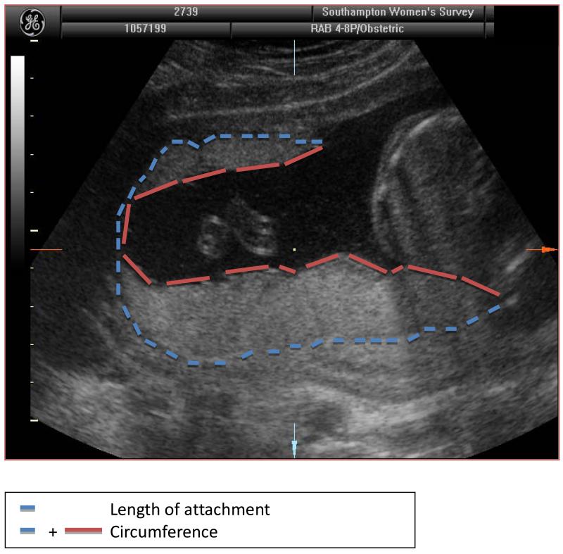
Study design: 914 mother-neonate pairs were included. Placental dimensions were measured via ultrasound at 19 weeks gestation. Dual X-ray absorptiometry (DXA) was performed on the neonates within the first two weeks of life.
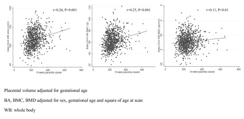
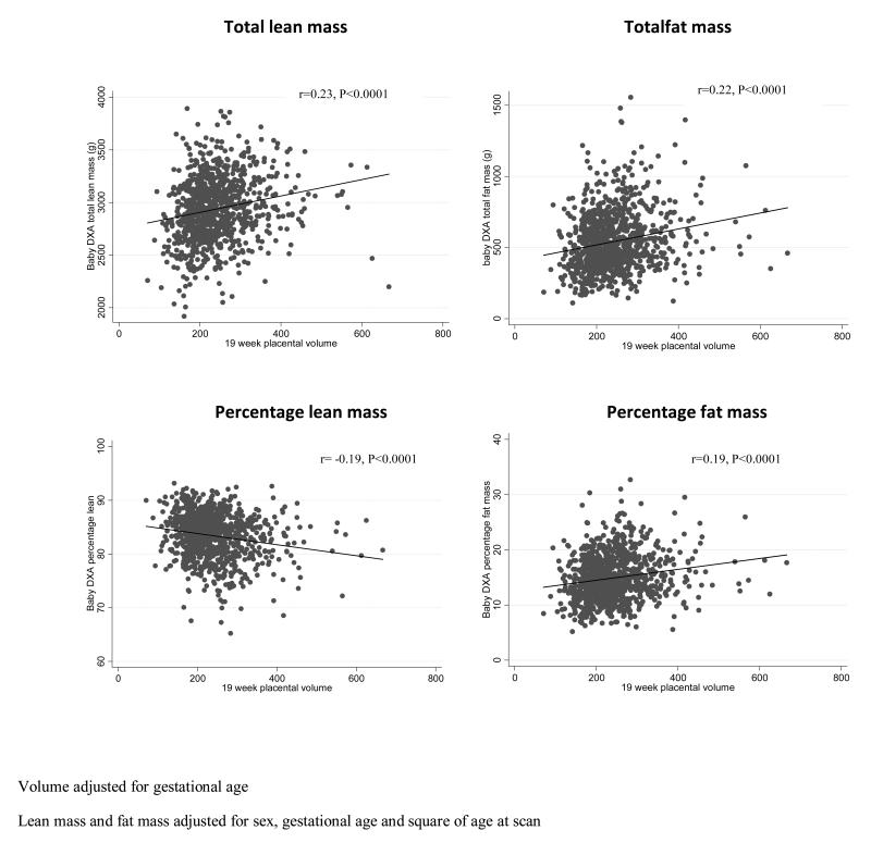
Results: We observed positive relationships between placental volume at 19 weeks, and neonatal bone area (BA; r = 0.26, p < 0.001), bone mineral content (BMC; r = 0.25, p < 0.001) and bone mineral density (BMD; r = 0.10, p = 0.001). Thus placental volume accounted for 6.25% and 1.2% of the variation in neonatal BMC and BMD respectively at birth. These associations remained after adjustment for maternal factors previously shown to be associated with neonatal bone mineral accrual (maternal height, smoking, walking speed in late pregnancy, serum 25(OH) vitamin D and triceps skinfold thickness).
Conclusions: We found that placental volume at 19 weeks gestation was positively associated with neonatal bone size and mineral content. These relationships appeared independent of those maternal factors known to be associated with neonatal bone mass, consistent with notion that such maternal influences might act through modulation of aspects of placental function, e.g. utero-placental blood flow or maternal nutrient concentrations, rather than placental size itself. Low placental volume early in pregnancy may be a marker of a reduced postnatal skeletal size and increased risk of later fracture.
Copyright © 2012 Elsevier Ltd. All rights reserved.
Figures
Similar articles
-
Placental volume at 11 weeks is associated with offspring bone mass at birth and in later childhood: Findings from the Southampton Women's Survey.Placenta. 2020 Sep 15;99:101-107. doi: 10.1016/j.placenta.2020.07.017. Epub 2020 Jul 22. Placenta. 2020. PMID: 32784052 Free PMC article.
-
Maternal serum retinol and β-carotene concentrations and neonatal bone mineralization: results from the Southampton Women's Survey cohort.Am J Clin Nutr. 2016 Oct;104(4):1183-1188. doi: 10.3945/ajcn.116.130146. Epub 2016 Sep 14. Am J Clin Nutr. 2016. PMID: 27629051 Free PMC article.
-
Placental Size Is Associated Differentially With Postnatal Bone Size and Density.J Bone Miner Res. 2016 Oct;31(10):1855-1864. doi: 10.1002/jbmr.2840. Epub 2016 Apr 22. J Bone Miner Res. 2016. PMID: 26999363 Free PMC article. Clinical Trial.
-
Pathophysiology of placental-derived fetal growth restriction.Am J Obstet Gynecol. 2018 Feb;218(2S):S745-S761. doi: 10.1016/j.ajog.2017.11.577. Am J Obstet Gynecol. 2018. PMID: 29422210 Review.
-
Bone in the pregnant mother and newborn at birth.Clin Chim Acta. 2003 Jul 1;333(1):1-11. doi: 10.1016/s0009-8981(02)00025-6. Clin Chim Acta. 2003. PMID: 12809730 Review.
Cited by
-
Placental volume at 11 weeks is associated with offspring bone mass at birth and in later childhood: Findings from the Southampton Women's Survey.Placenta. 2020 Sep 15;99:101-107. doi: 10.1016/j.placenta.2020.07.017. Epub 2020 Jul 22. Placenta. 2020. PMID: 32784052 Free PMC article.
-
Ultrasound Evaluation of Placental Thickness: Insights From an Observational Study and Implications for Fetal Growth Assessment.Cureus. 2024 Jun 20;16(6):e62760. doi: 10.7759/cureus.62760. eCollection 2024 Jun. Cureus. 2024. PMID: 39036116 Free PMC article.
-
Vertebral cross-sectional area: an orphan phenotype with potential implications for female spinal health.Osteoporos Int. 2017 Apr;28(4):1179-1189. doi: 10.1007/s00198-016-3832-z. Epub 2016 Dec 14. Osteoporos Int. 2017. PMID: 27975301 Review.
-
A head start: The relationship of placental factors to craniofacial and brain development.Dev Dyn. 2025 Mar 19:10.1002/dvdy.70018. doi: 10.1002/dvdy.70018. Online ahead of print. Dev Dyn. 2025. PMID: 40105397 Review.
-
Sexual Dimorphism and the Origins of Human Spinal Health.Endocr Rev. 2018 Apr 1;39(2):221-239. doi: 10.1210/er.2017-00147. Endocr Rev. 2018. PMID: 29385433 Free PMC article. Review.
References
-
- Hernandez CJ, Beaupre GS, Carter DR. A theoretical analysis of the relative influences of peak BMD, age-related bone loss and menopause on the development of osteoporosis. Osteoporos Int. 2003 Oct;14(10):843–7. - PubMed
-
- Cooper C, Cawley M, Bhalla A, Egger P, Ring F, Morton L, et al. Childhood growth, physical activity, and peak bone mass in women. J Bone Miner Res. 1995 Jun;10(6):940–7. - PubMed
-
- Dennison EM, Syddall HE, Sayer AA, Gilbody HJ, Cooper C. Birth weight and weight at 1 year are independent determinants of bone mass in the seventh decade: the Hertfordshire cohort study. Pediatr Res. 2005 Apr;57(4):582–6. - PubMed
-
- Cooper C, Eriksson JG, Forsen T, Osmond C, Tuomilehto J, Barker DJ. Maternal height, childhood growth and risk of hip fracture in later life: a longitudinal study. Osteoporos Int. 2001;12(8):623–9. JID - 9100105. - PubMed
-
- Godfrey K, Walker-Bone K, Robinson S, Taylor P, Shore S, Wheeler T, et al. Neonatal bone mass: influence of parental birthweight, maternal smoking, body composition, and activity during pregnancy. J Bone Miner Res. 2001 Sep;16(9):1694–703. JID - 8610640. - PubMed
Publication types
MeSH terms
Grants and funding
LinkOut - more resources
Full Text Sources
