. 2012 May 28;2(1):9.
doi: 10.1186/2044-5040-2-9.

Leaky ryanodine receptors in β-sarcoglycan deficient mice: a potential common defect in muscular dystrophy

Affiliations

Leaky ryanodine receptors in β-sarcoglycan deficient mice: a potential common defect in muscular dystrophy

Daniel C Andersson et al. Skelet Muscle. .

Abstract

Background: Disruption of the sarcolemma-associated dystrophin-glycoprotein complex underlies multiple forms of muscular dystrophy, including Duchenne muscular dystrophy and sarcoglycanopathies. A hallmark of these disorders is muscle weakness. In a murine model of Duchenne muscular dystrophy, mdx mice, cysteine-nitrosylation of the calcium release channel/ryanodine receptor type 1 (RyR1) on the skeletal muscle sarcoplasmic reticulum causes depletion of the stabilizing subunit calstabin1 (FKBP12) from the RyR1 macromolecular complex. This results in a sarcoplasmic reticular calcium leak via defective RyR1 channels. This pathological intracellular calcium leak contributes to reduced calcium release and decreased muscle force production. It is unknown whether RyR1 dysfunction occurs also in other muscular dystrophies.

Methods: To test this we used a murine model of Limb-Girdle muscular dystrophy, deficient in β-sarcoglycan (Sgcb-/-).

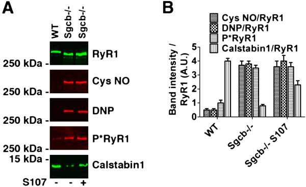
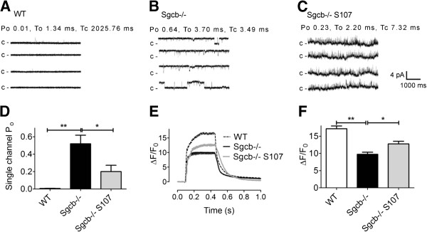
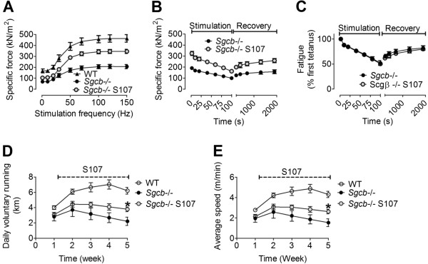
Results: Skeletal muscle RyR1 from Sgcb-/- deficient mice were oxidized, nitrosylated, and depleted of the stabilizing subunit calstabin1, which was associated with increased open probability of the RyR1 channels. Sgcb-/- deficient mice exhibited decreased muscle specific force and calcium transients, and displayed reduced exercise capacity. Treating Sgcb-/- mice with the RyR stabilizing compound S107 improved muscle specific force, calcium transients, and exercise capacity. We have previously reported similar findings in mdx mice, a murine model of Duchenne muscular dystrophy.

Conclusions: Our data suggest that leaky RyR1 channels may underlie multiple forms of muscular dystrophy linked to mutations in genes encoding components of the dystrophin-glycoprotein complex. A common underlying abnormality in calcium handling indicates that pharmacological targeting of dysfunctional RyR1 could be a novel therapeutic approach to improve muscle function in Limb-Girdle and Duchenne muscular dystrophies.


Figures

Figure 1
Figure 1
EDL muscles from β-sarcoglycan deficient mice exhibit dystrophic morphology and abnormal mitochondrial morphology. (A, B) EDL muscle cross-sections from wild-type (WT) and β-sarcoglycan mice stained with hematoxylin and eosin. (C) Percentage of fibers with the nucleus localized in the center (average ± SEM). (D) Percentages of normal, degenerated (weak eosin staining, examples indicated by asterisk) and necrotic (loss of eosin stain and swollen fiber, example indicated by a circle) muscle fibers. (E) Fiber size was more variable in Sgcb−/− EDL. This is indicated by the difference in the frequency distribution of fiber cross-sectional area. The inset in (E) is an expansion of the region indicated by the dashed rectangle in the main graph. Data were obtained from four mice and > 600 fibers in each group. The scale bar in images (A) and (B) indicate 250 μm. Representative electron microscopy images of EDL muscle from (F) WT and (G) Sgcb−/− mice. Arrows indicate normal mitochondria (F) or mitochondria with abnormal morphology, including low cristae density (G). Images from 11 fibers and two mice in each group were investigated under blinded conditions. The sample is magnified at × 25,000. Scale bar indicates 500 nm.
Figure 2
Figure 2
RyR1 in β-sarcoglycan deficient muscle is cysteine-nitrosylated, oxidized, and depleted of calstabin1. (A) Representative immunoblot of immunoprecipitated RyR1 from wild-type (WT) and β-sarcoglycan deficient (Sgcb−/−) EDL muscles. Antibodies against RyR1-S2844 phosphorylation (P*RyR1), cysteine-nitrosylated (Cys NO) proteins, calstabin1, and the protein oxidation marker 2,4- dinitrophenylhydrazone (DNP) was used. The muscle from a mouse treated with S107 is marked (+). (B) Bar graph showing average band intensities normalized to RyR1 expression (mean ± SEM, n = 3 for all groups).
Figure 3
Figure 3
β-sarcoglycan deficient muscle displays RyR1 dysfunction and defective SR Ca2+release that is restored by S107 treatment. (A-C) Representative RyR1 single channel current traces in samples from WT (A), Sgcb−/− (B), and Sgcb−/− S107 (C) treated mice. Channel activity was measured at 90 nmol/L (nM) free cytosolic [Ca2+]. Channel openings are shown as upward deflections; the closed (c -) state of the channel is indicated by horizontal bars in the beginning of each tracing. For each group, channel activity is illustrated by four different traces, each of 5 s length as indicated by dimension bars. The single channel open probability (Po), To (mean open time) and Tc (mean closed time) were calculated from a 2 min recording under 90 nmol/L free cytosolic [Ca2+] are shown above the upper trace. (D) Bar graph summarizing RyR1 single channel Po under 90 nmol/L free cytosolic [Ca2+] from WT (n = 4; white bar), Sgcb−/− (n = 3; black bar), and Sgcb−/− + S107 (n = 4; red bar) samples. Data presented as mean ± S.E.M; * P <0.05; ** P <0.01 (ANOVA). (E) Representative tetanic Ca2+ transients (normalized Fluo-4 fluorescence) in FDB muscle fibers from wild-type (WT), β-sarcoglycan-deficient control (Sgcb−/−), and S107-treated β-sarcoglycan-deficient (Sgcb−/− S107) mice. (F) Average Ca2+ transient amplitudes (±SEM, n = 6 (WT) n =20 (Sgcb−/−), n = 26 (Sgcb−/− S107) cells from three mice in each group, * P <0.05, ** P <0.01 (ANOVA)).
Figure 4
Figure 4
S107 treatment increases muscle force and exercise capacity in β-sarcoglycan deficient mice. (A) Force-frequency curves of EDL muscle from WT control, β-sarcoglycan-deficient (Sgcb−/−), and S107-treated Sgcb−/− (Sgcb−/− S107) mice. (B) Fatigue stimulation (50 tetani; each tetanic stimulation had a duration of 350 ms and was produced by stimulating the muscle with 0.5 ms pulses at 70 Hz frequency) on the same muscles as (A). (C) Relative decline in force production during fatigue in (B). EDL force measurements are presented as mean ± SEM, n = 6–9. (D, E) Exercise capacity in Sgcb−/− mice is improved by S107. Daily voluntary running distance (D) and average running speed (E). Pooled data are presented as mean ± SEM, n = 8–5, * P <0.05 (ANOVA).

Similar articles

Cited by

References

    1. Lim LE, Campbell KP. The sarcoglycan complex in limb-girdle muscular dystrophy. Curr Opin Neurol. 1998;11:443–452. doi: 10.1097/00019052-199810000-00006. - DOI - PubMed
    1. Petrof BJ, Shrager JB, Stedman HH, Kelly AM, Sweeney HL. Dystrophin protects the sarcolemma from stresses developed during muscle contraction. Proc Natl Acad Sci U S A. 1993;90:3710–3714. doi: 10.1073/pnas.90.8.3710. - DOI - PMC - PubMed
    1. Durbeej M, Cohn RD, Hrstka RF, Moore SA, Allamand V, Davidson BL, Williamson RA, Campbell KP. Disruption of the beta-sarcoglycan gene reveals pathogenetic complexity of limb-girdle muscular dystrophy type 2E. Mol Cell. 2000;5:141–151. doi: 10.1016/S1097-2765(00)80410-4. - DOI - PubMed
    1. Durbeej M, Campbell KP. Muscular dystrophies involving the dystrophin-glycoprotein complex: an overview of current mouse models. Curr Opin Genet Dev. 2002;12:349–361. doi: 10.1016/S0959-437X(02)00309-X. - DOI - PubMed
    1. Lim LE, Duclos F, Broux O, Bourg N, Sunada Y, Allamand V, Meyer J, Richard I, Moomaw C, Slaughter C, Tome FMS, Fardeau M, Jackson CE, Beckmann JS, Campbell KP. Beta-sarcoglycan: characterization and role in limb-girdle muscular dystrophy linked to 4q12. Nat Genet. 1995;11:257–265. doi: 10.1038/ng1195-257. - DOI - PubMed

LinkOut - more resources