

Clinicopathologic correlations of renal microthrombosis and inflammatory markers in proliferative lupus nephritis
- PMID: 22640796
- PMCID: PMC3446507
- DOI: 10.1186/ar3856
Clinicopathologic correlations of renal microthrombosis and inflammatory markers in proliferative lupus nephritis
Abstract
Introduction: Microthrombosis is often observed in lupus nephritis (LN) lesions, but its clinical significance is unknown. We evaluated the clinicopathologic correlations of renal microthrombosis and inflammatory markers in LN.
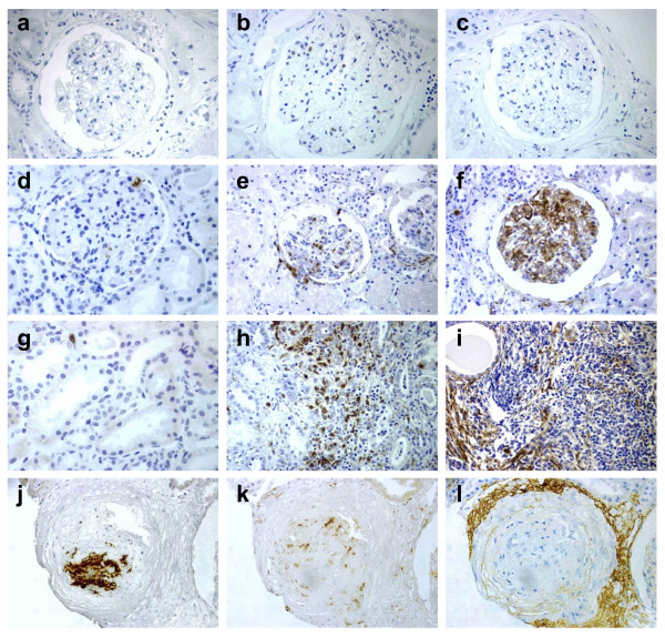
Methods: Kidney biopsies from 58 patients with systemic lupus erythematosus (SLE) proliferative nephritis were analyzed with immunohistochemistry (IHC) for intravascular platelet aggregates (CD61), macrophagic infiltration (CD68), and activated complement deposition (C4d). Clinical data at the time of kidney biopsy and follow-up were analyzed with regard to pathologic IHC data.
Results: Microthrombosis was present in 52% of the tissues. It was significantly more prevalent in patients with antiphospholipid antibodies (aPLs) (62% versus 42%). The presence of microthrombosis significantly correlated with higher macrophagic infiltration. Macrophagic infiltration but not microthrombosis was significantly correlated with C4d deposition. Only macrophagic infiltration showed a correlation with SLE and renal activity (proteinuria and active sediment), whereas neither the presence of CD61+ microthrombi nor the extent of C4d deposition correlated with LN severity or outcome.
Conclusions: Microthrombosis is associated with higher macrophagic infiltration in LN but does not seem to increase independently the severity of renal damage. Macrophagic infiltration was the best marker of SLE and renal activity in this LN series.
Figures
References
-
- Cameron JS. Lupus nephritis. J Am Soc Nephrol. 1999;10:413–424. - PubMed
-
- Austin HA III, Boumpas DT, Vaughan EM, Balow JE. High-risk features of lupus nephritis: importance of race and clinical and histological factors in 166 patients. Nephrol Dial Transplant. 1995;10:1620–1628. - PubMed
-
- Esdaile JM, Federgreen W, Quintal H, Suissa S, Hayslett JP, Kashgarian M. Predictors of one year outcome in lupus nephritis: the importance of renal biopsy. Q J Med. 1991;81:907–918. - PubMed
-
- Yang N, Isbel NM, Nikolic-Paterson DJ, Li Y, Ye R, Atkins RC, Lan HY. Local macrophage proliferation in human glomerulonephritis. Kidney Int. 1998;54:143–151. - PubMed
-
- Takemura T, Yoshioka K, Murakami K, Akano N, Okada M, Aya N, Maki S. Cellular localization of inflammatory cytokines in human glomerulonephritis. Virchows Arch. 1994;424:459–464. - PubMed
