Review
. 2013 Jan 1;185(1):94-104.
doi: 10.1016/j.resp.2012.05.019. Epub 2012 May 26.

Chronic hyperoxia and the development of the carotid body

Affiliations
Review

Chronic hyperoxia and the development of the carotid body

Ryan W Bavis et al. Respir Physiol Neurobiol. .

Abstract

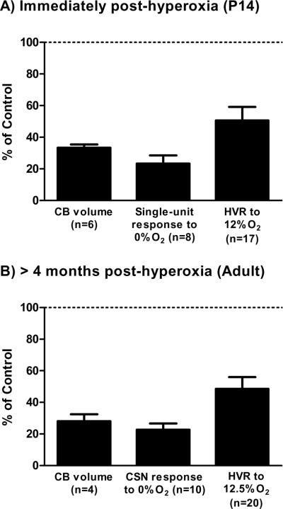
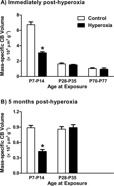
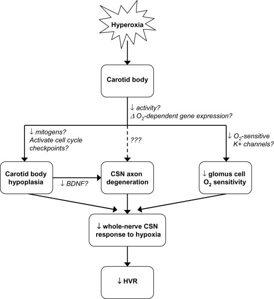
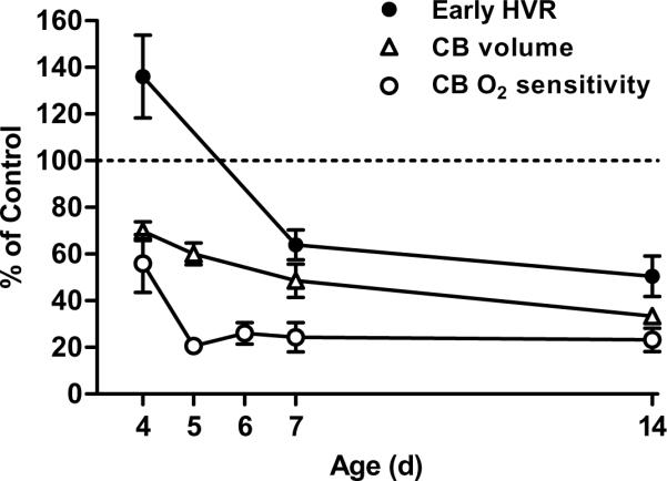
Preterm infants often experience hyperoxia while receiving supplemental oxygen. Prolonged exposure to hyperoxia during development is associated with pathologies such as bronchopulmonary dysplasia and retinopathy of prematurity. Over the last 25 years, however, experiments with animal models have revealed that moderate exposures to hyperoxia (e.g., 30-60% O(2) for days to weeks) can also have profound effects on the developing respiratory control system that may lead to hypoventilation and diminished responses to acute hypoxia. This plasticity, which is generally inducible only during critical periods of development, has a complex time course that includes both transient and permanent respiratory deficits. Although the molecular mechanisms of hyperoxia-induced plasticity are only beginning to be elucidated, it is clear that many of the respiratory effects are linked to abnormal morphological and functional development of the carotid body, the principal site of arterial O(2) chemoreception for respiratory control. Specifically, developmental hyperoxia reduces carotid body size, decreases the number of chemoafferent neurons, and (at least transiently) diminishes the O(2) sensitivity of individual carotid body glomus cells. Recent evidence suggests that hyperoxia may also directly or indirectly impact development of the central neural control of breathing. Collectively, these findings emphasize the vulnerability of the developing respiratory control system to environmental perturbations.


Figures

Fig. 1
Fig. 1
Representative effects of developmental hyperoxia on carotid body (CB) volume, CB hypoxic response (single-unit or whole-nerve CSN response), and hypoxic ventilatory response (HVR). Rats were exposed to 60% O2 for the first two postnatal weeks and studied (A) immediately (P13–14) or (B) >4 weeks (Adult) after return to room air. Note that the HVR for P13–14 rats represents the early phase of the response (i.e., first minute of hypoxia). Values (mean ± SEM) are expressed as a percentage of those measured for age-matched rats reared in room air (i.e., “Control” rats). All values are significantly reduced relative to Control (i.e., <100%; one-sample t-test, all P<0.001). Data were compiled from previously published studies (Donnelly et al., 2005; Bavis et al., 2007, 2008, 2010; Dmitrieff et al., 2012).
Fig 2
Fig 2
Postnatal growth of the carotid body in Sprague-Dawley rats in terms of carotid body volume (filled symbols, n=8–13 per age) and the number of glomus cells that underwent mitosis in the preceding 24 hours (i.e., number of BrdU-positive glomus cells) (open symbols, n=3–5 per age). Although this experiment focused on growth between 2 and 12 weeks of age, carotid body volumes for 24–26 week old rats from another experiment (i.e., Control rats from Fig. 3B) are plotted for comparison. No effect of sex was detected, so data for males and females have been pooled. All values are mean ±SEM.
Fig 3
Fig 3
Mass-specific carotid body (CB) volume in rats exposed to one week of 60% O2 at postnatal ages P7–P14, P28–P35, and P70–P77 (Hyperoxia) and in age-matched rats reared in room air (Control). Volumes were determined immediately following the hyperoxic exposure (panel A) or after a five month recovery in room air (panel B). All values are mean ±SEM; n=6 per treatment group at each age. * P<0.05 vs. age-matched Control.
Fig. 4
Fig. 4
Developmental hyperoxia impairs the hypoxic ventilatory response (HVR) through its effects the carotid body, including carotid body hypoplasia, carotid sinus nerve (CSN) axon degeneration, and diminished glomus cell O2 sensitivity; changes to carotid body size and innervation persist into adulthood, while chemoreceptor O2 sensitivity may recover after return to normoxia. Current evidence suggests that CSN axon degeneration is secondary to carotid body hypoplasia (and associated loss of trophic support), but more direct effects of hyperoxia on chemoafferent neurons cannot be excluded (dotted line). Hypothesized pathways by which hyperoxia influences carotid body development are also described (text adjacent to arrows); although some pathways have been supported experimentally (e.g., changes in BDNF expression), question marks indicate that causality has not been established yet for any of these pathways.
Fig. 5
Fig. 5
Time course for changes in carotid body (CB) volume, CB O2 sensitivity, and the hypoxic ventilatory response (HVR) in neonatal rats reared from birth in 60% O2. Carotid body O2 sensitivity is based on the peak single-unit chemoreceptor activity during severe hypoxia (0% O2), while HVR represents the early phase of the response to 12% O2 (i.e., first minute of hypoxia). Values (mean ± SEM) are expressed as a percentage of those measured for age-matched rats reared in room air (i.e., “Control” rats); n=6 per age for CB volume, n= 6–16 per age for CB O2 sensitivity, and n=15–17 per age for HVR. Data were compiled from previously published studies (Donnelly et al., 2005; Bavis et al., 2010, 2011b; Dmitrieff et al., 2012).

Similar articles

Cited by

References

    1. Balkowiec A, Katz DM. Activity-dependent release of endogenous brain-derived neurotrophic factor from primary sensory neurons detected by ELISA in situ. J. Neurosci. 2000;20:7417–7423. - PMC - PubMed
    1. Bavis RW. Developmental plasticity of the hypoxic ventilatory response after perinatal hyperoxia and hypoxia. Respir. Physiol. Neurobiol. 2005;149:287–299. - PubMed
    1. Bavis RW, Mitchell GS. Long-term effects of the perinatal environment on respiratory control. J. Appl. Physiol. 2008;104:1220–1229. - PubMed
    1. Bavis RW, Simons JC. Developmental hyperoxia attenuates the hypoxic ventilatory response in Japanese quail (Coturnix japonica) Respir. Physiol. Neurobiol. 2008;164:411–418. - PMC - PubMed
    1. Bavis RW, Olson EB, Jr., Mitchell GS. Critical developmental period for hyperoxia-induced blunting of hypoxic phrenic responses in rats. J. Appl. Physiol. 2002;92:1013–1018. - PubMed

Publication types

LinkOut - more resources