. 2012 May 29:7:58.
doi: 10.1186/1746-1596-7-58.

HLA-G and HLA-E specific mRNAs connote opposite prognostic significance in renal cell carcinoma

Affiliations

HLA-G and HLA-E specific mRNAs connote opposite prognostic significance in renal cell carcinoma

Leos Kren et al. Diagn Pathol. .

Abstract

Background: Renal cell carcinoma (RCC) is characterized by its resistance to radiotherapy and/or chemotherapy. On the other hand, it is an immunogenic tumor - it is able to stimulate antitumor responses. A prognostic significance of HLA-G expression by neoplastic cells in RCC is not well characterized; significance HLA-E expression in RCC is not characterized at all.

Methods: In our study, we evaluated the expression of HLA-G and HLA-E specific mRNA transcripts produced by neoplastic cells in 38 cases of RCC and in 10 samples of normal kidney parenchyma. The results were statistically correlated with various clinico-pathological parameters.

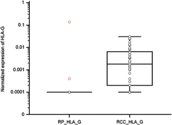
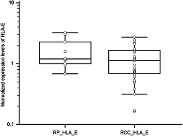
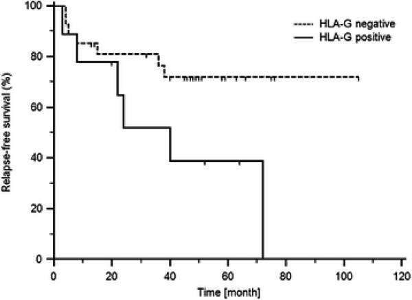
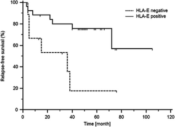
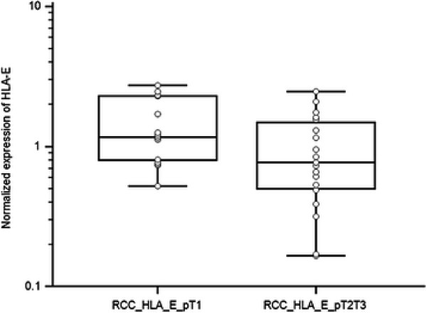
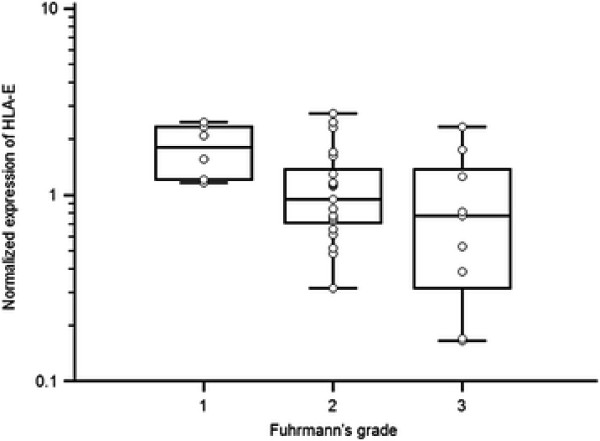
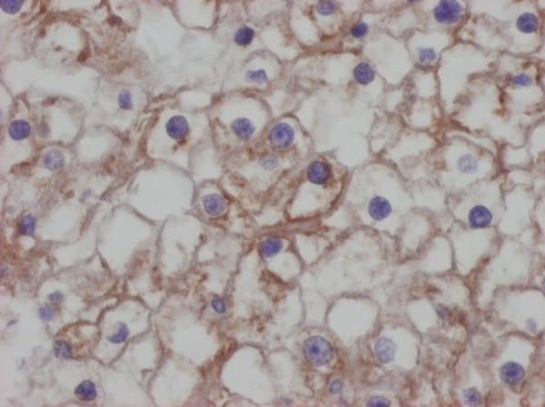
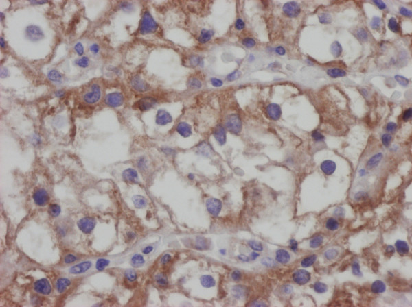
Results: We confirmed that HLA-G is downregulated in normal kidney tissue; if it is up-regulated in RCC, then it is connected to worse prognosis. On the other hand, HLA-E mRNA transcripts were present in both normal kidney tissue and RCC and their increasing concentrations counterintuitively carried better prognosis, more favorable pT stage and lower nuclear Fuhrmann's grade.

Conclusion: Considering the fact that there is known aberrant activation of HLA-G and HLA-E expression by interferons, identification of HLA-G and HLA-E status could contribute to better selection of RCC patients who could possibly benefit from more tailored neoadjuvant biological/immunological therapy. Thus, these molecules could represent useful prognostic biomarkers in RCC, and the expression of both these molecules in RCC deserves further study. THE VIRTUAL: Slide(s) for this article can be found here: http://www.diagnosticpathology.diagnomx.eu/vs/7383071387016614.


Figures

Figure 1
Figure 1
Comparison of HLA-G expression levels in RCC tissue and non-tumoral renal parenchyma (p = 0.0058, Mann–Whitney test).
Figure 2
Figure 2
Comparison of HLA-E expression levels in RCC tissue and non-tumoral renal parenchyma (p = 0.126, Mann–Whitney test).
Figure 3
Figure 3
Relapse-free survival of RCC patients based on the HLA-G expression (cut off = 75th percentile of HLA-G expression levels in RCC tissue; p = 0,0401, Long-rank test).
Figure 4
Figure 4
Relapse-free survival of RCC patients based on the HLA-E expression (cut off = 25th percentile of HLA-E expression levels in RCC tissue; p = 0,0115, Long-rank test).
Figure 5
Figure 5
HLA-E expression levels in RCC of different pT stage (p = 0.041, Mann–Whitney test).
Figure 6
Figure 6
Correlation of HLA-E expression with Fuhrmann’s grade of RCC (p = 0.0423, Kruskal-Wallis test).
Figure 7
Figure 7
Positivity of RCC for the HLA-G, immunohistochemistry, DAB method, clone MEM-G/1, Exbio Prague. Analysed by Olympus BX45 microscope equipped with Olympus DP50 digital camera. Olympus Viewfinder Lite™software was used to acquire and process the image, 400×.
Figure 8
Figure 8
Positivity of RCC for the HLA-E, immunohistochemistry, DAB method, clone MEM-E/02, Exbio Prague. Analysed by Olympus BX45 microscope equipped with Olympus DP50 digital camera. Olympus Viewfinder Lite™software was used to acquire and process the image, 400×.

References

    1. Kovats S, Main EK, Librach C. et al.A class I antigen, HLA-G, expressed in human trophoblasts. Science. 1990;248:220–223. doi: 10.1126/science.2326636. - DOI - PubMed
    1. King A, Allan DS, Bowen M. et al.HLA-E is expressedon trophoblast and interacts with CD94/NKG2 receptors on decidual NK cells. Eur J Immunol. 2000;30:1623–1631. doi: 10.1002/1521-4141(200006)30:6<1623::AID-IMMU1623>3.0.CO;2-M. - DOI - PubMed
    1. Pradhan D, Kakkar N, Bal A, Singh SK, Joshi K. Sub-typing of renal cell tumours; contribution of ancillary techniques. Diagn Pathol. 2009;4:21. doi: 10.1186/1746-1596-4-21. - DOI - PMC - PubMed
    1. Ronkainen H, Soini Y, Vaarala MH, Kauppila S, Hirvikoski P. Evaluation of neuroendocrine markers in renal cell carcinoma. Diagn Pathol. 2010;5:28. doi: 10.1186/1746-1596-5-28. - DOI - PMC - PubMed
    1. Eckel-Passow JE, Lohse CM, Sheinin Y, Crispen PL, Krco CJ, Kwon ED. Tissue microarrays: one size does not fit all. Diagn Pathol. 2010;5:48. doi: 10.1186/1746-1596-5-48. - DOI - PMC - PubMed

Publication types

MeSH terms