. 2012 Sep 6:219:183-91.
doi: 10.1016/j.neuroscience.2012.05.048. Epub 2012 May 26.

Mechanism of the sex difference in neuronal ischemic cell death

Affiliations

Mechanism of the sex difference in neuronal ischemic cell death

S L Fairbanks et al. Neuroscience. .

Abstract

Background: Stroke risk and outcome are different in men and women. We hypothesized that this is partly due to an inherent difference in susceptibility to ischemia between neurons from male vs. female brains. We tested whether neurons from male rodents are more susceptible to in-vitro ischemia than cells from females, and if this is related to increased expression of soluble epoxide hydrolase (sEH). sEH contributes to neuronal cell death by inactivating neuroprotective epoxyeicosatrienoic acids (EETs).

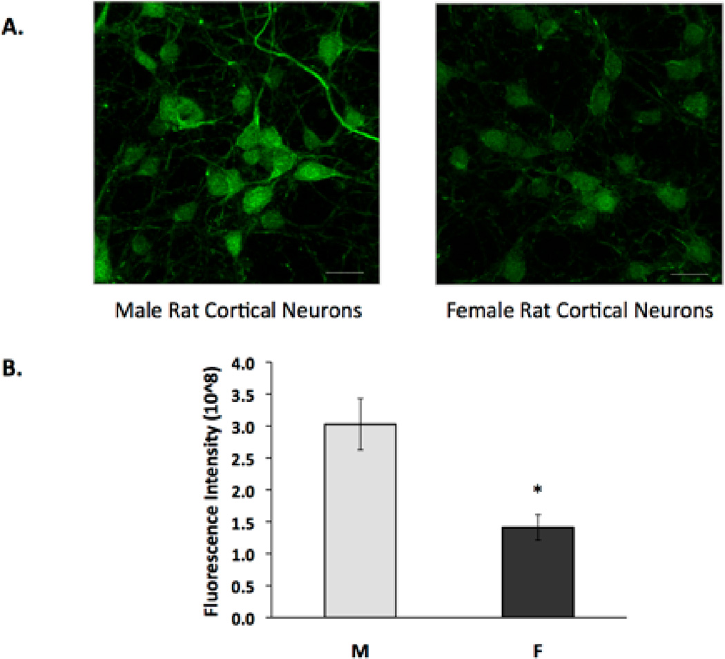
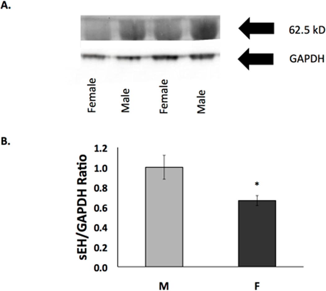
Methods: Rodent cortical neurons were cultured, and exposed to oxygen-glucose deprivation (OGD); then cell death was measured. EETs levels were determined by LC-MS/MS. Expression of sEH-encoding ephx2 was determined by qRT-PCR. Western blotting, immunocytochemistry, and hydrolase activity assay assessed protein expression and activity.

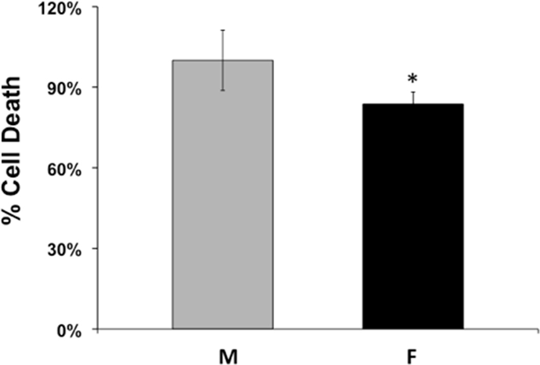
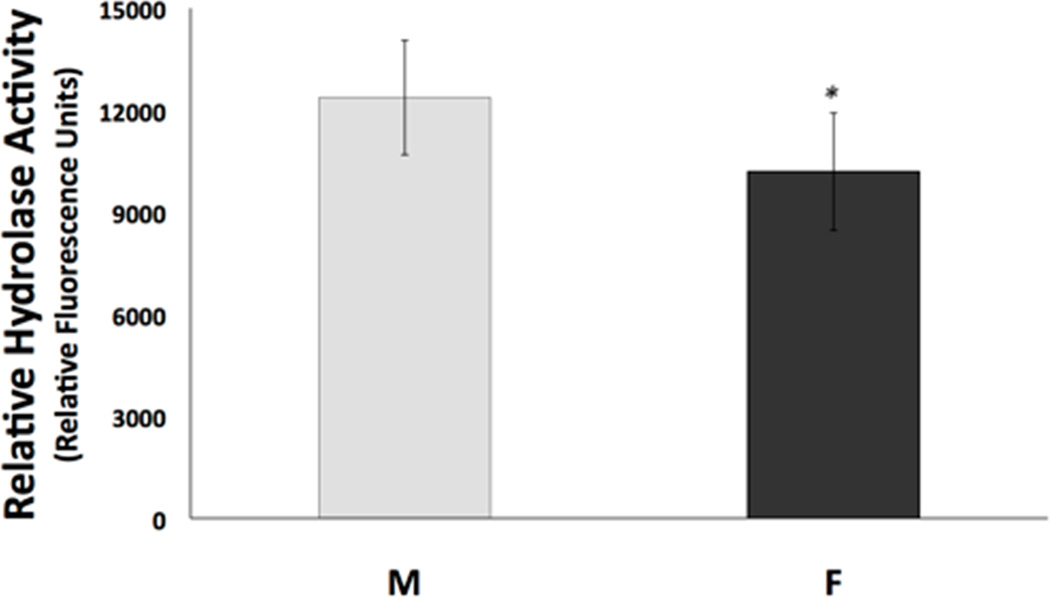
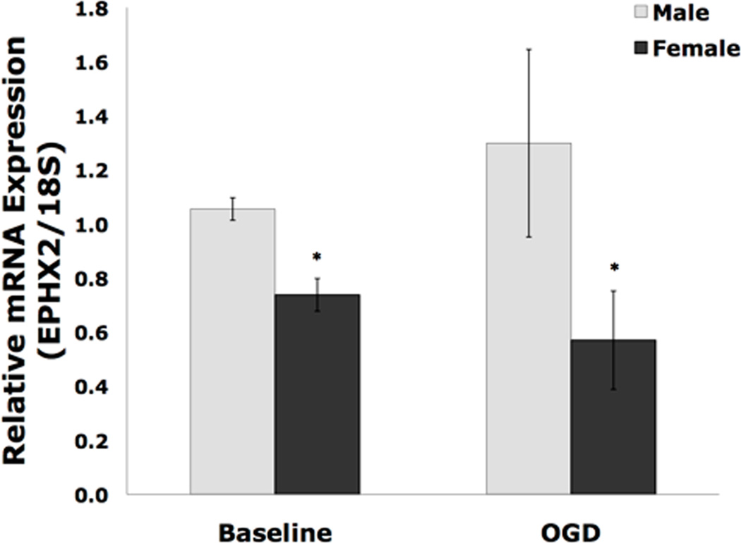
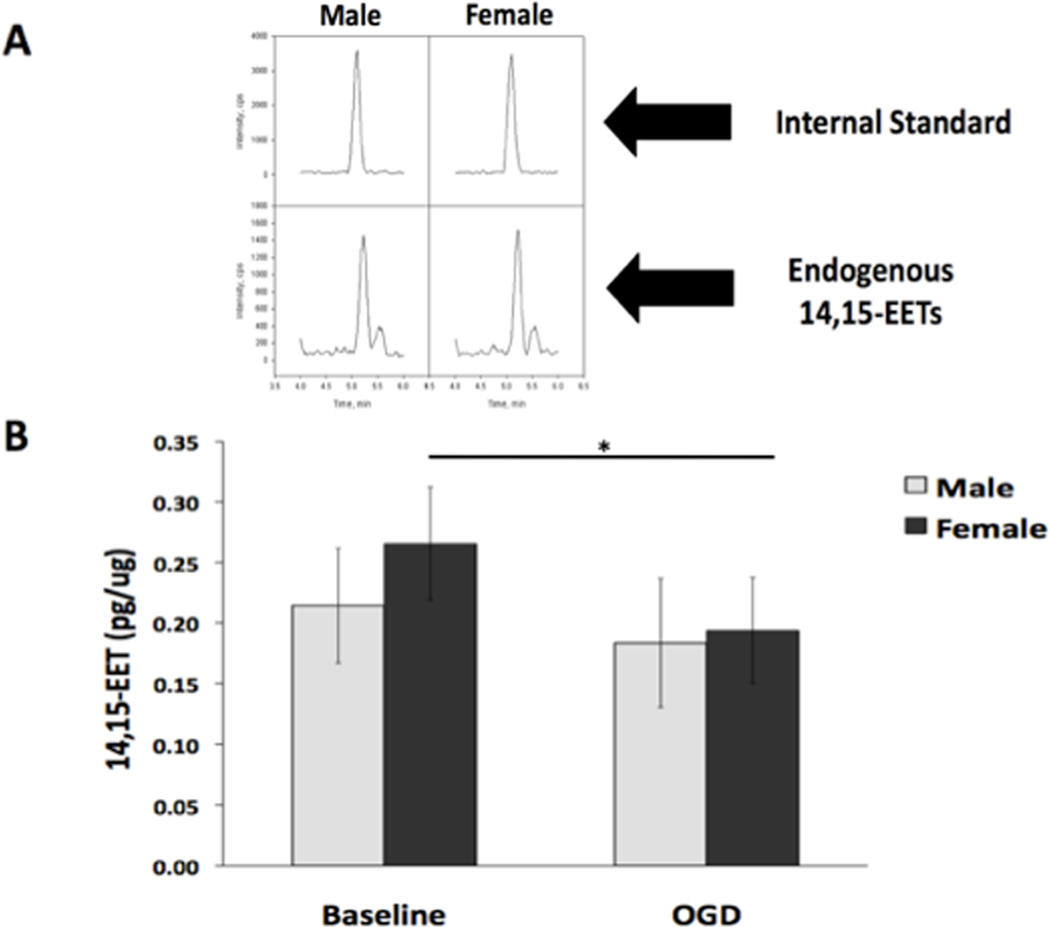
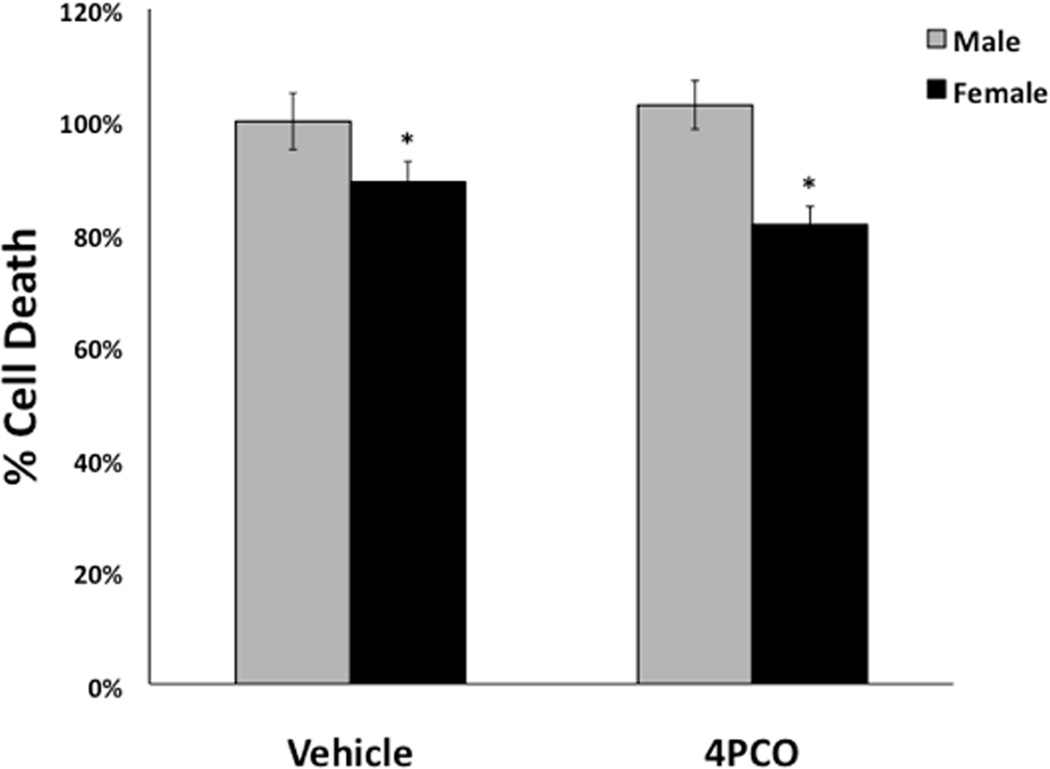
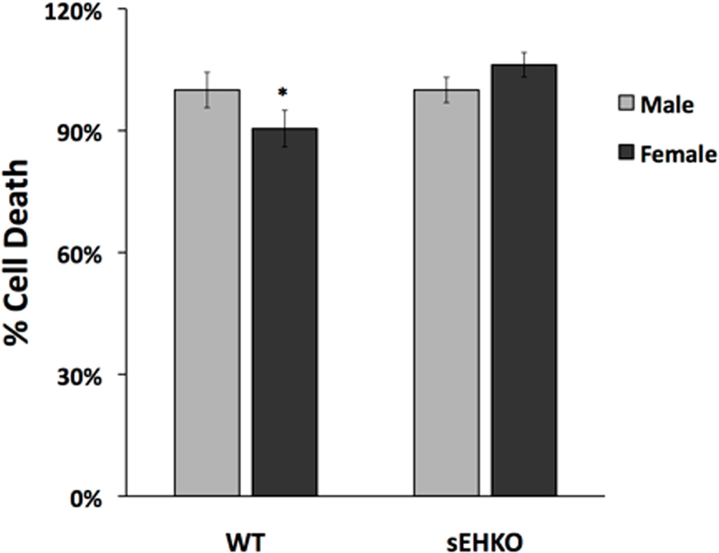
Results: Cell death after OGD was higher in neurons from males vs. females, which correlated with higher ephx2 mRNA and stronger sEH immunoreactivity. However, EETs levels were similar in both sexes and pharmacological inhibition of the hydrolase domain of sEH did not abolish the sex difference in cell death. Genetic knockout of sEH in mice abolished the sex difference observed in neurons isolated from these mice after OGD.

Conclusions: Cultured cortical neurons from females are more resistant to ischemia than neurons from males. Neurons from females have less sEH activity compared to neurons from males at baseline, although sEH levels were not measured after OGD. While pharmacological inhibition of the hydrolase domain of sEH does not affect cell death, knockout of the gene encoding sEH eradicates the sex difference seen in wild-type neurons, suggesting a role for further study of the lesser-known phosphatase domain of sEH and its role in sexual dimorphism in neuronal sensitivity to ischemia.


Figures

Figure 1
Figure 1. Cell death in primary cultured cortical neurons from male and female rat brains
Cortical neurons were cultured from embryonic day 18 male (M) and female (F) rat embryos and were subjected to OGD on day 10 in vitro. Cell death was measured by LDH release in neurons from male and female rats 24 hrs after reoxygenation. The graph depicts average cell death in neurons from male and female embryos expressed relative to the mean of cell death in males. (n=9, *p=0.0019).
Figure 2
Figure 2. Hydrolase activity assay in cultured neurons from male and female rat brains
On day 10 in vitro, neuronal cultures from male (M) and female (F) rats were homogenized and incubated with epoxyfluor 7, which, when hydrolyzed by sEH, becomes a fluorescent compound. Fluorescence was measured using a fluorescent plate reader, and activity was normalized to reaction time and amount of protein. (n=6, *p=0.01)
Figure 3
Figure 3. Immunocytochemistry for sEH in neuronal cultures
Cultured cortical neurons were immunolabeled for sEH, and images were obtained using confocal microscopy. All images were captured at identical time points with neurons cultured from the same litter and plated at equal densities. Microscope settings were identical for all imaged cells. (A) sEH is broadly expressed in cortical neurons as seen in representative confocal images. Scale bar, 20 µm. (B) The bar graph represents average total fluorescent intensity minus background intensity in neurons from males (M) and females (F). (n=4, *p=0.011) Images are representative of at least four biological replicates.
Figure 4
Figure 4. Western blot analysis of soluble epoxide hydrolase in cortical neurons from males and females
Western blot of protein extracts of neurons from males and females harvested after 10 days in vitro. sEH expression in cultured cortical neurons from males is significantly higher than in neurons from females. (A) Representative image of Western blot. Top panel is probed with anti-sEH antibody and bottom panel is probed with anti-GAPDH. (B) Quantification of Western blots of sEH normalized against GAPDH. Cells from males and females were paired within each experiment (n=4, *p=0.004)
Figure 5
Figure 5. Real-time quantitative PCR for EPHX2 in cultured cortical neurons
Real-time quantitative PCR was performed on the 11th day in vitro on cortical cultured neurons from males (M) and females (F). Baseline represents untreated cells, while OGD represents cells that have undergone 2 hours OGD and then 24 hours reoxygenation. (n=6, *p<0.05) There were no differences between baseline and post-OGD values in either group.
Figure 6
Figure 6. 14,15-EET was present in cells from males and females at baseline and after OGD
The concentration of EETs in neurons from males and females was evaluated using liquid chromatography-tandem mass spectrometry (LC-MS/MS) in untreated (baseline) cells and cells subjected to 2 hours of OGD followed by 24 hours reoxygenation. (A) Representative 14,15-EET peaks obtained via LC-MS/MS in both male (M) and female (F) cells. The top panel represents the d8 14,15-EET internal standard. The bottom panel shows the endogenous 14, 15-EET peaks from the cells. The left two are from male cells and the right two are from female cells. The SRM transitions monitored were m/z 327.2 to 182.2 for the internal standard and the m/z for the 14,15 EET is 319.2 to 175. (B) The concentration of total 14,15-EET is not significantly different in cells from males and females at baseline (n=5, p=0.07). After OGD, 14,15-EET significantly decreased in cells from females, but not males. * Indicates a significant decrease in 14,15-EET after OGD in cells from females (n=5, *p=0.015).
Figure 7
Figure 7. Inhibition of sEH using 4-PCO does not alter the difference in sensitivity to OGD between cells from males vs. females
On day 10 in vitro, cultured cortical neurons from males and females were treated with 4-Phenylchalcone oxide (4-PCO, 2 µM), an inhibitor of the hydrolase activity of soluble epoxide hydrolase, or vehicle (DMSO). Treatment was initiated 1 hour prior to OGD and continued throughout OGD and 24 hours of reoxygenation. Cells from females were more resistant to cell death in both vehicle and 4-PCO treated cells as expressed relative to the mean in cells from males (n=5, *p<0.05).
Figure 8
Figure 8. Knockout of sEH eliminates the difference in sensitivity to OGD between cells from males vs. females
Cortical neurons from wild-type (WT) and sEH knockout (sEHKO) mice were cultured and exposed to OGD on the 10th day in vitro. WT male mice had significantly more cell death after OGD compared with corresponding WT female mice, but this sex difference was not significant in the knockout mice. Average cell death in neurons from male and female embryos was expressed relative to the mean of cell death in males (n=6–8, *p<0.05).

References

    1. Alkayed NJ, Harukuni I, Kimes AS, London ED, Traystman RJ, Hurn PD. Gender-linked brain injury in experimental stroke. Stroke. 1998 Jan;29(1):159–165. discussion 166. - PubMed
    1. Alkayed NJ, Murphy SJ, Traystman RJ, Hurn PD, Miller VM. Neuroprotective effects of female gonadal steroids in reproductively senescent female rats. Stroke. 2000 Jan;31(1):161–168. - PubMed
    1. Bernstrom K, Kayganich K, Murphy RC, Fitzpatrick FA. Incorporation and distribution of epoxyeicosatrienoic acids into cellular phospholipids. The Journal of biological chemistry. 1992 Feb 25;267(6):3686–3690. - PubMed
    1. Du Lina, Bayir Hülya, Lai Yichen, Zhang Xiaopeng, Kochanek Patrick M, Watkins Simon C, Graham Steven H, Clark Robert S B. Innate gender-based proclivity in response to cytotoxicity and programmed cell death pathway. The Journal of biological chemistry. 2004 Sep 10;279(37):38563–38570. - PubMed
    1. Golomb MR, Fullerton HJ, Nowak-Gottl U, deVeber G for the International Pediatric Stroke Study Group. Male Predominance in Childhood Ischemic Stroke: Findings From the International Pediatric Stroke Study. Stroke. 2008 Dec 29;40(1):52–57. - PubMed

Publication types

MeSH terms

Substances