. 2012 May 28;197(5):605-11.
doi: 10.1083/jcb.201112141.

Talin activates integrins by altering the topology of the β transmembrane domain

Affiliations

Talin activates integrins by altering the topology of the β transmembrane domain

Chungho Kim et al. J Cell Biol. .

Abstract

Talin binding to integrin β tails increases ligand binding affinity (activation). Changes in β transmembrane domain (TMD) topology that disrupt α-β TMD interactions are proposed to mediate integrin activation. In this paper, we used membrane-embedded integrin β3 TMDs bearing environmentally sensitive fluorophores at inner or outer membrane water interfaces to monitor talin-induced β3 TMD motion in model membranes. Talin binding to the β3 cytoplasmic domain increased amino acid side chain embedding at the inner and outer borders of the β3 TMD, indicating altered topology of the β3 TMD. Talin's capacity to effect this change depended on its ability to bind to both the integrin β tail and the membrane. Introduction of a flexible hinge at the midpoint of the β3 TMD decoupled the talin-induced change in intracellular TMD topology from the extracellular side and blocked talin-induced activation of integrin αIIbβ3. Thus, we show that talin binding to the integrin β TMD alters the topology of the TMD, resulting in integrin activation.


Figures

Figure 1.
Figure 1.
Fluorescence-based assay for measuring TMD embedding. (A) The amino acid sequence of β3 TMD tail peptide. The red arrow indicates the site of acid cleavage, and black arrows indicate bimane labeling sites. (B) Embedding changes of TMD. Yellow pentagons represent bimanes, and the structure of monobromobimane and TMD tilt angle are also shown. (C) Structure of β3 TMD, with side chains of Leu694 and Ile721 indicated. (D) Negative-stain electron microscopic image of nanodiscs containing β3 TMD tail peptide. Arrows indicate nanodiscs. Bar, 50 nm. (E) Emission spectra of nanodiscs containing β3 TMD tail bimane labeled at L694C (L694C-bimane nanodisc) in the presence or absence of K716E mutation (left). The same peptides in the presence of 2% SDS are shown on the right. Data are representative of three independent experiments.
Figure 2.
Figure 2.
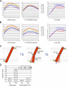
Talin induces increased embedding of both ends of the β3 TMD. (A) Emission spectra of L694C-bimane nanodisc in the presence of increasing concentrations of THD. Fluorescence intensities were normalized to the maximum fluorescence intensity when no THD was present. (B) The emission spectra of I721C-bimane nanodiscs were analyzed as in A. (C) Nanodiscs containing truncated β3 TMD tail peptide were bimane labeled at I721C (ΔI721C-bimane), and response to THD was analyzed as in A. (A–C) All data are representative of at least three independent experiments. (D) Talin-induced topographic change of β3 TMD that can account for altered embedding. Orange cylinders indicate a β3 cytoplasmic tail, which forms a stable helix upon talin binding.
Figure 3.
Figure 3.
A flexible kink in β3 TMD reduces transmission of talin-induced TMD topology change and integrin activation. (A) Emission spectra of β3(L694C)-bimane nanodisc (left) in the presence of increasing concentrations of THD compared with those of β3(A711P,L694C)-bimane nanodisc (middle). Similar fluorescence intensities the in presence of 2% SDS are shown on the right. Fluorescence normalized to the maximum fluorescence intensity of L694C-bimane in the absence of talin is shown. (B) Emission spectra of β3(I721C) and β3(A711P,I721C)-bimane nanodiscs were analyzed as in A. (A and B) Data are representative of at least three independent experiments. (C) Effects of β3(A711P) mutation in response to THD. β3(A711P) breaks the TMD helix into two helices connected by a flexible kink; the flexible kink inhibits the transmission of THD-induced increased embedding across the membrane. (D) CHO cells stably expressing αIIbβ3 or αIIbβ3(A711P) were transfected with THD or vector (vec), and αIIbβ3 activation was measured after 24 h. The arrowhead indicates the band corresponding to THD (∼50 kD). Error bars are SEM of three independent experiments.
Figure 4.
Figure 4.
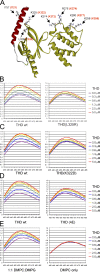
Mechanism of talin-induced topology change. (A) Crystal structure of talin2 F2-F3 (yellow) bound to integrin β1D tail (red; Protein Data Bank accession no. 3G9W). The amino acids in talin2 that bind to the membrane-proximal β1 tail (L328) and to lipid (K325, K258, K274, R276, and K280) are indicated with corresponding talin1 residues in parentheses. β1D(I757) (corresponds to β3(I721)), the position of bimane labeling, is indicated. (B) The emission spectra of L694C-bimane nanodiscs in the presence of increasing concentrations of THD (left) or THD(L325R) (right). wt, wild type. (C) Effect of THD(K322D), which blocks lipid binding, on the emission spectra was analyzed as in B. (D) Effect of mutations in lipid-binding residues of the THD F2 domain (K256E, K272E, K274E, and R277E) on the emission spectra was analyzed in as in B. (E) I721C-bimane nanodiscs were assembled with a 1:1 mixture of DMPC and DMPG (left) or with DMPC only (right), and their responses to the addition of THD were analyzed as in B. (B–E) Data are representative of at least three independent experiments.

Similar articles

Cited by

References

    1. Anthis N.J., Wegener K.L., Ye F., Kim C., Goult B.T., Lowe E.D., Vakonakis I., Bate N., Critchley D.R., Ginsberg M.H., Campbell I.D. 2009. The structure of an integrin/talin complex reveals the basis of inside-out signal transduction. EMBO J. 28:3623–3632 10.1038/emboj.2009.287 - DOI - PMC - PubMed
    1. Cordes F.S., Bright J.N., Sansom M.S. 2002. Proline-induced distortions of transmembrane helices. J. Mol. Biol. 323:951–960 10.1016/S0022-2836(02)01006-9 - DOI - PubMed
    1. Denisov I.G., Grinkova Y.V., Lazarides A.A., Sligar S.G. 2004. Directed self-assembly of monodisperse phospholipid bilayer nanodiscs with controlled size. J. Am. Chem. Soc. 126:3477–3487 10.1021/ja0393574 - DOI - PubMed
    1. Elliott P.R., Goult B.T., Kopp P.M., Bate N., Grossmann J.G., Roberts G.C., Critchley D.R., Barsukov I.L. 2010. The structure of the talin head reveals a novel extended conformation of the FERM domain. Structure. 18:1289–1299 10.1016/j.str.2010.07.011 - DOI - PMC - PubMed
    1. García-Guerra R., García-Domínguez J.A., González-Rodríguez J. 1996. A new look at the lipid composition of the plasma membrane of human blood platelets relative to the GPIIb/IIIa (integrin cxIIβ3) content. Platelets. 7:195–205 10.3109/09537109609023579 - DOI - PubMed

Publication types