. 2012 Jul;295(7):1181-91.
doi: 10.1002/ar.22501. Epub 2012 May 29.

Dorsorostral snout muscles in the rat subserve coordinated movement for whisking and sniffing

Affiliations

Dorsorostral snout muscles in the rat subserve coordinated movement for whisking and sniffing

Sebastian Haidarliu et al. Anat Rec (Hoboken). 2012 Jul.

Abstract

Histochemical examination of the dorsorostral quadrant of the rat snout revealed superficial and deep muscles that are involved in whisking, sniffing, and airflow control. The part of M. nasolabialis profundus that acts as an intrinsic (follicular) muscle to facilitate protraction and translation of the vibrissae is described. An intraturbinate and selected rostral-most nasal muscles that can influence major routs of inspiratory airflow and rhinarial touch through their control of nostril configuration, atrioturbinate and rhinarium position, were revealed.


Figures

Figure 1
Figure 1
Morphology of the rat rhinarium. (A) Anterior view of the rat rhinarium. Boxed area shows the borders of the region that corresponds to B after this rat snout was subjected to histological treatment. (B) Autofluorescence of a nasal tubercle superficial slice (60 μm thick) stained for cytochrome oxidase activity. 1, regio suprarhinarica; 2, nostrils; 3, sulcus medianus (philtrum); 4, paramedian ridges; 5, nasal tubercles (pads); 6, atrioturbinate; 7, epidermal (friction) ridges arranged as a whorl, with the centre of the whorl (8) in the ventrolateral part of each tubercle. M, medial; V, ventral. Scale bars are 1 mm in (A), and 0.2 mm in (B).
Figure 2
Figure 2
Light microscopy of the right lateral wall in the nasal cavity of an adult rat. 1, nostril; 2, atrioturbinate; 3, isthmus; 4, nasoturbinate; 5, maxilloturbinate; 6, ethmoturbinates. R, rostral; V, ventral. Scale bar = 1 mm.
Figure 3
Figure 3
A sagittal radiovisiogram of the rostral area of the head (A), and light microscopy of a parasagittal snout slice stained for cytochrome oxidase activity (B) in adult rats. Black lines in (B) show the borders of the DRQ. 1, rhinarium; 2, nostril; 3, upper lip; 4, upper incisives; 5, short pieces of stainless steel tubes threaded by the A4 and B4 vibrissae at their proximal ends; 6, nasal bones; 7, lower incisives; 8, atriturbinate; 9, rostral pole of the premaxilla. R, rostral; V, ventral. Scale bars = 1 mm.
Figure 4
Figure 4
Schematic representation of muscles related to the dorsorostral part of the rat snout. Encircled areas comprise origins of the following muscles: green area, rhinarial muscles; pink area, two extrinsic whisker protractors; and blue area, most of the parts of the M. nasolabialis profundus. 1, ventromedial part of the lateral nasal cartilage; 2, M. levator rhinarii; 3, septal cartilage; 4, atrioturbinate; 5, atrium; 6, insertion site of the M. dilator nasi; 7, ITM; 8, 10, 11, and 12, the most superficial, superficial, posterior, and pseudointrinsic portions, respectively, of the Pars interna of the M. nasolabialis profundus; 9, origin of the Pars interna profunda; 13, M. transversus nasi; 14, belly of the M. dilator nasi; 15, rhinarium; 16, Pars anterior of the M. nasolabialis profundus; 17, M. depressor septi nasi; 18, M. depressor rhinarii; 19 and 20, origins of the Partes mediae superior et inferior, and 21 and 22, of the Partes maxillares profunda et superficialis, respectively, of the M. nasolabialis profundus. Marked black circles represent mystacial vibrissae.
Figure 5
Figure 5
Light microscopy of a horizontal slice of the dorsal part of a 1-week-old rat snout. (A) A slice of the whole snout; (B) and (C), higher magnifications of the boxed areas in (A) and (B), respectively. α, the dorsal-most straddler; A1 - A4, the dorsal-most row of vibrissae. 1, septum; 2, olfactory bulb; 3, striated fibers of the M. transversus nasi. Scale bars are 1 mm in (A) and (B), and 0.1 mm in (C).
Figure 6
Figure 6
Light microscopy of a horizontal slice of an adult rat snout in the plane of the vibrissa follicles in row A. α, follicle of the dorsal-most straddler; A1 - A4, vibrissa follicles of row A; M, medial; R, rostral. 1, belly, and 2, tendon of the M. dilator nasi. Scale bar = 1 mm.
Figure 7
Figure 7
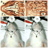
Anatomical location and effect of electrical stimulation of the M. dilator nasi. (A) Horizontal slice of the head of a 7-day-old rat. 1, belly, and 2, tendon of the M. dilator nasi; 3, septum; 4, vibrissa row A; 5, olfactory bulb. Scale bar = 1 mm. (B) Light microscopic view of a snout tangential slice that contains an electrolytic lesion (6) caused by stimulating electrode in the belly of the M dilator nasi (1) in an adult rat. Inset: higher magnification of the lesion site (6). R, rostral; V, ventral. Scale bar = 0.2 mm. (C) Anesthetized rat with the head fixed in a stereotactic device. 7, nasal piece of the headholder without ear bars; 8, guides that contain muscle stimulating electrodes. (D) Nose deflection during electrical stimulation (AC, 10 V, 50 Hz) of the right M. dilator nasi.
Figure 8
Figure 8
Appearance of the rostral-most muscles in the rat snout in different planes: oblique (A), horizontal (B), and coronal (C). Slice shown in (B) was obtained by cutting the snout at the level shown by a solid horizontal line in (A). 1, rhinarium; 2, M. depressor rhinarii; 3. M. depressor septi nasi; 4, Partes mediae superior et inferior of the M. nasolabialis profundus; 5, septal cartilage; 6, atrioturbinate; 7, incisive; 8, medial end of the lateral nasal cartilage; 9, philtrum. A4-C4, vibrissa follicles. Scale bars are 1 mm in (A) and (C), and 0.5 mm in (B).
Figure 9
Figure 9
Coronal (A) and oblique (B) view of the M. levator rhinarii (1) in adult rats. 2, atrium; 3, rostral splitted part of the septal cartilage; 4, M. depressor rhinarii; 5, M. depressor septi nasi; 6 rhinarium; 7, tuberculum nasi; 8, atrioturbinate; 9, roof cartilage; 10, lateral nasal cartilage; 11, M. transverses nasi. RM, rostromedial; V, ventral. Scale bars = 1 mm.
Figure 10
Figure 10
Light microscopy of tangential slices of the DRQ of an adult rat snout at ∼0.3 mm (A) and ∼0.45 mm (B) from the surface of the skin. 1, rostrum; 2, atrium; 3, nasal vibrissae; 4, Pars interna superficialis, 5, Pars anterior, and 6, posterior slips of the Pars interna of the M. nasolabialis profundus; 7, fascicles of the Mm. nasolabialis et maxillolabialis. A4 - C4, vibrissa follicles; R, rostral; V, ventral. Scale bars = 1 mm.
Figure 11
Figure 11
Light microscopy of a tangential slice of an adult rat snout cut at ∼0.6 mm from the surface of the skin. 1, PIM; 2, atrioturbinate; 3, dorsal meatus; 4, ventral meatus; 5, rostrum; 6, roof cartilage; 7, lateral nasal cartilage. A4, B4, vibrissa follicles; R, rostral; V, ventral. Scale bar = 1 mm.
Figure 12
Figure 12
Light microscopic view of the PIM (1) insertion sites on the capsule (3) of the vibrissa B4 in an adult rat parasagittal slice cut at ∼0.8 mm from the surface of the skin. (A) An entire slice. (B) Higher magnification of boxed area in (A). (C) Higher magnification of boxed area in (B). 2, IM. A4 - C4, vibrissa follicles; R, rostral; V, ventral. Scale bars are 1 mm in (A) and (B), and 0.1 mm in (C).
Figure 13
Figure 13
Light microscopy of a coronal slice prepared from a tissue block containing the lateral wall of the atrium. (A) An entire slice; (B) and (C) Higher magnifications of the boxed areas in (A) and (B), respectively. 1, atrioturbinate; 2, dorsal meatus; 3, ventral meatus; 4, ITM; 5, intraturbinate cartilage; 6, site of contact between collagenous structures of the muscle and cartilage. M, medial; V, ventral. Scale bars are 1 mm in (A), and 0.1 mm in (B) and (C).
Figure 14
Figure 14
Light microscopy of an adult rat coronal slice (A) that contains origins (1) of the Partes anterior et interna of the M. nasolabialis profundus. (B) and (C) Higher magnifications of the boxed areas in (A) and (B), respectively. 2, septal cartilage; 3, ductus nasolacrimalis; 4, maxilloturbinate; 5, roof cartilage; 6, lateral nasal cartilage; 7, sites of muscle attachment to the nasal cartilage. M, medial; V, ventral. Scale bars are 1mm in (A), 0.2 mm in (B), and 0.1 mm in (C).

Similar articles

Cited by

References

    1. Ahissar E, Knutsen PM. Object localization with whiskers. Biol Cybern. 2008;98:449–458. - PubMed
    1. Bahar A, Dudai Y, Ahissar E. Neural signature of taste familiarity in the gustatory cortex of the freely behaving rat. J Neurophysiol. 2004;92:3298–3308. - PubMed
    1. Banke J, Mess A, Zeller U. Functional morphology of the rostral head region of Cryptomys hottentotus (Bathyergidae, Rodentia) Proceedings of the 8th International Symposium on African Small Mammals. 2001:231–241.
    1. Berg RW, Kleinfeld D. Rhythmic whisking by rat: retraction as well as protraction of the vibrissae is under active muscular control. J Neurophysiol. 2003;89:104–117. - PubMed
    1. Bermejo R, Friedman W, Zeigler HP. Topography of whisking II: Interaction of whisker and pad. Somatosens Mot Res. 2005;22:213–220. - PubMed

Publication types

LinkOut - more resources