. 2012 May 29:9:102.
doi: 10.1186/1742-2094-9-102.

Telmisartan directly ameliorates the neuronal inflammatory response to IL-1β partly through the JNK/c-Jun and NADPH oxidase pathways

Affiliations

Telmisartan directly ameliorates the neuronal inflammatory response to IL-1β partly through the JNK/c-Jun and NADPH oxidase pathways

Tao Pang et al. J Neuroinflammation. .

Abstract

Background: Blockade of angiotensin II type 1 (AT1) receptors ameliorates brain inflammation, and reduces excessive brain interleukin-1 beta (IL-1β) production and release from cortical microglia. The aim of this study was to determine whether, in addition, AT1 receptor blockade directly attenuates IL-1β-induced inflammatory responses in neuronal cultures.

Methods: SK-N-SH human neuroblasts and primary rat cortical neurons were pretreated with telmisartan followed by exposure to IL-1β. Gene expression was determined by reverse transcriptase (RT)-PCR, protein expression and kinase activation by western blotting, NADPH oxidase activity by the lucigenin method, prostaglandin E2 (PGE2) release by enzyme immunoassay, reactive oxygen species (ROS) generation by the dichlorodihydrofluorescein diacetate fluorescent probe assay, and peroxisome proliferator-activated receptor gamma (PPARγ) involvement was assessed with the antagonists GW9662 and T0070907, the agonist pioglitazone and the expression of PPARγ target genes ABCG1 and CD36.

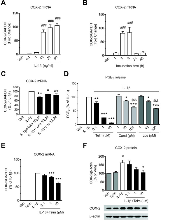
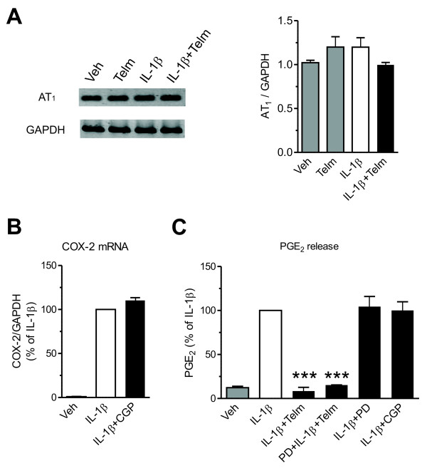
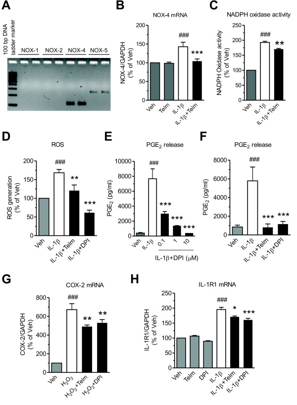
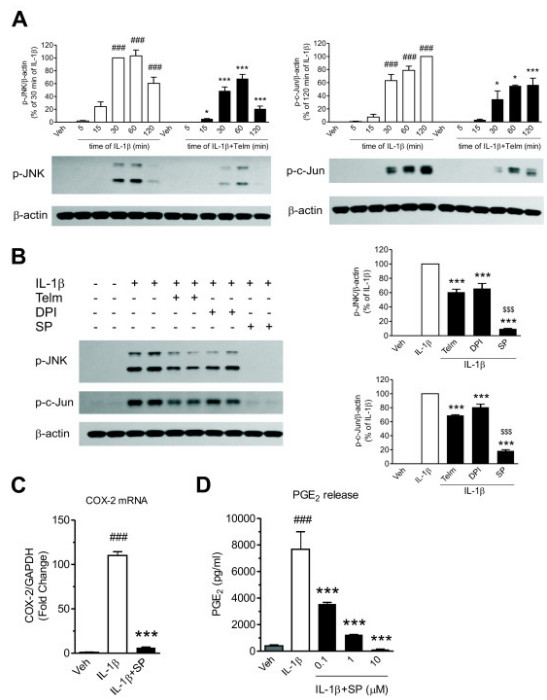
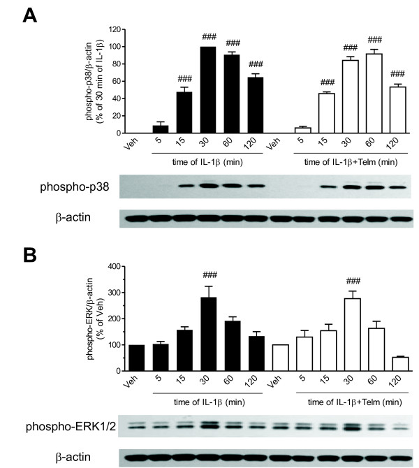
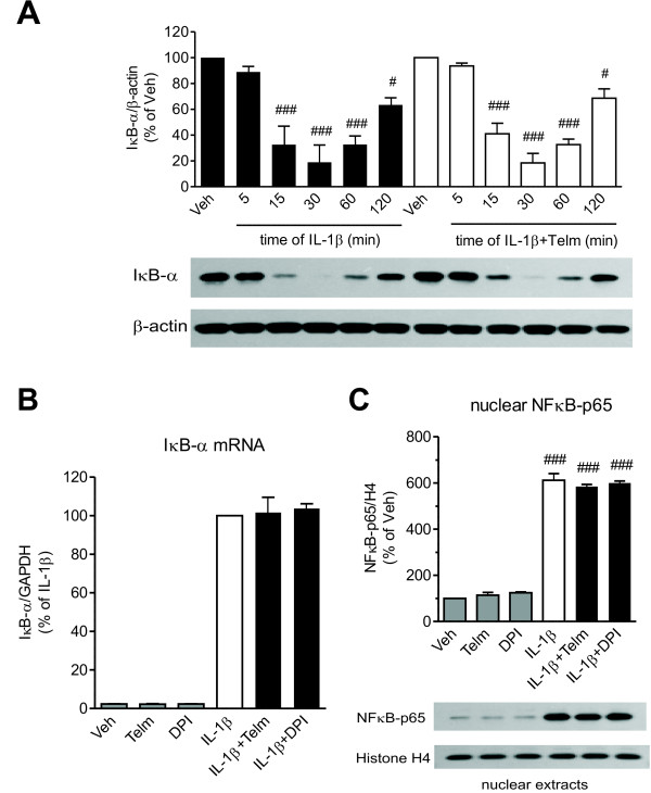
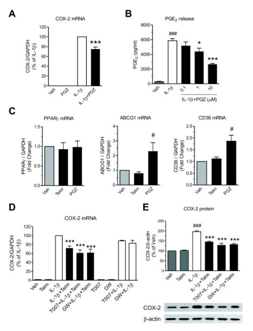
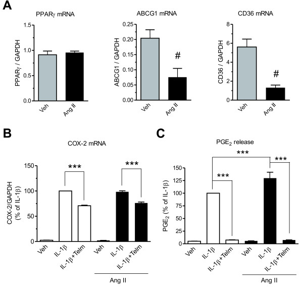
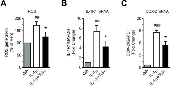
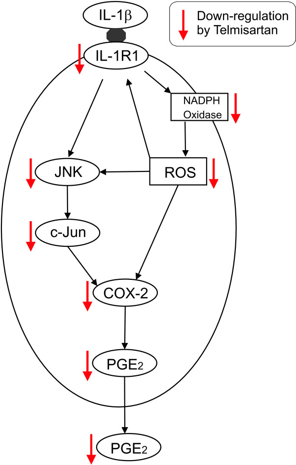
Results: We found that SK-N-SH neuroblasts expressed AT1 but not AT2 receptor mRNA. Telmisartan reduced IL-1β-induced cyclooxygenase-2 (COX-2) expression and PGE2 release more potently than did candesartan and losartan. Telmisartan reduced the IL-1β-induced increase in IL-1R1 receptor and NADPH oxidase-4 (NOX-4) mRNA expression, NADPH oxidase activity, and ROS generation, and reduced hydrogen peroxide-induced COX-2 gene expression. Telmisartan did not modify IL-1β-induced ERK1/2 and p38 mitogen-activated protein kinase (MAPK) phosphorylation or nuclear factor-κB activation but significantly decreased IL-1β-induced c-Jun N-terminal kinase (JNK) and c-Jun activation. The JNK inhibitor SP600125 decreased IL-1β-induced PGE2 release with a potency similar to that of telmisartan. The PPARγ agonist pioglitazone reduced IL-1β-induced inflammatory reaction, whereas telmisartan did not activate PPARγ, as shown by its failure to enhance the expression of the PPARγ target genes ABCG1 and CD36, and the inability of the PPARγ antagonists GW9662 and T0070907 to modify the effect of telmisartan on COX-2 induction. The effect of telmisartan on IL-1β-stimulated COX-2 and IL-1R1 mRNA expression and ROS production was replicated in primary rat cortical neurons.

Conclusions: Telmisartan directly ameliorates IL-1β-induced neuronal inflammatory response by inhibition of oxidative stress and the JNK/c-Jun pathway. Our results support the hypothesis that AT1 receptor blockers are directly neuroprotective, and should be considered for the treatment of inflammatory conditions of the brain.


Figures

Figure 1
Figure 1
Telmisartan inhibits interleukin-1 beta (IL-1β)-induced cyclooxygenase-2 (COX-2) mRNA and protein expression and prostaglandin E2(PGE2) release in SK-N-SH neuroblasts (A,B) IL-1β dose-dependently and time-dependently induces COX-2 mRNA expression in SK-N-SH neuroblasts. The cells were incubated with (A) the indicated concentrations of IL-1β for 3 hours, or with (B) 10 ng/ml IL-1β at indicated time intervals to determine COX-2 mRNA expression. Results are expressed as fold change relative to vehicle-treated group (Veh). (C) Telmisartan, candesartan, and losartan reduced IL-1β induced COX-2 mRNA expression with similar potency. The cells were pretreated with 10 μmol/l telmisartan (Telm), candesartan (Cand), or losartan (Los) for 2 hours before exposure for 3 hours to 10 ng/ml IL-1β. Results are expressed as the percentage of IL-1β. (D) Telmisartan was the most effective of AT1 receptor blockers at reducing the IL-1β-induced PGE2 release. Cells were pretreated with indicated concentrations of Telm, Cand, or Los for 2 hours before exposure for 24 hours to 10 ng/ml IL-1β to determine cumulative PGE2 release. Results are expressed as the percentage of IL-1β. (E,F) Telmisartan dose-dependently reduced IL-1β-induced COX-2 mRNA and protein expression. The cells were pretreated with indicated concentrations of Telm for 2 hours, then incubated with 10 ng/ml IL-1β for (E) 3 hours to determine COX-2 mRNA expression, or (F) 24 hours to determine COX-2 protein expression. The picture is a representative western blot. All results are means ± SEM from at least three independent experiments. * P < 0.05, ** P < 0.01, *** P < 0.001 vs. IL-1β; # P < 0.05, ### P < 0.001 vs. Veh; $$$ P < 0.001 vs. IL-1β + 10 μmol Telm.
Figure 2
Figure 2
Role of angiotensin II receptor type 1 (AT1) and type 2 (AT2) receptors in interleukin-1 beta (IL-1β)-induced neuronal inflammatory response in SK-N-SH cells. (A) Expression of Angiotensin II AT1 receptor mRNA in SK-N-SH neuroblasts. The cells were pretreated with 10 μmol/l telmisartan (Telm) for 2 hours before exposure for 3 hours to 10 ng/ml IL-1β. (Right) AT1 receptor expression normalized to GAPDH mRNA. The picture shows visualized products of the RT-PCR reaction after separation on agarose gel. (B) Angiotensin II AT2 receptor agonist CGP 42112 (CGP) does not modify IL-1β-induced COX-2 mRNA expression. The cells were pretreated with 1 μmol/l CGP 42112 for 2 hours before exposur for 3 hourse to 10 ng/ml IL-1β. (C) Angiotensin II AT2 receptor antagonist PD 123319 or AT2 receptor agonist CGP 42112 do not affect IL-1β-induced PGE2 release or the inhibitory effect of telmisartan. The cells were pretreated for 2 hours with 10 μmol/l PD 123319 (PD), 10 μmol/l CGP, or 10 μmol/l Telm alone or in combination with PD, before exposure for 24 hours to 10 ng/ml IL-1β to determine cumulative PGE2 release. Results are expressed as a percentage of IL-1β. All results are means ± SEM from at least three independent experiments. *** P < 0.001 vs. IL-1β.
Figure 3
Figure 3
Telmisartan reduces interleukin-1 beta (IL-1β)-induced NADPH oxidase activation, reactive oxygen species formation and IL-1 receptor 1 (IL-1R1) mRNA expression in SK-N-SH neuroblasts. (A) Untreated SK-N-SH cells were analyzed for mRNA expression of different NADPH oxidase isoforms. NADPH oxidase-4 (NOX-4) is the dominant isoform of NADPH oxidase in SK-N-SH neuroblasts. The picture shows visualized products of a RT-PCR reaction after separation in an agarose gel. (B,C) Telmisartan reduced IL-1β-induced NOX-4 mRNA expression and NADPH oxidase activity. The cells pretreated with 10 μmol/l telmisartan (Telm) for 2 hours, were incubated with 10 ng/ml IL-1β for 3 hours to determine (B) NOX-4 mRNA expression and (C) NADPH oxidase activity. (D) Telmisartan reduced IL-1β-induced reactive oxygen species (ROS) generation to a lesser extent than does diphenyleneiodonium (DPI). The cells were pretreated with 10 μmol/l Telm or 5 μmol/l DPI for 2 hours before 1 hours exposure to 10 ng/ml IL-1β to determine ROS generation. (E,F) DPI dose-dependently inhibited IL-1β-induced PGE2 release with a potency similar to telmisartan. The cells pretreated with indicated concentrations of DPI or Telm for 2 hour were incubated with IL-1β for 24 hours to determine cumulative PGE2 release. (G) Both telmisartan and DPI reduce hydrogen peroxide-induced COX-2 mRNA expression. The cells were pretreated with 10 μmol/l Telm or 5 μmol/l DPI for 2 hours before exposure for 3 hours to 100 μmol/l hydrogen peroxide (H2O2) to determine COX-2 mRNA expression. (H) Both telmisartan and DPI reduce IL-1β-induced expression of IL-1β receptor IL-1R1 mRNA. The cells were pretreated with 10 μmol/l Telm or 5 μmol/l DPI for 2 hours before exposure for 3 hours to 10 ng/ml IL-1β to determine IL-1R1 mRNA expression. Results are presented as a percentage of Veh. All results are means ± SEM from at least three independent experiments. * P < 0.05, ** P < 0.01, *** P < 0.001 vs. IL-1β or H2O2; ### P < 0.001 vs. Veh.
Figure 4
Figure 4
Neuroprotective effect of telmisartan is partially mediated through inhibition of the c-Jun N-terminal kinase (JNK)/c-Jun pathway in SK-N-SH neuroblasts. (A) Telmisartan attenuated the time-dependent activation of JNK and c-Jun in response to interleukin-1 beta (IL-1β). The cells were pretreated for 2 hours with 10 μmol/l telmisartan (Telm) before exposure to 10 ng/ml IL-1β for the indicated time intervals. Phosphorylation of JNK and c-Jun was determined by western blotting. Representative blots are shown under the corresponding bar graphs. ### P < 0.001 vs. Veh; * P < 0.05, *** P < 0.001 vs. corresponding IL-1β group. (B) Telmisartan inhibited IL-1β-stimulated JNK and c-Jun activation with a potency similar to that of diphenyleneiodonium (DPI) but to a lesser extent than the JNK inhibitor SP600125. The cells were pretreated for 2 hours with 10 μmol/l Telm, 5 μmol/l DPI, or 10 μmol/l SP600125 (SP) before exposure for 30 minutes to 10 ng/ml IL-1β. The phosphorylation of JNK and c-Jun was detected as above. Results are shown as a percentage of the IL-1β-treated group. (C,D) The JNK inhibitor SP600125 abrogated the IL-1β-induced COX-2 mRNA expression and PGE2 release. The cells were pretreated for (C) 2 hours with 10 μmol/l SP before exposure for 3 hours to 10 ng/ml IL-1β to determine COX-2 mRNA expression, or with (D) the indicated concentrations of SP600125 before exposure for 24 hours to 10 ng/ml IL-1β to determine cumulative PGE2 release. All results are presented as means ± SEM from three independent experiments. *** P < 0.001 vs. IL-1β; ### P < 0.001 vs. Veh; $$$ P < 0.001 vs. IL-1β + Telm.
Figure 5
Figure 5
Telmisartan did not alter the interleukin-1 beta (IL-1β)-stimulated activation of p38 mitogen-activated protein kinase (MAPK) or extracellular signal-regulated kinase (ERK)1/2 in SK-N-SH neuroblasts. (A,B) Cells were pretreated for 2 hours with 10 μmol/l telmisartan (Telm) before exposure to 10 ng/ml IL-1β for the indicated time intervals. The phosphorylations of (A) p38 MAPK and (B) ERK1/2 were determined by western blotting and normalized to the levels of β-actin. All data are presented as means ± SEM from three independent experiments. Representative blots are shown under the corresponding bar graphs. ### P < 0.001 vs. Veh.
Figure 6
Figure 6
The nuclear factor-kappa B (NF-κB) pathway is not involved in the neuroprotective effect of telmisartan in SK-N-SH neuroblasts. (A) Telmisartan does not prevent time-dependent IκB-α protein degradation in cells in response to interleukin-1 beta (IL-1β). Cells were pretreated for 2 hours with 10 μmol/l telmisartan (Telm) before exposure to 10 ng/ml IL-1β for the indicated time intervals. IκB-α protein levels were determined in whole-cell extracts, and normalized to β-actin. (B) Neither telmisartan nor diphenyleneiodonium (DPI) modified IL-1β-induced expression of IκB-α mRNA. The cells were pretreated for 2 hours with 10 μmol/l Telm or 5 μmol/l DPI before exposure for 3 hours to 10 ng/ml IL-1β to determine IκB-α mRNA expression. (C) Neither telmisartan nor DPI affected IL-1β-induced nuclear translocation of the NF-κB p65 subunit. The cells were pretreated for 2 hours with 10 μmol/l Telm or 5 μmol/l DPI before exposure for 30 minutes to 10 ng/ml IL-1β. The NF-κB p65 subunit protein was determined in nuclear extracts and normalized to the level of the nuclear protein histone H4. Representative western blots are shown below the corresponding bar graphs. Results are presented as means ± SEM from three independent experiments. # P < 0.05, ### P < 0.001 vs. Veh.
Figure 7
Figure 7
Peroxisome proliferator-activated receptor gamma (PPARγ) activation is not involved in the neuroprotective effect of telmisartan in SK-N-SH neuroblasts. (A,B) The PPARγ agonist pioglitazone inhibited interleukin-1 beta (IL-1β)-induced cyclooxygenase-2 (COX-2) gene expression and prostaglandin E2 (PGE2) release. The cells were pretreated for (A) 2 hours with 10 μmol/l pioglitazone (PGZ) before exposure for 3 hours to 10 ng/ml IL-1β to determine COX-2 mRNA expression or (B) with indicated concentrations of PGZ before exposure for 24 hours to 10 ng/ml IL-1β to determine cumulative PGE2 release. (C) Pioglitazone, but not telmisartan, induced gene expression of the PPARγ target genes ABCG1 and CD36. The cells were incubated for 3 hours with 10 μmol/l PGZ or 10 μmol/l Telm to determine gene expression of PPARγ and its target genes ABCG1 and CD36. Results are shown as fold change relative to the vehicle-treated group (Veh). (D,E) PPARγ antagonists did not change the inhibitory effect of telmisartan on IL-1β-induced COX-2 expression. The cells were pretreated for 1 hour with 1 μmol/l T0070907 (T007) or 20 μmol/l GW9662 (GW), followed by 10 μmol/l Telm for 2 hours before exposure for (D) 3 hours to 10 ng/ml IL-1β to determine COX-2 mRNA, or (E) 24 hours of IL-1β to determine COX-2 protein expression. The picture below is a representative western blot. All results are means ± SEM from at least three independent experiments. * P < 0.05, *** P < 0.001 vs. IL-1β; # P < 0.05, ### P < 0.001 vs. Veh.
Figure 8
Figure 8
Effect of Angiotensin II (Ang II) on SK-N-SH neuroblasts. (A) Ang II did not affect peroxisome proliferator-activated receptor gamma (PPARγ) gene expression, but strongly inhibited the expression of the PPARγ target genes ABCG1 and CD36. The cells were treated with 1 μmol/l Ang II for 24 hours to determine PPARγ, ABCG1 and CD36 mRNA expression. # P < 0.05 vs. Veh. (B) Ang II affected neither interleukin-1 beta (IL-1β)-induced cyclooxygenase-2 (COX-2) mRNA expression nor the inhibitory effects of telmisartan. Cells cultured for 24 hours in the presence of 1 μmol/l Ang II were pretreated for 2 hours with 10 μmol/l telmisartan (Telm) before exposure for 3 hours to 10 ng/ml IL-1β to determine COX-2 mRNA expression. Results are presented as a percentage of the IL-1β-treated group. (C) Ang II augmented IL-1β-induced PGE2 release but did not modify the inhibitory effect of telmisartan. The cells, cultured for 24 hours in the presence of 1 μmol/l Ang II, were pretreated for 2 hours with 10 μmol/l Telm before exposure for 24 hours to 10 ng/ml IL-1β to determine cumulative PGE2 release. Results are presented as a percentage of IL-1β. All results are means ± SEM from at least three independent experiments. *** P < 0.001.
Figure 9
Figure 9
Telmisartan inhibits interleukin-1 beta (IL-1β)-induced reactive oxygen species (ROS) generation, interleukin-1 receptor type 1 (IL-1R1) and cyclooxygenase-2 (COX-2) gene expression in primary rat cortical neurons. Cells were pretreated for 2 hours with 10 μmol/l telmisartan (Telm) before exposure for 2 hours to 1 ng/ml IL-1β to determine (A) ROS generation, and (B, C) IL-1R1 and COX-2 mRNA expression. Results are means ± SEM from at least three independent experiments. * P < 0.05 vs. IL-1β; ## P < 0.01, ### P < 0.001 vs. Veh.
Figure 10
Figure 10
Proposed pathways involved in the neuroprotective effects of telmisartan. The pro-inflammatory effects of interleukin-1 beta (IL-1β) in neurons are the consequence of binding to its IL-1R1 receptor, which in turn increases NADPH oxidase activation, reactive oxygen species (ROS) formation, c-Jun N-terminal kinase (JNK) and c-Jun activation, and IL-1 receptor 1 (IL-1R1) gene expression. This results in enhanced expression of COX-2 and prostaglandin E2 (PGE2). Telmisartan abrogates these pro-inflammatory effects of IL-1β by mechanisms involving inhibition of NADPH oxidase activation and the JNK/c-Jun pathway.

Similar articles

Cited by

References

    1. Vitkovic L, Bockaert J, Jacque C. “Inflammatory” cytokines: neuromodulators in normal brain? J Neurochem. 2000;74:457–471. - PubMed
    1. Pinteaux E, Trotter P, Simi A. Cell-specific and concentration-dependent actions of interleukin-1 in acute brain inflammation. Cytokine. 2009;45:1–7. doi: 10.1016/j.cyto.2008.10.008. - DOI - PubMed
    1. Dinarello CA. The IL-1 family and inflammatory diseases. Clin Exp Rheumatol. 2002;20:S1–S13. - PubMed
    1. Rothwell NJ, Luheshi GN. Interleukin 1 in the brain: biology, pathology and therapeuric target. Trends Neurosci. 2000;23:618–625. doi: 10.1016/S0166-2236(00)01661-1. - DOI - PubMed
    1. Rothwell N. Interleukin-1 and neuronal injury: mechanisms, modification, and therapeutic potential. Brain Behav Immun. 2003;17:152–157. doi: 10.1016/S0889-1591(02)00098-3. - DOI - PubMed

Publication types

MeSH terms