. 2012 May 29;7(1):11.
doi: 10.1186/1750-9378-7-11.

Exacerbated metastatic disease in a mouse mammary tumor model following latent gammaherpesvirus infection

Affiliations

Exacerbated metastatic disease in a mouse mammary tumor model following latent gammaherpesvirus infection

Vinita S Chauhan et al. Infect Agent Cancer. .

Abstract

Background: Controversy exists as to the ability of human gammaherpesviruses to cause or exacerbate breast cancer disease in patients. The difficulty in conducting definitive human studies can be overcome by investigating developing breast cancer in a mouse model. In this study, we utilized mice latently infected with murine gammaherpesvirus 68 (HV-68) to question whether such a viral burden could exacerbate metastatic breast cancer disease using a mouse mammary tumor model.

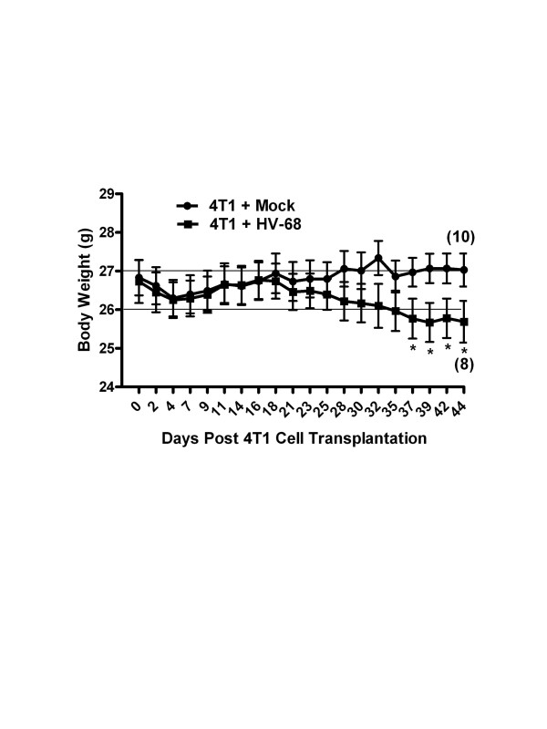
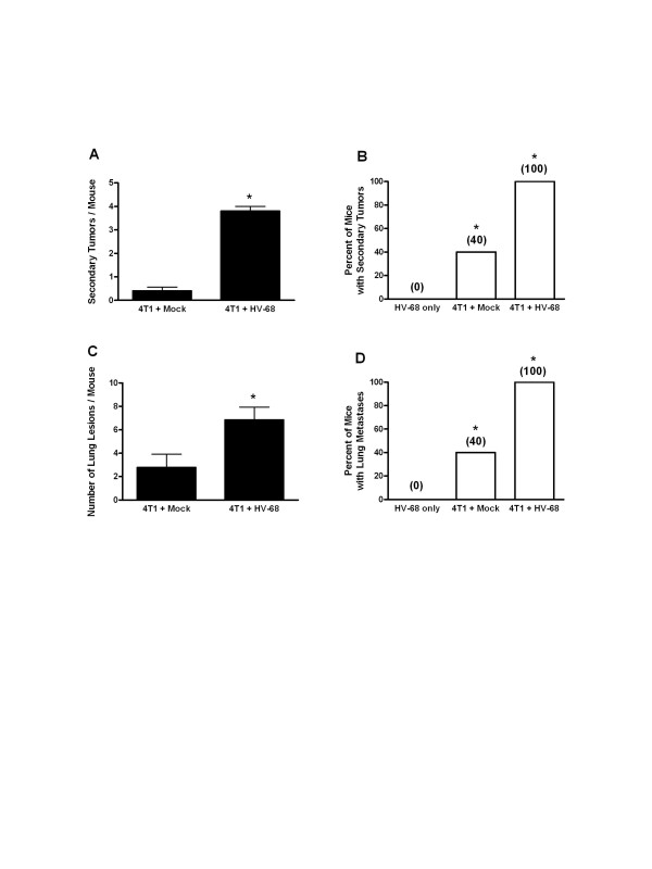
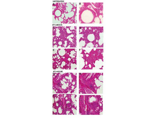
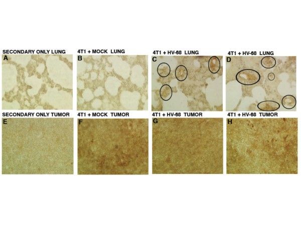
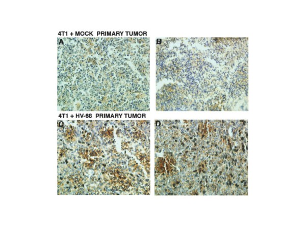
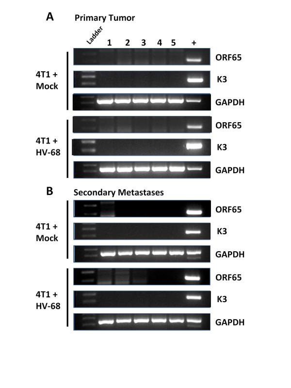
Results: Mice latently infected with HV-68 had a similar primary tumor burden, but much greater metastatic disease, when compared to mock treated mice given the transplantable tumor, 4 T1. This was true for lung lesions, as well as secondary tumor masses. Increased expression of pan-cytokeratin and VEGF-A in tumors from HV-68 infected mice was consistent with increased metastatic disease in these animals. Surprisingly, no viral particles could be cultured from tumor tissues, and the presence of viral DNA or RNA transcripts could not be detected in primary or secondary tumor tissues.

Conclusions: Latent HV-68 infection had no significant effect on the size of primary 4 T1 mammary tumors, but exacerbated the number of metastatic lung lesions and secondary tumors when compared to mock treated mice. Increased expression of the tumor marker, pan-cytokeratin, and VEGF-A in tumors of mice harboring latent virus was consistent with an exacerbated metastatic disease. Mechanisms responsible for this exacerbation are indirect, since no virus could be detected in cancerous tissues.


Figures

Figure 1
Figure 1
Morbidity of HV-68 infected mice as 4 T1 breast tumors develop. Groups of mice (N = 10) were mock treated (circles) or HV-68 infected (squares). Six months later mice were injected with syngeneic 4 T1 mammary tumor cells in the mammary fat pad. Mice were weighed and scored for morbidity following cancer cell transplantation. Results are shown as mean values (± standard deviations) with asterisks indicating a statistically significant difference between groups. Numbers in parentheses indicate the animals that remained alive when the experiment was terminated. This study was repeated twice with similar results.
Figure 2
Figure 2
Primary mammary tumor burden in mock and HV-68 infected mice. At death or at day 44 following tumor cell transplantation, groups of mice (N = 10) were euthanized and primary mammary tumor tissue excised and weighed. Panel A shows mean tumor weights (± standard deviations). Panel B shows the ratio of average body weight to average tumor weight (± standard deviations) as a measure of tumor burden.
Figure 3
Figure 3
Metastatic tumor burden in mock and HV-68 infected mice. At day 44 following tumor cell transplantation, groups of mice (N = 8–10) were euthanized. Secondary tumors, lungs, and blood were taken from each animal. Panels A and B, respectively, show the average number of secondary tumors and percentage of mice with secondary tumors for each group (± standard deviations). Panel C and D, respectively, show the average number of lung metastases and percentage of mice with lung metastases for each group (+ standard deviations). Asterisks indicate statistically significant differences between groups.
Figure 4
Figure 4
H & E staining to visualize metastatic lung lesions in untreated, mock and HV-68 infected mice. At day 44 following tumor cell transplantation, groups of mice (N = 8–10) were euthanized. Lungs were taken from each animal, fixed in formalin, and paraffin embedded for H & E staining. Left panels ( A, C, E, G, and I) show 20X magnification and right panels ( B, D, F, H, and J) show 40X magnifications of the same micrographs. Panels A and B show representative microscopic lung sections from an untreated mouse. Panels C, D, E and F show representative microscopic lung sections from two mock treated mice that were transplanted with 4 T1 tumor cells. Panels G, H, I and J show representative microscopic lung sections from two HV-68 infected mice transplanted with 4 T1 tumor cells. Circles show the location of clearly discernable metastatic lesions in HV-68 infected mice.
Figure 5
Figure 5
Pan-cytokeratin staining of lung and secondary tumor sections. At day 44 following tumor cell transplantation, groups of mice (N = 8–10) were euthanized. Lungs and primary tumor masses were excised from the animals. Tissue was fixed in formalin, paraffin embedded, and sectioned for staining with an antibody against pan-cytokeratin as a marker for 4 T1 cells. The chromogen, DAB, stains brown and was used to detect the presence of anti-pan-cytokeratin antibody binding to tumor cells. Panel B shows a representative anti-pan-cytokeratin stained microscopic lung section from a mouse transplanted with 4 T1 tumor cells. Panels C and D show representative anti-pan-cytokeratin stained microscopic lung sections from two HV-68 infected mice transplanted with 4 T1 tumor cells. Circled regions in Panels C and D indicate areas of increased staining for cytokeratins in infected mice. Pan-cytokeratin staining was also used to identify secondary metastatic tumor masses as being composed of 4 T1 cells. Tumor sections from a representative mock treated mouse (Panel F) and from two representative HV-68 infected mice (Panels G and H) showed similar staining for this tumor marker. It should be noted that due to increased mucus in lung tissues of tumor bearing mice, there was higher background staining as evident by the secondary antibody-only control (Figure 5A). This increased background staining was not observed in the control for primary tumors (Figure 5E).
Figure 6
Figure 6
VEGF-A staining of primary mammary tumor sections. At death or at day 44 following tumor cell transplantation, groups of mice (N = 10) were euthanized. Primary mammary tumors were excised from each animal, fixed in formalin, paraffin embedded, and sectioned for staining with an antibody against VEGF-A as a marker for angiogenesis. The chromogen, DAB, stains brown and was used to detect the presence of anti-VEGF-A antibody binding. Panels A and B show representative anti-VEGF-A stained microscopic mammary tumor sections from two mock treated mice transplanted with 4 T1 tumor cells. Panels C and D show representative anti-VEGF-A stained microscopic mammary tumor sections from two HV-68 infected mice transplanted with 4 T1 tumor cells.
Figure 7
Figure 7
No detectable HV-68 RNA transcripts in primary mammary tumors and secondary metastases. At death, or at day 44 following tumor cell transplantation, groups of mice (N = 10) were euthanized. Primary mammary tumors and secondary metastases were excised from each animal, and RNA extracted from each tissue. Quantitative RT-PCR was performed to detect the presence of one replicating HV-68 transcript (ORF65) and one latent HV-68 transcript (K3). Results from 5 different animals (1, 2, 3, 4, 5) are shown as amplified fragments electrophoresed on ethidium bromide stained polyacryamide gels. The correct size of each amplified fragment is indicated by comparison to DNA ladders run on the same gel (Ladder). Positive controls for the amplification of each gene were also included in each reaction (+). The presence of GAPDH RNA was used as a positive control in each RT-PCR reaction.

Similar articles

Cited by

References

    1. Cohen JI. The biology of Epstein-Barr virus: lessons learned from the virus and the host. Curr Opin Immunol. 1999;11(4):365–370. doi: 10.1016/S0952-7915(99)80062-4. - DOI - PubMed
    1. Amarante MK, Watanabe MA. The possible involvement of virus in breast cancer. J Cancer Res Clin Oncol. 2009;135(3):329–337. doi: 10.1007/s00432-008-0511-2. - DOI - PubMed
    1. Glaser SL, Hsu JL, Gulley ML. Epstein-Barr virus and breast cancer: state of the evidence for viral carcinogenesis. Cancer Epidemiol Biomarkers Prev. 2004;13(5):688–697. - PubMed
    1. Hippocrate A, Oussaief L, Joab I. Possible role of EBV in breast cancer and other unusually EBV-associated cancers. Cancer Lett. 2011;305(2):144–149. doi: 10.1016/j.canlet.2010.11.007. - DOI - PubMed
    1. Joshi D, Buehring GC. Are viruses associated with human breast cancer? Breast Cancer Res Treat: Scrutinizing the molecular evidence; 2012. - PubMed

LinkOut - more resources