. 2012 May;3(5):546-58.
doi: 10.18632/oncotarget.503.

Dramatic reduction of sequence artefacts from DNA isolated from formalin-fixed cancer biopsies by treatment with uracil- DNA glycosylase

Affiliations

Dramatic reduction of sequence artefacts from DNA isolated from formalin-fixed cancer biopsies by treatment with uracil- DNA glycosylase

Hongdo Do et al. Oncotarget. 2012 May.

Abstract

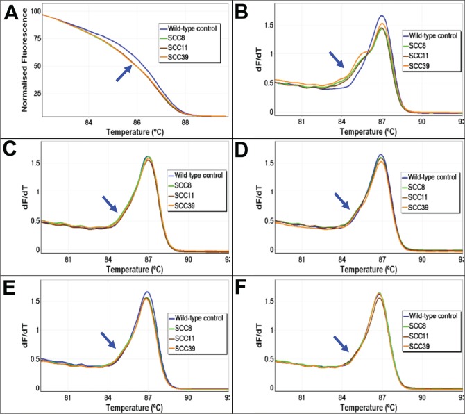
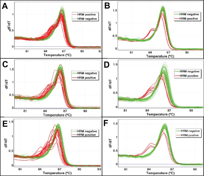
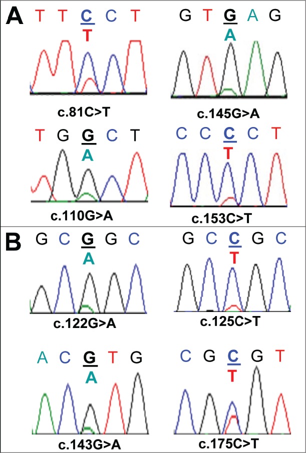
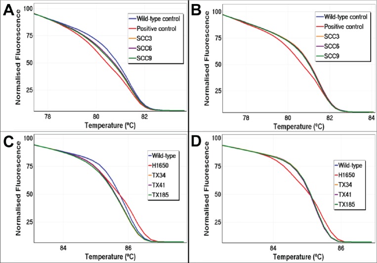
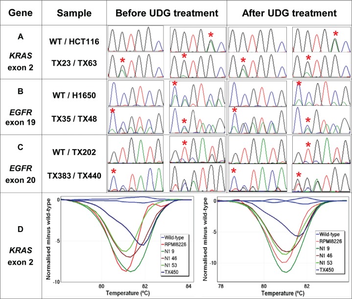
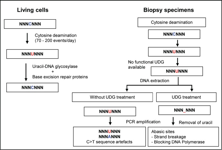
Non-reproducible sequence artefacts are frequently detected in DNA from formalinfixed and paraffin-embedded (FFPE) tissues. However, no rational strategy has been developed for reduction of sequence artefacts from FFPE DNA as the underlying causes of the artefacts are poorly understood. As cytosine deamination to uracil is a common form of DNA damage in ancient DNA, we set out to examine whether treatment of FFPE DNA with uracil-DNA glycosylase (UDG) would lead to the reduction of C>T (and G>A) sequence artefacts. Heteroduplex formation in high resolution melting (HRM)-based assays was used for the detection of sequence variants in FFPE DNA samples. A set of samples that gave false positive HRM results for screening for the E17K mutation in exon 4 of the AKT1 gene were chosen for analysis. Sequencing of these samples showed multiple non-reproducible C:G>T:A artefacts. Treatment of the FFPE DNA with UDG prior to PCR amplification led to a very marked reduction of the sequence artefacts as indicated by both HRM and sequencing analysis, indicating that uracil lesions are the major cause of sequence artefacts. Similar results were shown for the BRAF V600 region in the same sample set and EGFR exon 19 in another sample set. UDG treatment specifically suppressed the formation of artefacts in FFPE DNA as it did not affect the detection of true KRAS codon 12 and true EGFR exon 19 and 20 mutations. We conclude that uracil in FFPE DNA leads to a significant proportion of sequence artefacts. These can be minimised by a simple UDG pretreatment which can be readily carried out, in the same tube, as the PCR immediately prior to commencing thermal cycling. HRM is a convenient way of monitoring both the degree of damage and the effectiveness of the UDG treatment. These findings have immediate and important implications for cancer diagnostics where FFPE DNA is used as the primary genetic material for mutational studies guiding personalised medicine strategies and where simple effective strategies to detect mutations are required.


Figures

Figure 1
Figure 1. The melting profiles of FFPE DNA before and after UDG treatment
The melting profiles of the AKT1 HRM assay for three representative FFPE DNA samples (SCC8, SCC11, and SCC39) without (Panels A and B) and with UDG treatment using four different UDG concentrations (Panels C – F) are shown. The early melting profiles that are indicative of heteroduplex formation were seen in all three samples without UDG treatment. UDG treatment prior to PCR amplification resulted in a marked reduction of heteroduplex formation. Panel A: Normalised plot without UDG treatment. Panel B: First negative derivative plot without UDG treatment. Panels C – F: First negative derivative plots with a concentration of 0.1, 0.25, 0.5, and 1 UDG unit/reaction, respectively. The early melting region of the heteroduplexes is indicated with a blue arrow.
Figure 2
Figure 2. The effect of UDG treatment on sequence artefacts in AKT1 as assessed using LCN-HRM
The frequency of sequence artefacts in the AKT1 sequence were assessed in three FFPE DNA samples (SCC7, SCC8, and SCC14) with and without UDG treatment using LCN-HRM. The melting profiles of 60 individual LCN-HRM products are presented in the negative first derivative plot. Positive LCN-HRM reactions are shown in red and wild-type reactions are shown in green. There is a marked reduction in the number of LCN-HRM positive reactions after UDG treatment in all three samples. In SCC7, a total of 34 reactions were positive without UDG treatment (Panel A), which is markedly reduced to 5 after UDG treatment (Panel B). In SCC8, 24 and 10 LCN-HRM reactions were positive without (Panel C) and with UDG treatment (Panel D), and 20 and 3 LCN-HRM positives are found without (Panel E) and with UDG treatment (Panel F) in SCC14.
Figure 3
Figure 3. Sequence artefacts detected in FFPE DNA by Sanger sequencing
Multiple non-reproducible sequence artefacts detected in the AKT1 sequence from FFPE DNA are shown. Panel A: Four sequence artefacts detected in the SCC8 sample without UDG treatment. Three of the sequence artefacts (c.81C>T, c.145G>A and c.153C>T) were found in the same amplicon from one replicate and the c.110G>A change was detected in the second replicate. Panel B: Four sequence artefacts detected in three FFPE DNA samples (SCC7, SCC11, and SCC14) after UDG treatment. c.122G>A and c.143G>A changes were detected in different replicates from the SCC7 sample. A c.125C>T (SCC11) and a c.175C>T (SCC14) change was found in a replicate of SCC11 and SCC14 respectively. All of the C:G>T:A changes that were found after UDG treatment were detected in the sequence context of CpG dinucleotides.
Figure 4
Figure 4. UDG treatment reduces artefactual false positives by HRM
Sequence artefacts arising from uracil lesions can cause false HRM positives by formation of heteroduplexes. Treatment of FFPE DNA prior to PCR amplification removes uracil lesions, resulting in markedly reducing false HRM positives. BRAF exon 15 and EGFR exon 19 HRM results of three representative samples are shown. Panel A: Normalised plot for BRAF exon 15 without UDG treatment. Panel B: Normalised plot for BRAF exon 15 with UDG treatment. Panel C: Normalised plot for EGFR exon 19 without UDG treatment. Panel D: Normalised plot for EGFR exon 19 with UDG treatment.
Figure 5
Figure 5. Detection of true KRAS and EGFR mutations after UDG treatment
The effect of UDG treatment on detection of various types of true mutations are examined using a set of FFPE DNA samples harbouring either KRAS or EGFR exon 19 deletions and exon 20 insertion mutations. All KRAS-mutant and EGFR-mutant samples are correctly identifiable by HRM or Sanger sequencing regardless of UDG treatment. The positions of KRAS mutations and representative nucleotides of EGFR mutations are indicated by a red asterisk. Panel A: Sequence traces of KRAS exon 2 before and after UDG treatment. Both TX23 and TX63 samples harbour KRAS c.35G>A mutations and HCT116 cell line DNA contains a KRAS c.38G>A mutation. Panel B: Sequence traces of EGFR exon 19 before and after UDG treatment. Both TX35 and H1650 harbour EGFR p.E746_A750del mutations and TX48 harbours a p.T751_I759delinsN mutation. Panel C: Sequence traces of EGFR exon 20 before and after UDG treatment. TX202, TX383 and TX440 samples harbour EGFR p.C775_R776insPA, p.H773_R776insYNPY, and p.D770_H773insGSVD, respectively. Panel D: Difference plots of low-level KRAS-mutant samples before (left) and after UDG treatment (right). KRAS mutations detected are c.35G>T (N1 9), c.35G>T (N1 46), c.35G>C (N1 53), c.34G>T (TX450). RPMI8226 cell line DNA contains a KRAS c.35G>C mutation.
Figure 6
Figure 6. Uracil lesions in FFPE DNA leading to sequence artefacts and in vitro removal of uracil by uracil-DNA glycosylase
Spontaneous cytosine deamination is a frequent DNA damage that takes place at a rate of 70 - 200 events per day in the human genome. In normal cells, the resulting uracil lesions are effectively removed by UDG. The resulting abasic sites are then repaired by the base excision DNA repair system. However, in biopsy specimen, if cytosine deamination occurs during sample collection, formalin fixation, and fixed tissue storage, the resulting uracil lesions cannot be repaired due to the absence of functional DNA repair proteins. When DNA is extracted from the tissue with uracil lesions and then used as template for PCR amplification, transitional C:G>T:A sequence artefacts are generated as uracil efficiently pairs with adenine. The generation of artefactual C:G>T:A transitions from the uracil lesions in FFPE DNA can be effectively eliminated by treating FFPE DNA with UDG in vitro prior to PCR amplification. Abasic sites generated by the removal of uracil bases may reduce the extension by DNA polymerase and strand breakage during the repetitive exposure to high temperature during PCR cycling. Thus, treatment of FFPE DNA with UDG prior to PCR amplification eliminates the generation of artefactual C:G>T:A transitions arising from uracil lesions.

References

    1. de Bono JS, Ashworth A. Translating cancer research into targeted therapeutics. Nature. 2010;467(7315):543–549. - PubMed
    1. Ferte C, Andre F, Soria JC. Molecular circuits of solid tumors: prognostic and predictive tools for bedside use. Nature reviews clinical oncology. 2010;7(7):367–380. - PubMed
    1. Quach N, Goodman MF, Shibata D. In vitro mutation artifacts after formalin fixation and error prone translesion synthesis during PCR. BMC clinical pathology. 2004;4(1):1. - PMC - PubMed
    1. Do H, Dobrovic A. Limited copy number-high resolution melting (LCN-HRM) enables the detection and identification by sequencing of low level mutations in cancer biopsies. Molecular cancer. 2009;8:82. - PMC - PubMed
    1. Gallegos Ruiz MI, Floor K, Rijmen F, Grunberg K, Rodriguez JA, Giaccone G. EGFR and K-ras mutation analysis in non-small cell lung cancer: comparison of paraffin embedded versus frozen specimens. Cellular oncology. 2007;29(3):257–264. - PMC - PubMed

Publication types

MeSH terms