. 2012 Aug;20(8):1550-8.
doi: 10.1038/mt.2012.103. Epub 2012 May 29.

Targeting the hemoglobin scavenger receptor CD163 in macrophages highly increases the anti-inflammatory potency of dexamethasone

Affiliations

Targeting the hemoglobin scavenger receptor CD163 in macrophages highly increases the anti-inflammatory potency of dexamethasone

Jonas H Graversen et al. Mol Ther. 2012 Aug.

Abstract

Synthetic glucocorticoids are potent anti-inflammatory drugs but serious side effects such as bone mobilization, muscle mass loss, immunosuppression, and metabolic alterations make glucocorticoid therapy a difficult balance. The therapeutic anti-inflammatory effect of glucocorticoids relies largely on the suppressed release of tumor-necrosis factor-α and other cytokines by macrophages at the sites of inflammation. We have now developed a new biodegradable anti-CD163 antibody-drug conjugate that specifically targets the glucocorticoid, dexamethasone to the hemoglobin scavenger receptor CD163 in macrophages. The conjugate, that in average contains four dexamethasone molecules per antibody, exhibits retained high functional affinity for CD163. In vitro studies in rat macrophages and in vivo studies of Lewis rats showed a strong anti-inflammatory effect of the conjugate measured as reduced lipopolysaccharide-induced secretion of tumor-necrosis factor-α. The in vivo potency of conjugated dexamethasone was about 50-fold that of nonconjugated dexamethasone. In contrast to a strong systemic effect of nonconjugated dexamethasone, the equipotent dose of the conjugate had no such effect, measured as thymus lymphocytes apoptosis, body weight loss, and suppression of endogenous cortisol levels. In conclusion, the study shows antibody-drug conjugates as a future approach in anti-inflammatory macrophage-directed therapy. Furthermore, the data demonstrate CD163 as an excellent macrophage target for anti-inflammatory drug delivery.


Figures

Figure 1
Figure 1
Chemical structure of dexamethasone-hemisuccinate (MW 459 Da) linked to a primary amino group of the anti-CD163 antibody. The conjugate contained in average four dexamethasone molecules per IgG as determined by reverse phase HPLC determination of total and free dexamethasone and determination of protein concentration.
Figure 2
Figure 2
Binding of anti-CD163-dexamethasone to CD163-expressing CHO cells and rat macrophages. (a) Western blot analysis of the binding of anti-CD163 and anti-CD163-dexamethasone to lysate from CHO cells transfected with rat CD163 or mock-transfected CHO cells. (b) Flow cytometry of CHO cells expressing rat CD163 were incubated with anti-CD163 (solid curve) or anti-CD163-dexamethasone conjugate (dashed curve), and stained with anti-mouse IgG-FITC. The dotted curve represents negative control staining with anti-mouse IgG-FITC. (c–e) Flow cytometry (dot plot) of rat splenocytes were stained with anti-rat CD172a-PE (Ed-9), anti-rat CD4-APC, anti dexamethasone-PerCP, and anti-CD163-dexamethasone-Alexa Fluor 488 as shown in c,d or mouse isotype control IgG as shown in e. The percentage of CD4+ monocytes/macrophages are indicated in the quadrants. CHO, Chinese hamster ovary.
Figure 3
Figure 3
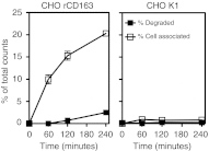
Uptake of 125I-labeled anti-CD163 in rat CD163-expressing cells. Specific uptake of 125I-anti-CD163 in CHO cells expressing rat CD163 (left) or mock-transfected CHO cells (right), showing both cell associated anti-CD163 (open squares) and rate of degraded anti-CD163 measured as free 125I (filled squares). Values are mean ± 1 SD of triplicates. CHO, Chinese hamster ovary; SD, standard deviation.
Figure 4
Figure 4
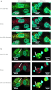
Confocal fluorescence microscopy of the endocytotic uptake of anti-CD163-dexamethasone in CD163-expressing CHO transfectants and rat macrophages. A mixture of nontransfected and CD163-transfected CHO cells (a) or rat spleen cell suspensions (b) were incubated with anti-CD163-dexamethasone for 30 minutes and 2 hours before staining with anti-mouse IgG-Alexa Fluor 488 (green), anti-dexamethasone-PerCP (red), and Hoechst 33342 (light blue) nuclei staining. Colocation of anti-CD163 and dexamethasone is indicated by yellow color in bottom panels of a and b displays, marked by white arrow. Specificity was confirmed by lack of anti-CD163-dexamethasone binding in CD163 nonexpressing cells in the single displays. CHO, Chinese hamster ovary; TNF-α, tumor-necrosis factor-α.
Figure 5
Figure 5
Anti-CD163-dexamethasone suppression of LPS-mediated TNF-α stimulation in vitro. Rat spleen cells were cultured and incubated with serial dilutions of anti-CD163-dexamethasone conjugate, mouse IgG-dexamethasone, and free dexamethasone for 15 minutes, washed and incubated overnight. Cell supernatants were analyzed for TNF-α after 4 hours of LPS stimulation. The graph shows the normalized mean values ± 1 SD from six independent studies using six different rats for splenocyte preparation (range of maximum TNF-α levels was 100–200 pg/ml). Anti-CD163-dexamethasone was consistently significantly different from free dexamethasone (P < 0.05) for all individual studies. SD, standard deviation; TNF-α, tumor-necrosis factor-α.
Figure 6
Figure 6
Increased anti-inflammatory efficacy of anti-CD163-dexamethasone. Female Lewis rats were pretreated with anti-CD163-dexamethasone, free dexamethasone, mouse IgG-dexamethasone or vehicle (n = 6 per group) 19 hours before stimulation with LPS. The concentration of (a) TNF-α and (b) IL-1 in serum samples were determined 2 hours post-LPS injection in sandwich ELISA assays. Values are mean ± 1 SD. ELISA, enzyme-linked immunosorbent assay; SD, standard deviation; TNF-α, tumor-necrosis factor-α.
Figure 7
Figure 7
Time course of injected anti-CD163-dexamethasone. Anti-CD163-dexamethasone was administrated intravenously and blood samples were collected at serial time-points post injection (n = 4). Serum concentrations of anti-CD163 (open circle) and dexamethasone (filled square) were determined in sandwich ELISA assays. Values are mean ± 1 SD. ELISA, enzyme-linked immunosorbent assay; SD, standard deviation.
Figure 8
Figure 8
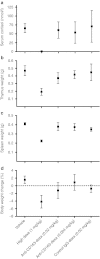
Systemic nonmacrophage effect of anti-CD163-dexamethasone in rats. Systemic effect was measured as suppression of (a) serum cortisol concentration and reduction organ ((b) thymus and (c) spleen) and (d) body weight. The cortisol concentration and the weight of the thymus, spleen, and whole body were measured upon killing the rats 2 days after injection of anti-CD163-dexamethasone, mouse control IgG-dexamethasone, free dexamethasone or vehicle at day 0 and 1 (n = 4 per group). Values are mean ± 1 SD. SD, standard deviation.

References

    1. Rhen T., and, Cidlowski JA. Antiinflammatory action of glucocorticoids–new mechanisms for old drugs. N Engl J Med. 2005;353:1711–1723. - PubMed
    1. McColl A, Michlewska S, Dransfield I., and, Rossi AG. Effects of glucocorticoids on apoptosis and clearance of apoptotic cells. ScientificWorldJournal. 2007;7:1165–1181. - PMC - PubMed
    1. Clark IA. How TNF was recognized as a key mechanism of disease. Cytokine Growth Factor Rev. 2007;18:335–343. - PubMed
    1. Saklatvala J, Dean J., and, Clark A. Control of the expression of inflammatory response genes. Biochem Soc Symp. 2003;70:95–106. - PubMed
    1. Dayer JM. The process of identifying and understanding cytokines: from basic studies to treating rheumatic diseases. Best Pract Res Clin Rheumatol. 2004;18:31–45. - PubMed

Publication types

MeSH terms