Comparative Study
. 2013 Aug;35(4):1143-56.
doi: 10.1007/s11357-012-9439-1. Epub 2012 May 28.

Caloric restriction-associated remodeling of rat white adipose tissue: effects on the growth hormone/insulin-like growth factor-1 axis, sterol regulatory element binding protein-1, and macrophage infiltration

Affiliations
Comparative Study

Caloric restriction-associated remodeling of rat white adipose tissue: effects on the growth hormone/insulin-like growth factor-1 axis, sterol regulatory element binding protein-1, and macrophage infiltration

Yoshikazu Chujo et al. Age (Dordr). 2013 Aug.

Abstract

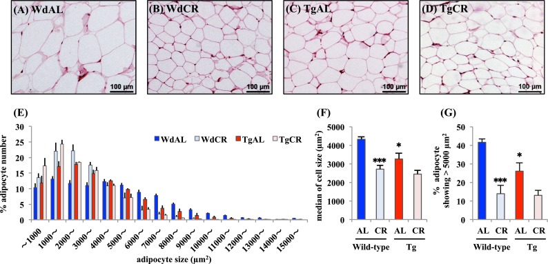
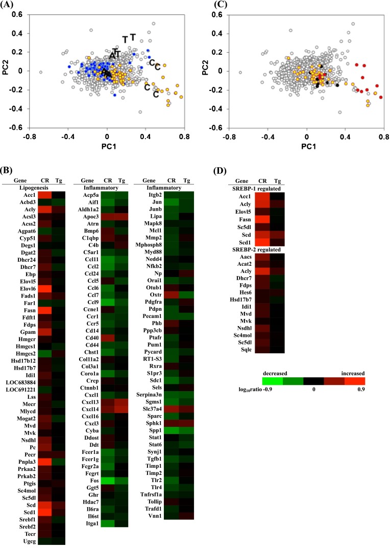
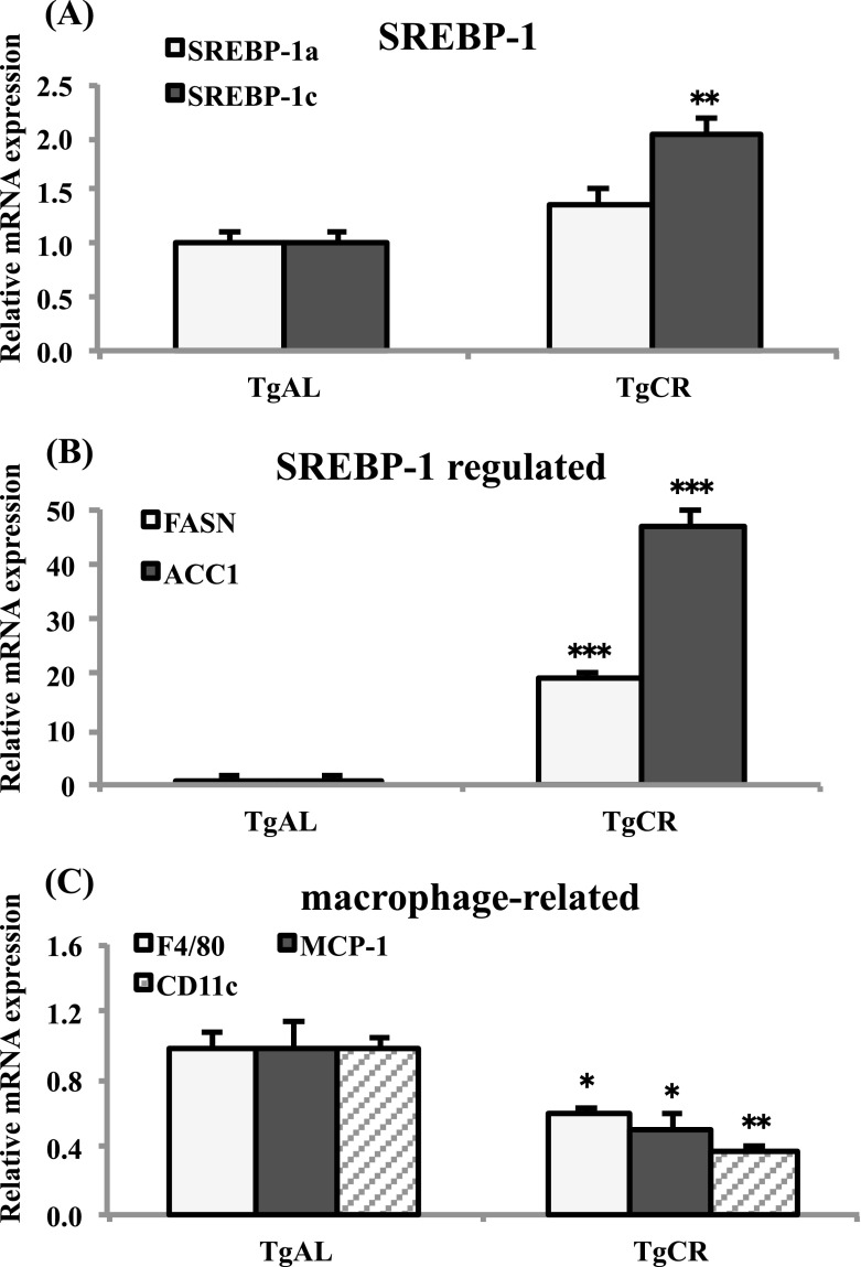
The role of the growth hormone (GH)-insulin-like growth factor (IGF)-1 axis in the lifelong caloric restriction (CR)-associated remodeling of white adipose tissue (WAT), adipocyte size, and gene expression profiles was explored in this study. We analyzed the WAT morphology of 6-7-month-old wild-type Wistar rats fed ad libitum (WdAL) or subjected to CR (WdCR), and of heterozygous transgenic dwarf rats bearing an anti-sense GH transgene fed ad libitum (TgAL) or subjected to CR (TgCR). Although less effective in TgAL, the adipocyte size was significantly reduced in WdCR compared with WdAL. This CR effect was blunted in Tg rats. We also used high-density oligonucleotide microarrays to examine the gene expression profile of WAT of WdAL, WdCR, and TgAL rats. The gene expression profile of WdCR, but not TgAL, differed greatly from that of WdAL. The gene clusters with the largest changes induced by CR but not by Tg were genes involved in lipid biosynthesis and inflammation, particularly sterol regulatory element binding proteins (SREBPs)-regulated and macrophage-related genes, respectively. Real-time reverse-transcription polymerase chain reaction analysis confirmed that the expression of SREBP-1 and its downstream targets was upregulated, whereas the macrophage-related genes were downregulated in WdCR, but not in TgAL. In addition, CR affected the gene expression profile of Tg rats similarly to wild-type rats. Our findings suggest that CR-associated remodeling of WAT, which involves SREBP-1-mediated transcriptional activation and suppression of macrophage infiltration, is regulated in a GH-IGF-1-independent manner.


Figures

Fig. 1
Fig. 1
Effects of CR and Tg on morphologic features of white adipose tissue. Representative hematoxylin/eosin-stained histological sections of WAT from WdAL (a), WdCR (b), TgAL (c), and TgCR (d) rats (magnification ×40; scale bar 100 μm). e Distribution of adipocyte sizes in wild-type and tg rats. f Median of adipocyte sizes in wild-type and Tg rats. g The percentage of adipocytes showing >5,000 μm2 in wild-type and Tg rats. Adipocyte size was calculated based on a quantitative morphometric method using ImageJ 1.43u/Java1.6.0_22 software. Error bars are SEM for each group (n = 4). Approximately 1,000–2,100 adipocytes were counted per rat. *P < 0.05 and ***P < 0.001 vs. WdAL (Tukey’s t test)
Fig. 2
Fig. 2
Principal component analysis of gene expression. a PC ordination of 6,641 ANOVA-positive genes based on the DNA microarray data of WdAL, WdCR, and TgAL (four biological repeats per group). A, C, and T represent WdAL, WdCR, and TgAL rats, respectively. The yellow and blue dots represent genes involved in lipid biosynthesis and inflammation, respectively. Gene function was defined based on annotations in the Gene Ontology (GO) Biological Process database (release 31) provided by the manufacturer. Genes involved in lipid biosynthesis were derived from the gene lists (GO: 0008610, 0006633, and 0019432). Genes involved in inflammation were derived from the gene lists (GO: 0006955, 0006954, 0034097, and 0030593). b Heat map of genes involved in lipid biosynthesis and inflammation. Genes involved in lipid biosynthesis were mostly upregulated by CR, while those involved in inflammation were predominantly downregulated by CR. The expression of these genes was not influenced by Tg. c Scatter plots of SREBP-1- (red) and SREBP-2- (black) regulated genes listed by Horton et al. (2003). All of the genes were included in the genes listed in GO 0008610, 0006633, and 0019432 (yellow). d Heat map of SREBP-1- and SREBP-2-regulated genes identified by Horton et al. (2003). Most of the SREBP-1-regulated genes were upregulated by CR, while most of the SREBP-2-regulated genes were not affected by CR
Fig. 3
Fig. 3
Quantitative analysis of the mRNA expression of SREBPs, SREBP-1-regulated genes, SREBP-2-regulated genes, and macrophage-related genes in WdAL, WdCR, and TgAL rats. The mRNA expression levels in WAT of SREBPs (A: SREBP-1a, SREBP-1c, and SREBP-2), SREBP-1-regulated genes [B: fatty acid synthase (FASN) and acyl CoA carboxylase 1 (ACC1)], SREBP-2-regulated genes [C: squalene epoxidase (Sqle) and mevalonate kinase (Mvk)], and macrophage-related genes [D monocyte chemotactic protein-1 (MCP-1), F4/80, integrin alpha X (Itgax, CD11c), and CD163] were analyzed by real-time RT-PCR. Data were normalized against TBP expression. Values are means ± SEM (n = 3–6). *P < 0.05, **P < 0.01, and ***P < 0.001 vs. WdAL; #P < 0.05, ##P < 0.01, and ###P < 0.001 vs. WdCR (Tukey’s t test)
Fig. 4
Fig. 4
Quantitative analysis of the mRNA expression of SREBPs, SREBP-1-regulated genes, and macrophage-related genes in TgAL and TgCR rats. The mRNA expression levels in WAT of SREBPs (aSREBP-1a and SREBP-1c), SREBP-1-regulated genes [bfatty acid synthase (FASN) and acyl CoA carboxylase 1 (ACC1)], and macrophage-related genes [cF4/80, monocyte chemotactic protein-1 (MCP-1), integrin alpha X (Itgax, CD11c)] were analyzed by real-time RT-PCR. Data were normalized against TBP expression. Values are means ± SEM (n = 4–5). *P < 0.05, **P < 0.01, and ***P < 0.001 vs. TgAL (Tukey’s t test)

Similar articles

Cited by

References

    1. Al-Regaiey KA, Masternak MM, Bonkowski M, Sun L, Bartke A. Long-lived growth hormone receptor knockout mice: interaction of reduced insulin-like growth factor I/insulin signaling and caloric restriction. Endocrinology. 2005;146:851–860. doi: 10.1210/en.2004-1120. - DOI - PubMed
    1. Anderson R, Prolla T (2009) PGC-1alpha in aging and anti-aging interventions. Biochem Biophys Acta 1790:1059–1066 - PMC - PubMed
    1. Argmann C, Dobrin R, Heikkinen S, Auburtin A, Pouilly L, Cock TA, Koutnikova H, Zhu J, Schadt EE, Auwerx J. Ppargamma2 is a key driver of longevity in the mouse. PLoS Genet. 2009;5:e1000752. doi: 10.1371/journal.pgen.1000752. - DOI - PMC - PubMed
    1. Bartke A. Minireview: role of the growth hormone/insulin-like growth factor system in mammalian aging. Endocrinology. 2005;146:3718–3723. doi: 10.1210/en.2005-0411. - DOI - PubMed
    1. Bartke A, Wright JC, Mattison JA, Ingram DK, Miller RA, Roth GS. Extending the lifespan of long-lived mice. Nature. 2001;414:412. doi: 10.1038/35106646. - DOI - PubMed

Publication types

MeSH terms