

Review
doi: 10.1007/s11427-012-4318-7.
Epub 2012 May 27.
The role of Eph receptors in lens function and disease
Affiliations
- PMID: 22645087
- PMCID: PMC4026180
- DOI: 10.1007/s11427-012-4318-7
Item in Clipboard
Review
The role of Eph receptors in lens function and disease
Sci China Life Sci.
2012 May.
Abstract
Cataract is the single largest contributor to blindness in the world, with the disease having a strong genetic component. In recent years the Eph family of receptor tyrosine kinases has been identified as a key regulator in lens clarity. In this review we discuss the roles of the Eph receptors in lens biology and cataract development.
Figures
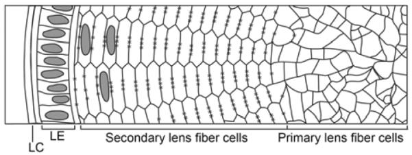
A cross-sectional view of the mature lens. The core of the lens consists of irregularly-shaped primary lens fiber cells formed during the early stages of lens development. After the initial development of primary fiber cells, secondary lens fiber cells form continuously from this core and are arranged in regular hexagonal structures. The anterior face of the lens contains a monolayer of lens epithelial cells (LE) that continually divide and differentiate into secondary lens fiber cells. The entire structure is encapsulated by a lens capsule (LC). * denotes gap junction complexes along the long edges of secondary fiber cells.
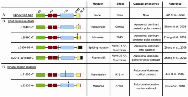
Human EphA2 cataract mutations. A, Structure of wild-type EphA2. BD, ephrin ligand binding domain; FN, fibronectin repeats; Kin, kinase domain; SAM, sterile alpha motif; PDZ, PDZ domain. B, EphA2 SAM Domain cataract mutants. C, EphA2 kinase domain cataract mutants. Arrows denote relative location of point mutations in indicated mutations. Black boxes denote novel amino acid changes.
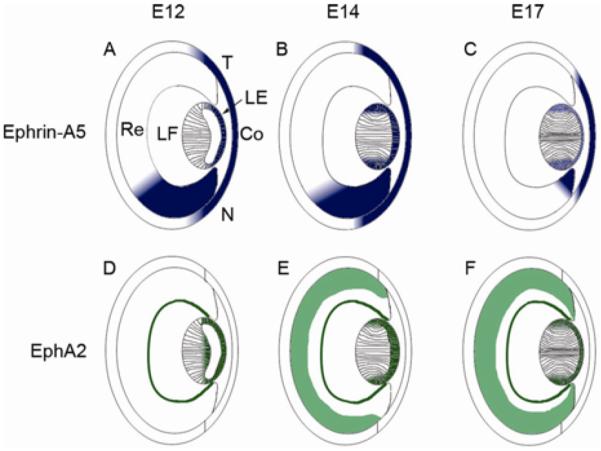
Expression of ephrin-A5 and EphA2 during ocular development. A–C, Ephrin-A5 expression at E12 (A), E14 (B), and E17 (C) is observed in the lens epithelium, lens fiber layer around the bow region, cornea, and the retina. Expression within the retinal region is graded with higher expression towards the nasal retina. Ephrin-A5 levels within the developing eye are reduced at later stages. D–F, EphA2 expression at E12 (D), E14 (E), and E17 (F) is observed in the lens epithelium, fiber cell layers, and parts of the retina during ocular development (data unpublished). Within the lens, EphA2 is observed in several of the same regions as ephrin-A5 has been found to be expressed (data unpublished). LE, lens epithelium; LF, lens fiber cells layer; Re, retina; Co, cornea; N, nasal retina; T, temporal retina.
Similar articles
-
Eph receptor tyrosine kinases in angiogenesis: from development to disease.Angiogenesis. 2004;7(1):17-28. doi: 10.1023/B:AGEN.0000037340.33788.87. Angiogenesis. 2004. PMID: 15302992 Review.
-
The role of Eph receptors and ephrin ligands in colorectal cancer.Int J Cancer. 2010 May 1;126(9):2003-11. doi: 10.1002/ijc.25147. Int J Cancer. 2010. PMID: 20039322 Review.
-
Eph, a protein family coming of age: more confusion, insight, or complexity?Sci Signal. 2008 Apr 15;1(15):re2. doi: 10.1126/stke.115re2. Sci Signal. 2008. PMID: 18413883 Review.
-
The role of Eph/ephrin molecules in stromal–hematopoietic interactions.Int J Hematol. 2016 Feb;103(2):145-54. doi: 10.1007/s12185-015-1886-x. Int J Hematol. 2016. PMID: 26475284 Review.
-
Eph receptor deficiencies lead to altered cochlear function.Hear Res. 2003 Apr;178(1-2):118-30. doi: 10.1016/s0378-5955(03)00068-6. Hear Res. 2003. PMID: 12684184
Cited by
-
BCR Signaling Inhibitors: an Overview of Toxicities Associated with Ibrutinib and Idelalisib in Patients with Chronic Lymphocytic Leukemia.Mediterr J Hematol Infect Dis. 2016 Feb 10;8(1):e2016011. doi: 10.4084/MJHID.2016.011. eCollection 2016. Mediterr J Hematol Infect Dis. 2016. PMID: 26977270 Free PMC article. Review.
-
N-cadherin regulates signaling mechanisms required for lens fiber cell elongation and lens morphogenesis.Dev Biol. 2017 Aug 1;428(1):118-134. doi: 10.1016/j.ydbio.2017.05.022. Epub 2017 May 26. Dev Biol. 2017. PMID: 28552735 Free PMC article.
-
Further analysis of the lens of ephrin-A5-/- mice: development of postnatal defects.Mol Vis. 2013;19:254-66. Epub 2013 Feb 3. Mol Vis. 2013. PMID: 23401654 Free PMC article.
-
Association of the ephreceptor tyrosinekinase-type A2 (EPHA2) gene polymorphism rs3754334 with age-related cataract risk: a meta-analysis.PLoS One. 2013 Aug 16;8(8):e71003. doi: 10.1371/journal.pone.0071003. eCollection 2013. PLoS One. 2013. PMID: 23976972 Free PMC article.
-
Breakdown of interlocking domains may contribute to formation of membranous globules and lens opacity in ephrin-A5(-/-) mice.Exp Eye Res. 2016 Apr;145:130-139. doi: 10.1016/j.exer.2015.11.017. Epub 2015 Nov 28. Exp Eye Res. 2016. PMID: 26643403 Free PMC article.
References
Publication types
MeSH terms
Substances
Grants and funding
LinkOut - more resources
Full Text Sources
Medical
Miscellaneous
