. 2012 Aug 17;287(34):28675-85.
doi: 10.1074/jbc.M112.378810. Epub 2012 May 29.

Mechanistic insights into regulated cargo binding by ACAP1 protein

Affiliations

Mechanistic insights into regulated cargo binding by ACAP1 protein

Ming Bai et al. J Biol Chem. .

Abstract

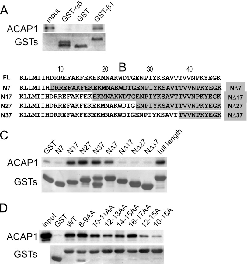
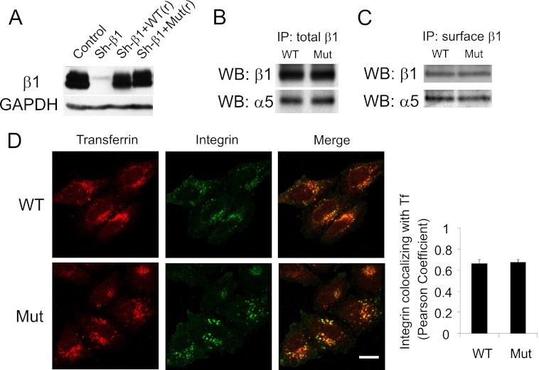
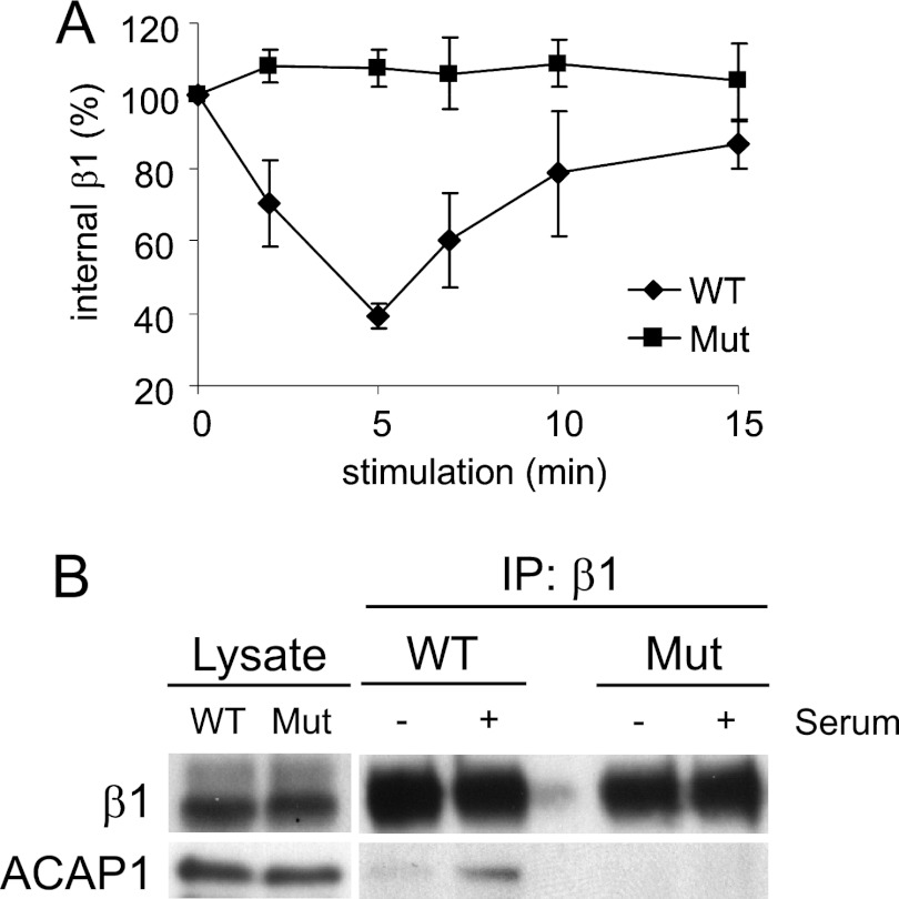
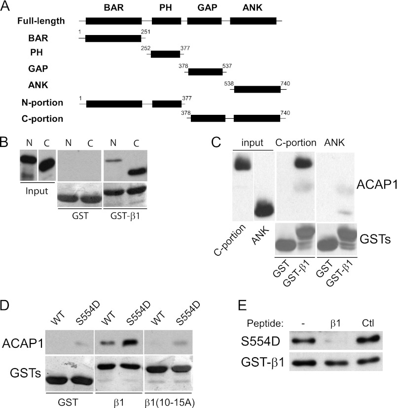
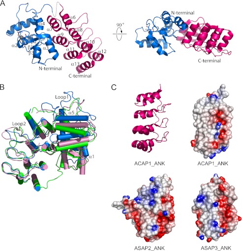
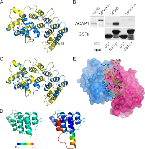
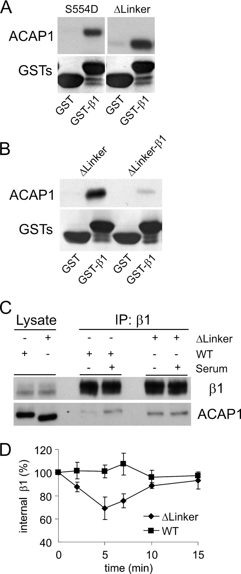
Coat complexes sort protein cargoes into vesicular transport pathways. An emerging class of coat components has been the GTPase-activating proteins (GAPs) that act on the ADP-ribosylation factor (ARF) family of small GTPases. ACAP1 (ArfGAP with coiled-coil, ankyrin repeat, and PH domains protein 1) is an ARF6 GAP that also acts as a key component of a recently defined clathrin complex for endocytic recycling. Phosphorylation by Akt has been shown to enhance cargo binding by ACAP1 in explaining how integrin recycling is an example of regulated transport. We now shed further mechanistic insights into how this regulation is achieved at the level of cargo binding by ACAP1. We initially defined a critical sequence in the cytoplasmic domain of integrin β1 recognized by ACAP1 and showed that this sequence acts as a recycling sorting signal. We then pursued a combination of structural, modeling, and functional studies, which suggest that phosphorylation of ACAP1 relieves a localized mechanism of autoinhibition in regulating cargo binding. Thus, we have elucidated a key regulatory juncture that controls integrin recycling and also advanced the understanding of how regulated cargo binding can lead to regulated transport.


Figures

FIGURE 1.
FIGURE 1.
Identifying a sequence in integrin β1 critical for its direct binding to ACAP1. A, ACAP1 binds directly to the cytoplasmic domain of integrin β1. The cytoplasmic domain of either α5 or β1 was fused to GST, and the resulting fusion proteins were bound to beads for incubation with soluble ACAP1 in pulldown experiments. B, truncation mutants of the cytoplasmic domain of integrin β1. Residues are numbered from the membrane-proximal end. FL, full-length ACAP1. C, identifying a region in the cytoplasmic domain of β1 responsible for its direct binding to ACAP1. Different truncations of β1 as GST fusion proteins were bound to beads for incubation with full-length ACAP1 as soluble recombinant protein in pulldown experiments. Beads were immunoblotted for ACAP1 and Coomassie Blue-stained for GST fusion proteins. D, alanine-scanning mutagenesis identifies specific residues within the cytoplasmic domain of β1 responsible for its direct binding to ACAP1. Residues within the cytoplasmic domain of β1 were mutated to alanines as indicated. The mutants as GST fusion proteins were then bound to beads for incubation with soluble ACAP1 in pulldown experiments. Beads were immunoblotted for ACAP1 and Coomassie Blue-stained for GST.
FIGURE 2.
FIGURE 2.
Replacing endogenous β1 with a β1 mutant that cannot bind efficiently to ACAP1. A, cell lines stably expressing different forms of integrin β1. The lentiviral system was used to replace endogenous β1 with transfected forms, which involved stably expressing shRNA against integrin β1 (Sh-β1), followed by stable expression of wild-type or mutant β1, which was resistant to shRNA through the introduced silent mutations. Expression was assessed by immunoblotting of whole cell lysates, with the level of GAPDH serving as a loading control. B, mutant β1 (Mut) assembles with α5 similarly as wild-type β1. Stable cell lines as described above were lysed, followed by immunoprecipitation (IP) for β1 and then immunoblotting for proteins as indicated. WB, Western blot. C, integrin heterodimers with mutant β1 are expressed at similar levels on the cell surface as those with wild-type β1. Stable cell lines as described above were bound with anti-β1 antibody at the cell surface. This pool of surface β1 was then immunoprecipitated, followed by immunoblotting for proteins as indicated. D, the mutant and wild-type integrins accumulate similarly at the recycling endosome under basal conditions. After allowing the surface pools of integrin and transferrin (Tf) to internalize for 2 h under basal conditions, cells were assessed by confocal microscopy comparing the distribution of integrin (green) and transferrin (red). Scale bar = 15 μm. The graph shows quantitative colocalization analysis, which reveals that wild-type and mutant integrins accumulate to a similar degree with internalized transferrin. The mean ± S.E. from three experiments is shown.
FIGURE 3.
FIGURE 3.
Mutant β1 that cannot bind efficiently to ACAP1 also cannot recycle efficiently. A, integrin β1 with a mutation (Mut) of residues critical for its direct binding to ACAP1 shows a reduced ability to recycle. A lentiviral system was used to deplete endogenous β1, followed by stable expression of transfected forms as indicated. Surface β1 integrins (tracked by antibody binding) were then allowed to accumulate at the recycling endosome under basal conditions, followed by stimulation at the times indicated for their recycling. The mean ± S. E. from three experiments is shown. B, stimulation-dependent association of endosomal β1 with ACAP1 is reduced by mutations in β1 that reduce its binding to ACAP1. Endosomal β1, tracked as described above, was assessed for association with ACAP1 by co-precipitation. IP, immunoprecipitation.
FIGURE 4.
FIGURE 4.
C-terminal portion of ACAP1 reproduces regulated cargo binding. A, schematic showing different domain constructs of ACAP1 generated as recombinant proteins. B, binding to the cytoplasmic domain of integrin β1 by different portions of ACAP1. The cytoplasmic domain of β1 as GST fusions bound to beads was incubated with the N- or C-terminal portion of ACAP1 in pulldown experiments. C, the ANK domain has reduced ability in binding to the β1 cargo. Recombinant forms of ACAP1 as indicated were incubated with GST-β1 on beads in pulldown experiments. D, regulation of cargo binding by Ser-554 is reproduced by the C-terminal portion. Different GST fusions on beads as indicated were incubated with the C-terminal portion of ACAP1 (WT or mutant S554D) in pulldown experiments. WB, Western blot. E, the recycling sorting signal in β1 as a free peptide competes for binding of the mutant (S554D) C-terminal portion to GST-β1 on beads. The pulldown experiment was performed. A free peptide containing an irrelevant sequence of similar length (derived from the cytoplasmic domain of Wbp1) was used as control (Ctl).
FIGURE 5.
FIGURE 5.
Structure of the C-terminal portion of ACAP1. A, the structure of the C-terminal portion of ACAP1 shown at two different orientations. The GAP domain is colored blue. The ANK domain is colored red. The zinc ion is shown as a gray sphere. The secondary structural elements (α-helices and β-sheets) are also labeled. B, comparing the GAP domain of ACAP1 with the corresponding domains in ASAP2 and ASAP3. The three structures are shown in schematic representation and superimposed based on their ANK domains. ACAP1 is colored blue, ASAP2 is colored green, and ASAP3 is colored pink. C, comparison of the predicted electrostatic surfaces of the different ANK domains. The ribbon representation of the ANK domain of ACAP1 is shown for orientation guidance. The electrostatic surfaces of the different ANK domains are shown, with negatively charged regions colored red and positively charged regions colored blue.
FIGURE 6.
FIGURE 6.
Linker region between the GAP and ANK domains predicted to mediate regulation of cargo binding by Ser-554. A, structure of the S554D-β1 fusion construct and comparison with the wild-type C-terminal portion. The wild-type form is colored blue, and the fusion construct is colored yellow. B, fusion of the β1 cargo peptide to the S554D form of the C-terminal portion prevents the resulting fusion construct from binding intermolecularly to the same cargo peptide on beads in a pulldown assay. The fusion construct was compared with the non-fusion counterpart in binding to GST-β1 in a pulldown experiment. Input shows proteins stained with Coomassie Blue, whereas pulldown results were immunoblotted for the proteins indicated. C, structure of the C-terminal portion with the S554D mutation and comparison with the S554D-β1 fusion construct. The S554D mutant is colored blue, and the fusion construct is colored yellow. D, temperature factor distribution of C-terminal helices in the ANK domain. The temperature factor (B factor) distribution was compared between S554D (left) and the fusion construct (right). The average B factor value was defined to 1.0, and the relative B factor value was derived from the ratio of every B factor value of Cα to the average value. The color bar shows the different values along a gradient, from 0.5 Å2 (dark blue) to 2.3 Å2 (dark red). A lower B factor predicts more stability. E, molecular modeling of the linker region. The GAP domain is colored blue, and the ANK domain is colored red. The linker region is modeled onto the surface representation of these domains, with the wild-type form shown in green, S554D shown in yellow, and S554A shown in blue. The regions of the ANK domain predicted to be covered by the linker are also labeled.
FIGURE 7.
FIGURE 7.
Linker region inhibits cargo binding. A, excision of the linker region results in enhanced cargo binding by ACAP1 in vitro. The different forms of ACAP1 were incubated with GST-β1 in a pulldown experiment. B, fusion of the β1 peptide to the linker mutant prevents the resulting fusion construct from binding intermolecularly to the same cargo peptide on beads in a pulldown assay. The fusion construct was compared with the non-fusion counterpart in binding to GST-β1 in a pulldown experiment. The input shows proteins stained with Coomassie Blue, whereas pulldown results were immunoblotted for the proteins indicated. C, excision of the linker regions results in enhanced cargo binding by ACAP1 in vivo. ACAP1 (either wild-type or with the linker region excised) was expressed in HeLa cells. The association of endosomal β1 with either form of ACAP1 was then assessed through co-precipitation (IP). D, excision of the linker region induces β1 recycling under basal (no stimulation) conditions. The integrin recycling assay was performed under basal conditions on HeLa cells that stably expressed either wild-type or mutant (with the linker region deleted) ACAP1. The mean ± S.E. from three experiments is shown.

References

    1. Bonifacino J. S., Glick B. S. (2004) The mechanisms of vesicle budding and fusion. Cell 116, 153–166 - PubMed
    1. Pucadyil T. J., Schmid S. L. (2009) Conserved functions of membrane active GTPases in coated vesicle formation. Science 325, 1217–1220 - PMC - PubMed
    1. Caswell P., Norman J. (2008) Endocytic transport of integrins during cell migration and invasion. Trends Cell Biol. 18, 257–263 - PubMed
    1. Pellinen T., Ivaska J. (2006) Integrin traffic. J. Cell Sci. 119, 3723–3731 - PubMed
    1. Margadant C., Monsuur H. N., Norman J. C., Sonnenberg A. (2011) Mechanisms of integrin activation and trafficking. Curr. Opin. Cell Biol. 23, 607–614 - PubMed

Publication types

MeSH terms