. 2012 Aug;80(8):2761-70.
doi: 10.1128/IAI.00385-12. Epub 2012 May 29.

Localization of Aggregatibacter actinomycetemcomitans cytolethal distending toxin subunits during intoxication of live cells

Affiliations

Localization of Aggregatibacter actinomycetemcomitans cytolethal distending toxin subunits during intoxication of live cells

Monika Damek-Poprawa et al. Infect Immun. 2012 Aug.

Abstract

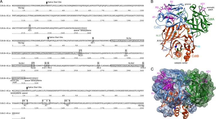
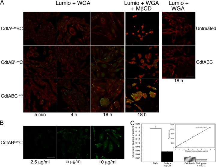
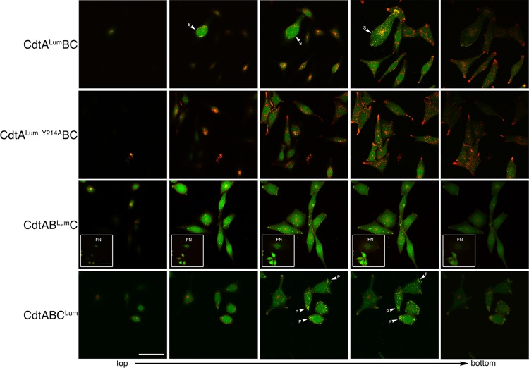
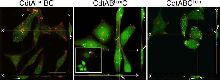
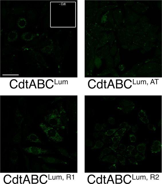
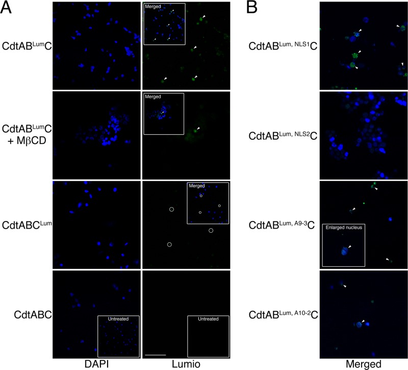
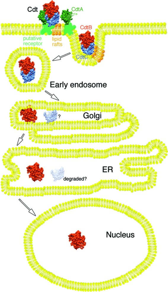
The cytolethal distending toxin (Cdt), produced by some clinically important Gram-negative bacterial species, is related to the family of AB-type toxins. Three heterologous proteins (CdtA, CdtB, and CdtC) and a genotoxin mode of action distinguish the Cdt from others in this toxin class. Crystal structures of several species-specific Cdts have provided a basis for predicting subunit interactions and functions. In addition, empirical studies have yielded significant insights into the in vivo interactions of the Cdt subunits. However, there are still critical gaps in information about the intoxication process. In this study, a novel protein tagging technology was used to localize the subunits in Chinese hamster ovary cells (CHO-K1). A tetracysteine motif was engineered in each subunit, and in subunits with mutations in predicted functional domains, to permit detection with the fluorescein arsenical hairpin binding (FlAsH) dye Lumio green. Live-cell imaging, in conjunction with confocal microscopy, was used to capture the locations of the individual subunits in cells intoxicated, under various conditions, with hybrid heterotrimers. Using this approach, we observed the following. (i) The CdtA subunit remains on the cell surface of CHO cells in association with cholesterol-containing and cholesterol-depleted membrane. (ii) The CdtB subunit is exclusively in the cytosol and, after longer exposure times, localizes to the nucleus. (iii) The CdtC subunit is present on the cell surface and, to a greater extent, in the cytosol. These observations suggest that CdtC, but not CdtA, functions as a chaperone for CdtB entry into cells.


Figures

Fig 1
Fig 1
Summary of genetic modifications. (A) Positions of amino acid additions and substitutions (boxed residues) in recombinant A. actinomycetemcomitans CdtA-His6, CdtB-His6, and CdtC-His6. The locations of the histidine and tetracysteine tags and active site in CdtB are underlined. The spacer sequence to improve Lumio green labeling is in italics. Predicted NLSs (25, 30) are in labeled boxes. The location where the wild-type amino acid sequence begins is marked with an arrow. (B) Ribbon backbone structure of the A. actinomycetemcomitans Cdt showing the locations of the mutated sites designated in panel A and in Table 1. The structure was modeled in Chimera 1.6 using Protein Data Bank file accession number 2F2F (37). Side chains are shown only for the amino acids targeted for mutation in sites AD1 in CdtA and AT, R1, and R2 in CdtC. Amino acids designated for mutation in sites A9, A10, and CH-1 in CdtB cluster around a catalytic center marked by a thin orange polygon. Amino acids targeted for mutation in the two NLS sites align along the back side of the structure as designated by the thin green line. (C) The same structure as shown in panel B except that CdtA and CdtC are depicted as a surface model and the heterotrimer is rotated approximately 90° vertically. Only the surface-exposed sites AD1, R1, and R2 are labeled.
Fig 2
Fig 2
Localization of the Cdt subunits in intoxicated cells. (A) Cultures of CHO-K1 were treated with 10 μg/ml (120 nM) of heterotrimer reconstituted with each of the subunits, containing tetracysteine and spacer sequences. Cells were labeled with Lumio green (green fluorescence) and WGA-Alexa Fluor 555 (red fluorescence) at 5 min, 4 h, and 18 h postintoxication. Cells were incubated with either no toxin (panel labeled Untreated) or wild-type heterotrimer (panel labeled CdtABC) as controls. In a subset of experiments, CHO-K1 cells were treated with MβCD, immediately prior to intoxication for 18 h, as described in Materials and Methods. Cells were colabeled as described above. The merged images are shown for all experiments. Scale bar = 50 μm. (B) To examine dose response, CHO-K1 cultures were exposed to 2.5, 5, and 10 μg/ml of heterotrimer reconstituted with CdtABLumC for 18 h. The cells were then labeled with Lumio green. Scale bar = 50 μm. (C) Quantification of cholesterol in isolated membrane rafts and total cell lysate before and after treatment with MβCD for 18 h. The results were expressed as μg of cholesterol per 1 μg of total protein in the sample. Statistically significant differences are marked by asterisks (*, P = 0.001; **, P = 0.0002). The cholesterol standard curve is shown in the inset.
Fig 3
Fig 3
Live-cell imaging of CHO-K1 cells exposed to 10 μg/ml of the heterotrimer CdtALumBC, CdtABLumC, or CdtABCLum. Successive Z sections from representative fields were taken from top to bottom. Cells were colabeled with Lumio green (green fluorescence) and WGA-Alexa Fluor 555 (red fluorescence) at 18 h postintoxication. Cells treated with a heterotrimer containing a binding-deficient CdtA subunit (CdtALum, Y214ABC) were used as a control. The insets in panel CdtABLumC show a representative field of cells containing fragmented nuclei (FN). S, cell surface; P, polar end. Scale bar = 50 μm.
Fig 4
Fig 4
Localization of the Cdt subunits in X-Y projection views of live CHO-K1 cells exposed to 10 μg/ml of the heterotrimer CdtALumBC, CdtABLumC, or CdtABCLum. XY projections were reconstructed from assembled single z sections from the live-cell experiment shown in Fig. 3. The inset in panel CdtABLumC shows a representative field of cells containing fragmented nuclei (FN). S, cell surface; C, cytosol. Scale bar = 50 μm.
Fig 5
Fig 5
Localization of mutated CdtC subunits in live cells. Cells were treated with 10 μg/ml of toxin reconstituted with either CdtCLum, AT and CdtCLum, R1 or CdtCLum, R2. Cells were labeled with Lumio green 18 h postintoxication. Lumio green-labeled cells not exposed to toxin are shown in the inset. Scale bar = 50 μm.
Fig 6
Fig 6
Nuclear localization of the Cdt subunits in live CHO-K1 cells treated with 10 μg/ml of CdtABLumC and CdtABCLum. (A) Nuclei were isolated from cells exposed to the hybrid toxins for 48 h as described in Materials and Methods. The nuclei were costained with DAPI and Lumio green. In some experiments, cells were treated with MβCD immediately prior to intoxication. The insets show merged DAPI and Lumio green images. The arrowheads mark nuclei positively labeled with Lumio green (green fluorescence) and their corresponding positions in the merged images. The circles show background staining (most likely membrane fragments that do not colocalize with DAPI-stained nuclei). Scale bar = 50 μm. (B) Cells were treated with Cdt containing the various mutated CdtB and CdtC subunits. Nuclei were isolated and labeled as in panel A. Only the merged images are shown.
Fig 7
Fig 7
Model of Cdt intoxication of CHO-K1 cells. The CdtA subunit anchors the heterotrimer to a putative specific receptor in the cell membrane, while CdtC plays an accessory role in the binding process. Interaction of the CdtC with lipid rafts, through a cholesterol recognition amino acid consensus (CRAC) domain, enhances the binding and may facilitate enclosure of a CdtB-CdtC dimer in early endosomes. A CdtB-CdtC dimer retrogradely passes to the endoplasmic reticulum from the Golgi apparatus. During this process, CdtC most likely dissociates from the CdtB subunit and is degraded by the ER-associated degradation (ERAD) pathway (depicted by the faded CdtC subunit and the question mark). Evidence suggests that CdtB is subsequently processed through the ER and delivered to the nucleus bypassing the ERAD pathway (12).

References

    1. Akifusa S, Heywood W, Nair SP, Stenbeck G, Henderson B. 2005. Mechanism of internalization of the cytolethal distending toxin of Actinobacillus actinomycetemcomitans. Microbiology 151:1395–1402 - PubMed
    1. Boesze-Battaglia K, et al. 2006. Cholesterol-rich membrane microdomains mediate cell cycle arrest induced by Actinobacillus actinomycetemcomitans cytolethal distending toxin. Cell. Microbiol. 8:823–836 - PMC - PubMed
    1. Boesze-Battaglia K, et al. 2009. Cytolethal distending toxin-induced cell cycle arrest of lymphocytes is dependent upon recognition and binding to cholesterol. J. Biol. Chem. 284:10650–10658 - PMC - PubMed
    1. Cao L, Bandelac G, Volgina A, Korostoff J, DiRienzo JM. 2008. Role of aromatic amino acids in receptor binding activity and subunit assembly of the cytolethal distending toxin of Aggregatibacter actinomycetemcomitans. Infect. Immun. 76:2812–2821 - PMC - PubMed
    1. Cao L, Volgina A, Huang CM, Korostoff J, DiRienzo JM. 2005. Characterization of point mutations in the cdtA gene of the cytolethal distending toxin of Actinobacillus actinomycetemcomitans. Mol. Microbiol. 58:1303–1321 - PMC - PubMed

Publication types

Substances

LinkOut - more resources