. 2012 Jun 12;109(24):9438-41.
doi: 10.1073/pnas.1200678109. Epub 2012 May 29.

Templates are available to model nearly all complexes of structurally characterized proteins

Affiliations

Templates are available to model nearly all complexes of structurally characterized proteins

Petras J Kundrotas et al. Proc Natl Acad Sci U S A. .

Abstract

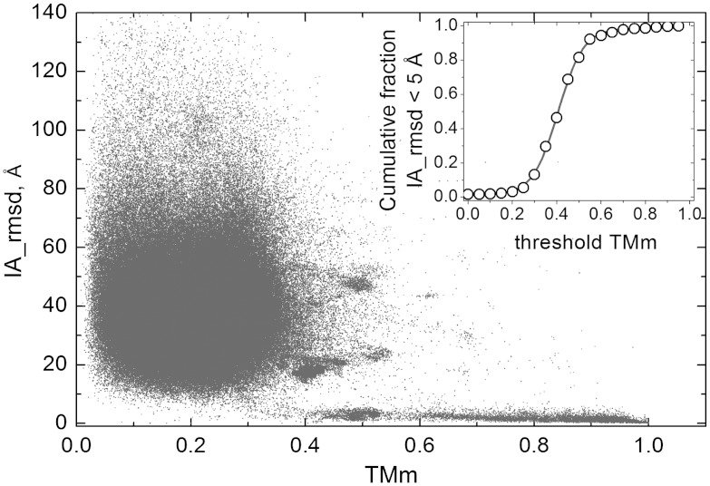
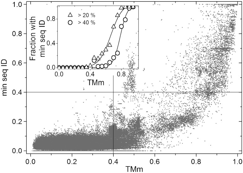
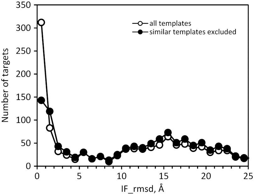
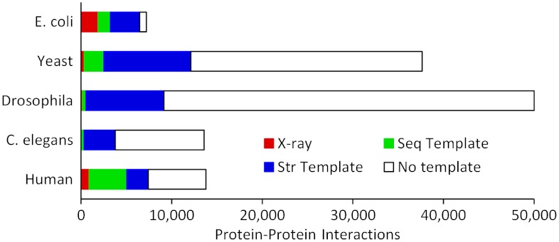
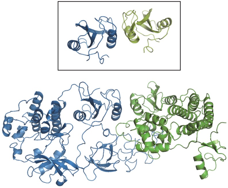
Traditional approaches to protein-protein docking sample the binding modes with no regard to similar experimentally determined structures (templates) of protein-protein complexes. Emerging template-based docking approaches utilize such similar complexes to determine the docking predictions. The docking problem assumes the knowledge of the participating proteins' structures. Thus, it provides the possibility of aligning the structures of the proteins and the template complexes. The progress in the development of template-based docking and the vast experience in template-based modeling of individual proteins show that, generally, such approaches are more reliable than the free modeling. The key aspect of this modeling paradigm is the availability of the templates. The current common perception is that due to the difficulties in experimental structure determination of protein-protein complexes, the pool of docking templates is insignificant, and thus a broad application of template-based docking is possible only at some future time. The results of our large scale, systematic study show that, surprisingly, in spite of the limited number of protein-protein complexes in the Protein Data Bank, docking templates can be found for complexes representing almost all the known protein-protein interactions, provided the components themselves have a known structure or can be homology-built. About one-third of the templates are of good quality when they are compared to experimental structures in test sets extracted from the Protein Data Bank and would be useful starting points in modeling the complexes. This finding dramatically expands our ability to model protein interactions, and has far-reaching implications for the protein docking field in general.


Conflict of interest statement

The authors declare no conflict of interest.

Figures

Fig. 1.
Fig. 1.
Correlation of the structural difference in binding modes with the structure alignment score. The interaction rmsd (IA) is plotted against the lowest of the two components protein TM-scores (TMm), in all-to-all pairwise comparison of 989 complexes extracted from Dockground at 95% sequence identity. In the inset, the cumulative fraction of the complex pairs with IA < 5  and TMm > threshold TMm is plotted as a function of threshold TMm to show the transition that occurs near the threshold TMm = 0.4.
Fig. 2.
Fig. 2.
Correlation of structure and sequence similarity. The lowest of the sequence identity fractions for two aligned components of a complex is plotted against the corresponding TMm, in all-to-all pairwise comparison of 989 complexes extracted from Dockground at 95% sequence identity. The lines separate quadrants below and above a sequence and a structure-based threshold (see Text). In the inset, the fraction of the complex pairs with lowest sequence identity > 20% and 40% is plotted in 0.05 bins of TMm values to show that many pairs with a similar binding mode (TMm > 0.4) have a low sequence identity, below 20 or 40%.
Fig. 3.
Fig. 3.
Benchmarking of template-based docking. The distribution of targets is shown according to the interface rmsd (IF) from the cocrystallized structure. The benchmarking of PDB complexes released in 2009–2011 was based on template structures from 2008 and earlier.
Fig. 4.
Fig. 4.
Structural coverage of PPI. The data for five genomes with the largest number of known PPI shows different categories of complexes structures: (red) complexes with a X-ray structure, (green) complexes with a sequence template, (blue) complexes for which the structure of the monomers is known or can be built by homology; a structural template is found for 99% of the latter complexes.
Fig. 5.
Fig. 5.
A complex built by structural alignment of the homology models of its components and a non-homologous structural template. The modeled complex comprises the human Lyn A tyrosine kinase (sequence GI code 198941, blue ribbon) and the tyrosine kinase binding domain (TKB) of the mouse Cbl (Casitas B-lineage) lymphoma protein (sequence GI code 6680858, green ribbon). The inset shows the homo-dimeric SH2 domain of the human Grb10 protein (PDB entry 1NRV), that was used as a template to build the model (TM-scores 0.90 and 0.69). Sequence identities between the target and the template monomer are 26 and 2.8%. The homology model of Lyn A was based on the human Src tyrosine kinase (chain A of PDB entry 2H8H, sequence identity 52%), that of the mouse Cbl TKB, on the human homolog (chain A of PDB entry 1FBV, sequence identity 94%).

References

    1. Janin J, Bahadur RP, Chakrabarti P. Protein–protein interaction and quaternary structure. Q Rev Biophys. 2008;41:133–180. - PubMed
    1. Kozakov D, Brenke R, Comeau SR, Vajda S. PIPER: An FFT-based protein docking program with pairwise potentials. Proteins. 2006;65:392–406. - PubMed
    1. Mintseris J, et al. Integrating statistical pair potentials into protein complex prediction. Proteins. 2007;69:511–520. - PubMed
    1. de Vries SJ, et al. HADDOCK versus HADDOCK: New features and performance of HADDOCK2.0 on the CAPRI targets. Proteins. 2007;69:726–733. - PubMed
    1. Wollacott AM, Zanghellini A, Murphy P, Baker D. Prediction of structures of multidomain proteins from structures of the individual domains. Protein Sci. 2007;16:165–175. - PMC - PubMed

Publication types

LinkOut - more resources