. 2011 Dec 23:2:96.
doi: 10.3389/fendo.2011.00096. eCollection 2011.

Surgical strategies in childhood craniopharyngioma

Affiliations

Surgical strategies in childhood craniopharyngioma

Jörg Flitsch et al. Front Endocrinol (Lausanne). .

Abstract

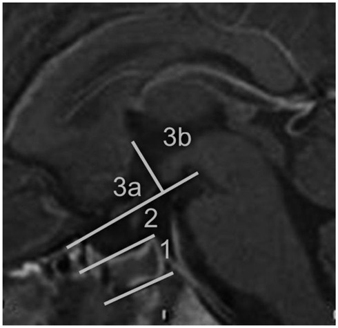
Craniopharyngiomas are biologically benign lesions (WHO Grade 1) of the sellar and suprasellar region, associated with a serious morbidity. About 50% of these tumors become clinically apparent during childhood. Clinical symptoms include headaches, chiasm syndrome, hydrocephalus, pituitary insufficiencies, and obesity. Growth arrest is a typical symptom in children. The treatment of craniopharyngiomas includes surgery as well as radiotherapy. The goal of surgery varies according to the tumor location and extension and may range from complete resection to biopsy. Surgical complications are well known and cause constant evaluation of surgical strategies. Diencephalic obesity is related to surgical manipulation of hypothalamic tissue. Therefore, a classification system for craniopharyngiomas based on preoperative MRI is suggested by the authors. Recurrences are frequent in craniopharyngiomas, even after complete or gross-total resection. Radiotherapy is therefore recommended to patients with incomplete resections. However, the ideal time for radiotherapy after surgery is under discussion. The treatment of craniopharyngiomas requires an interdisciplinary and multimodal approach. Each patient should receive an individually tailored treatment. Surgically, different approaches as well as different degrees of resection can be considered, depending on tumor location and tumor extension.

Keywords: childhood; craniopharyngioma; hypothalamic obesity; pituitary insufficiency; surgery.


Figures

Figure 1
Figure 1
Sagittal MRI of the midline. Suggestion of a classification system of craniopharyngiomas by preoperative MRI criteria. The intra- and suprasellar region can be divided into three sections. Section 1 is limited by the diaphragm sella, section 2 is below the optic chiasm and the mammillary bodies, section 3 is above the chiasm and mammillary bodies, subdivided into an area anterior and posterior of the mammillary bodies. In this particular patient, a transsphenoidal surgery of a Type 1 craniopharyngioma was performed previously, as can be seen by fluid within the sphenoid sinus. Section 1 is usually reached by the transsphenoidal route, whereas section 3a and 3b is mostly reserved for transcranial procedures. Depending on the tumor extension, section 2 can be reached by transcranial as well as transsphenoidal procedures.
Figure 2
Figure 2
Sagittal MRI of a child with an intra- and suprasellar craniopharyngioma reaching the optic nerves (Type 1). Special attention has to be given to the partial pneumatization of the sphenoid sinus, since the thick bone has to be drilled during the access to the sella. This can cause problems in orientation for the surgeon and endanger the carotid arteries, which may be solved by the use of a microdoppler and/or neuronavigation.
Figure 3
Figure 3
(A,B) Pre- and postoperative MRI of a subtotal resection of a craniopharyngioma extending into the third ventricle up to the mammillary bodies in a 4-year-old child. The postoperative MRI shows remaining intrasellar cystic tumor. Because of missing pneumatization of the sphenoid sinus a transcranial, pterional approach was chosen. The pituitary stalk could be preserved in this case. The patient is under close follow-up, in case of tumor growth prior to pneumatization of the sphenoid sinus, radiotherapy will be considered. (C) The intraoperative photo shows the optic nerves up to the chiasm as well as the spread pituitary stalk (arrow) within the cystic tumor (see parallel running small vessels of the pituitary stalk).
Figure 4
Figure 4
(A) Preoperative contrast enhanced MRI showing an infradiaphragmatic tumor rest after a previous transcranial surgery of a large craniopharyngioma. (B) Intraoperative fluoroscopy showing the surgical approach prior to opening of the sellar floor. Notice the catheter connected to a Rickham reservoir reaching the suprasellar region, which was implanted during the previous procedure. (C) Intraoperative microscopic view showing the catheter tip from the sella using a special mirror technique (Lüdecke technique). (D) Early postoperative MRI showing the extent of tumor resection. Up to today, no recurrence is reported.
Figure 5
Figure 5
(A,B) Preoperative solid and cystic lesion with large cyst within the third ventricle, causing headaches. T2 weighed images. (C,D) Postoperative MRI after endoscopic drainage of the cyst and insertion of a catheter into the cyst, connected with a frontal Rickham reservoir. Unfortunately, the infrachiasmatic cysts enlarged in size during follow-up prior to radiotherapy, requiring additional surgery (not shown).

Similar articles

Cited by

References

    1. Abe T., Ludecke D. K. (1997). Recent results of primary transnasal surgery for infradiaphragmatic craniopharyngioma. Neurosurg. Focus 3, E4.10.3171/foc.1997.3.6.5 - DOI - PubMed
    1. Barriger R. B., Chang A., Lo S. S., Timmerman R. D., DesRosiers C., Boaz J. C., Fakiris A. J. (2011). Phosphorus-32 therapy for cystic craniopharyngiomas. Radiother. Oncol. 98, 207–21210.1016/j.radonc.2010.12.001 - DOI - PubMed
    1. Becker G., Kortmann R. D., Skalej M., Bamberg M. (1999). The role of radiotherapy in the treatment of craniopharyngeoma – indications, results, side effects. Front. Radiat. Ther. Oncol. 33, 100–11310.1159/000061227 - DOI - PubMed
    1. Burghaus S., Hölsken A., Buchfelder M., Fahlbusch R., Riederer B. M., Hans V., Blümcke I., Buslei R. (2010). A tumor-specific cellular environment at the brain invasion border of adamantinomatous craniopharyngiomas. Virchows Arch. 456, 287–30010.1007/s00428-009-0873-0 - DOI - PubMed
    1. Cavalheiro S., Di Rocco C., Valenzuela S., Dastoli P. A., Tamburrini G., Massimi L., Nicacio J. M., Faquini I. V., Ierardi D. F., Silva N. S., Pettorini B. L., Toledo S. R. (2010). Craniopharyngiomas: intratumoral chemotherapy with interferon-alpha: a multicenter preliminary study with 60 cases. Neurosurg. Focus 28, E12.10.3171/2009.12.FOCUS09265 - DOI - PubMed

LinkOut - more resources