. 2012 Apr 5:3:67.
doi: 10.3389/fpls.2012.00067. eCollection 2012.

Endoplasmic reticulum-associated degradation of glycoproteins in plants

Affiliations

Endoplasmic reticulum-associated degradation of glycoproteins in plants

Silvia Hüttner et al. Front Plant Sci. .

Abstract

In all eukaryotes the endoplasmic reticulum (ER) has a central role in protein folding and maturation of secretory and membrane proteins. Upon translocation into the ER polypeptides are immediately subjected to folding and modifications involving the formation of disulfide bridges, assembly of subunits to multi-protein complexes, and glycosylation. During these processes incompletely folded, terminally misfolded, and unassembled proteins can accumulate which endanger the cellular homeostasis and subsequently the survival of cells and tissues. Consequently, organisms have developed a quality control system to cope with this problem and remove the unwanted protein load from the ER by a process collectively referred to as ER-associated degradation (ERAD) pathway. Recent studies in Arabidopsis have identified plant ERAD components involved in the degradation of aberrant proteins and evidence was provided for a specific role in abiotic stress tolerance. In this short review we discuss our current knowledge about this important cellular pathway.

Keywords: endoplasmic reticulum; protein degradation; protein glycosylation; protein quality control; ubiquitin–proteasome.


Figures
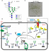
Figure 1
Figure 1
(A) Schematic presentation of the N-linked glycan core structure. The enzymes (GCSI, GCSII, MNS3) involved in the first processing steps are indicated. Upon removal of the terminal α1,2-linked mannose by a so far unknown plant α-mannosidase (MNS?) a free α1,6-mannose residue is exposed at the C-branch of the oligosaccharide, which presumably represents the glycan-specific degradation signal (Hong et al., 2009). (B) A proposed model for the role of N-glycans in ER-quality control and ERAD in plants. Upon transfer of the oligosaccharide (Glc3Man9GlcNAc2) from the lipid-linked precursor to asparagine residues of nascent polypeptides by the oligosaccharyltransferase complex (OST) the two terminal glucose residues are removed by α-glucosidase I and II (GCSI, GCSII). The monoglucosylated N-glycan is a signal for recognition and binding by the lectins calreticulin (CRT – not shown here) and calnexin (CNX). Together with other folding catalysts like members of the protein disulfide isomerase (PDI) family, CNX/CRT promote folding. Properly folded glycoproteins are subsequently released from the CNX/CRT cycle and further processed by α-mannosidases in the Golgi (Liebminger et al., 2009). Incompletely folded proteins can be re-glucosylated by UDP-glucose:glycoprotein glucosyltransferase (UGGT) and are subjected to another round of CNX/CRT-mediated folding. Terminally misfolded glycoproteins are recognized by a poorly described mechanism, which involves the detection of a non-native protein conformation and mannose trimming by MNS proteins to generate a specific glycan code that is then recognized by OS9. Efficient disposal of glycoproteins requires the HRD1–SEL1L/HRD3–OS9 complex, which results in ubiquitylation (Ub) and subsequent degradation in the cytosol. UBC32 might participate in this complex or is part of a plant DOA10-like ERAD complex (not indicated here). As the precise glycan signal for degradation is still unknown, different oligomannosidic structures (Man5GlcNAc2–Man8GlcNAc2) are shown. (C) Knockout of ERAD components results in reduced salt stress tolerance. However, the sel1l, os9, and hrd1a hrd1b mutants display different degrees of sensitivity. HRD1-deficient plants display the most severe phenotype. Seeds were directly germinated on 0.5× MS plates supplemented with 130 mM NaCl and grown for 16 days at 22°C with a 16-h-light photoperiod. The salt-sensitivity of sel1l and os9 has been described previously (Liu et al., ; Hüttner et al., 2012).

Similar articles

Cited by

References

    1. Aebi M., Bernasconi R., Clerc S., Molinari M. (2010). N-glycan structures: recognition and processing in the ER. Trends Biochem. Sci. 35, 74–8210.1016/j.tibs.2009.10.001 - DOI - PubMed
    1. Bernasconi R., Galli C., Calanca V., Nakajima T., Molinari M. (2010). Stringent requirement for HRD1, SEL1L, and OS-9/XTP3-B for disposal of ERAD-LS substrates. J. Cell Biol. 188, 223–23510.1083/jcb.200910042 - DOI - PMC - PubMed
    1. Bernasconi R., Pertel T., Luban J., Molinari M. (2008). A dual task for the Xbp1-responsive OS-9 variants in the mammalian endoplasmic reticulum: inhibiting secretion of misfolded protein conformers and enhancing their disposal. J. Biol. Chem. 283, 16446–1645410.1074/jbc.M802272200 - DOI - PMC - PubMed
    1. Bhamidipati A., Denic V., Quan E. M., Weissman J. S. (2005). Exploration of the topological requirements of ERAD identifies Yos9p as a lectin sensor of misfolded glycoproteins in the ER lumen. Mol. Cell 19, 741–75110.1016/j.molcel.2005.07.027 - DOI - PubMed
    1. Brandizzi F., Hanton S., DaSilva L., Boevink P., Evans D., Oparka K., Denecke J., Hawes C. (2003). ER quality control can lead to retrograde transport from the ER lumen to the cytosol and the nucleoplasm in plants. Plant J. 34, 269–28110.1046/j.1365-313X.2003.01728.x - DOI - PubMed

LinkOut - more resources